CHAPTER 12 Pancreaticojejunostomy (puestow procedure)
Step 1. Surgical anatomy
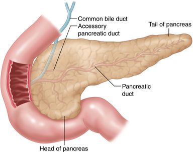
♦ The exocrine pancreas is drained via a main and an accessory duct (Figure 12-1).
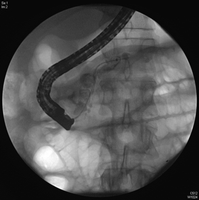
♦ The main pancreatic duct is often affected in chronic pancreatitis. Preoperative ERCP (Endoscopic Retrograde Cholangiopancreatography) is effective in demonstrating ductal dilatation and the presence of stones (Figure 12-2).
Step 2. Preoperative considerations
Patient preparation
♦ To be considered for this procedure, patients must have failed conservative medical therapy and be managed by a multidisciplinary team, including primary care and gastroenterology.
♦ Patients should undergo appropriate pre-operative imaging, including a high-resolution computed tomography (CT) and ERCP, or Magnetic Resonance Cholangiopancreatography (MRCP) (Figure 12-2).
♦ It is critical to assess pancreatic duct dilatation and ductal strictures, and to rule out the presence of a potentially malignant mass.
♦ The pancreatic duct should be dilated to greater than 7 mm in diameter.
♦ Preoperative placement of a pancreatic stent for temporary pain relief may be appropriate. This is not a contraindication to surgery.
♦ Many patients will have nutritional deficiencies and diabetes. Appropriate treatment should be initiated for these comorbidities before these patients undergo surgery.
♦ Informed consent should be obtained. It is important to set expectations appropriately. The patients should not expect cure, but a 70% to 80% improvement in pain.
Room setup and patient positioning
♦ The patient is placed in split-leg position, and foot plates are used to prevent sliding.
♦ Monitors are placed above the patient’s head.
♦ The operative surgeon usually stands on the patient’s right side with the assistant at the patient’s left side. The surgeon may find it helpful to move to the position between the legs during certain parts of the procedure (e.g., dissection of the pancreas medially).
Step 3. Operative steps
Access and port placement
♦ A Veress needle is introduced along the left subcostal margin, in the midclavicular line, and a pneumoperitoneum is created to a pressure of 15 mmHg using carbon dioxide.
♦ A 12-mm camera port is placed 3 cm to the left of midline and approximately 8 cm above the umbilicus. A 10-mm, 30-degree laparoscope is then introduced into the peritoneal space.
♦ Under direct vision, a 12-mm working port is placed 4 cm to the right of the midline and 6 to 8 cm above the umbilicus. A 5-mm working port is placed in the right upper quadrant. An assistant 5-mm port is placed in the left upper quadrant.
♦ A 5-mm incision is made in the subxiphoid space, and the Nathanson retractor is introduced to elevate the left lobe of the liver.
Stay updated, free articles. Join our Telegram channel

Full access? Get Clinical Tree


