Light Microscopy
With light microscopy, the pattern of glomerular inflammation is often described as mesangial, endocapillary, and/or extracapillary (crescentic); focal or diffuse; and segmental or global.
Mesangial/Endocapillary/Extracapillary (Crescentic). Inflammation can occur in several different regions of the glomerulus, leading to characteristic structural changes.
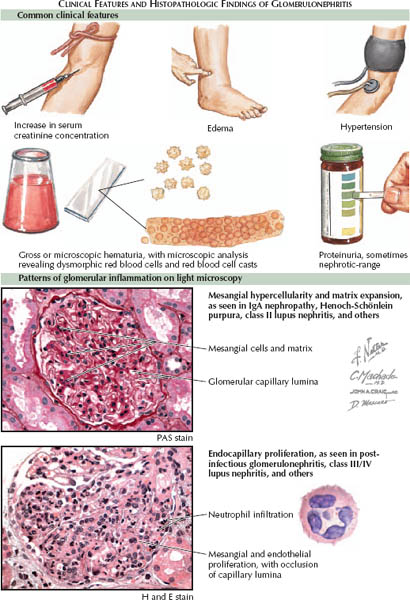
Mesangial involvement can manifest as mesangial hypercellularity (defined as more than three mesangial cells per mesangial area) and/or mesangial matrix expansion. These structural changes are often associated with microscopic hematuria and/or mild proteinuria, with preservation of normal filtration function. Common causes include IgA nephropathy and class II lupus nephritis.
Endocapillary involvement can manifest as occlusion of glomerular capillaries by endothelial and mesangial cell proliferation, as well as by leukocyte infiltration. These changes are often associated with hematuria, proteinuria, and reduction of filtration function. Common causes include postinfectious GN and class III or IV lupus nephritis.
< div class='tao-gold-member'>


