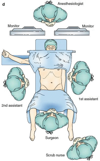
Fig. 6.1
(a) Barium swallow shows 3-cm sliding hiatal hernia. (b) High-resolution esophageal manometry shows hypotensive lower esophageal sphincter (LES) and normal esophageal peristalsis. (c) On ambulatory pH monitoring, two sensors show distal and proximal reflux. (d) Arrangement of the operating room for laparoscopic total fundoplication
6.2 Laparoscopic Total Fundoplication (360°)
6.2.1 Positioning of the Patient and Placement of Trocars
The patient lies supine on the operating table in low lithotomy position with the lower extremities extended on stirrups with knees flexed 20–30°
A bean bag is inflated to avoid sliding of the patient as a consequence of the steep reverse Trendelenburg position used during the entire procedure.
Pneumatic compression stockings are used to reduce the risk of deep venous thrombosis that is associated with both increased abdominal pressure secondary to pneumoperitoneum and the decreased venous return secondary to the steep reverse Trendelenburg position.
An orogastric tube is placed to decompress the stomach, and it is removed at the end of the procedure.
The surgeon stands between the patient’s legs, while the first and second assistant stand on the right and left sides of the operating table (Fig. 6.1d).
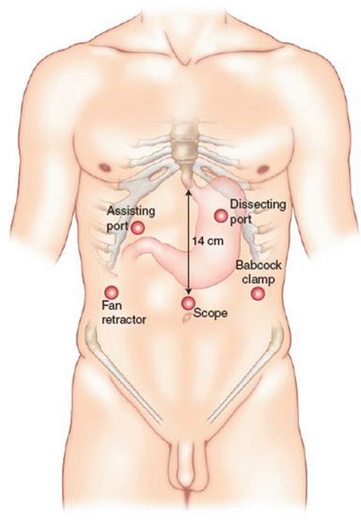
Fig. 6.2
Placement of trocars for laparoscopic fundoplication
6.2.2 Operative Procedure
Step 1
The gastrohepatic ligament is divided, beginning above the caudate lobe of the liver, where the ligament is usually very thin, until the right crus of the diaphragm is identified. An accessory left hepatic artery originating from the left gastric artery is frequently present in the gastrohepatic ligament. If it limits the exposure, it may be divided with no clinical consequences.
The right crus is separated from the right side of the esophagus by blunt dissection, the posterior vagus nerve is identified, and the right crus is dissected inferiorly toward the junction with the left crus. The use of a bipolar instrument allows right crus dissection to be performed more safely than with electrocautery, with a reduced risk of injury to the posterior vagus nerve due to the lateral spread of the electrical current from a monopolar instrument.
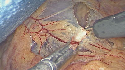
Fig. 6.3
Division of the gastrohepatic ligament
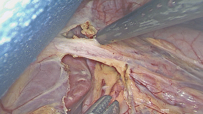
Fig. 6.4
Division of the gastrohepatic ligament
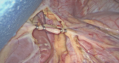
Fig. 6.5
The right crus of the diaphragm is identified
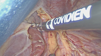
Fig. 6.6
Dissection of the right crus
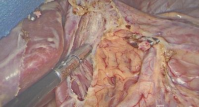
Fig. 6.7
Dissection of the right crus
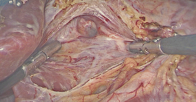
Fig. 6.8
Dissection of the right crus
Step 2
The peritoneum and the phrenoesophageal membrane above the esophagus are transected with the electrocautery, with identification of the anterior vagus nerve. To avoid injury to the anterior vagus nerve or the esophageal wall, the nerve should be left attached to the esophageal wall, and the peritoneum and the phrenoesophageal membrane should be lifted from the wall by blunt dissection before they are divided.
The left crus of the diaphragm is dissected bluntly downward toward the junction with the right crus.
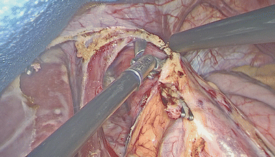
Fig. 6.9
Transection of the peritoneum and the phrenoesophageal membrane above the esophagus
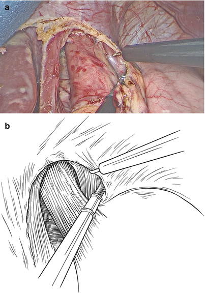
Fig. 6.10
(a) and (b), Dissection of the left crus of the diaphragm
The short gastric vessels are taken down all the way to the left pillar of the crus, starting at the level of the middle portion of the gastric body and continuing upward until the most proximal short gastric vessel is divided.
Possible complications during this step of the procedure are bleeding, either from the short gastric vessels or from the spleen, and damage to the gastric wall.
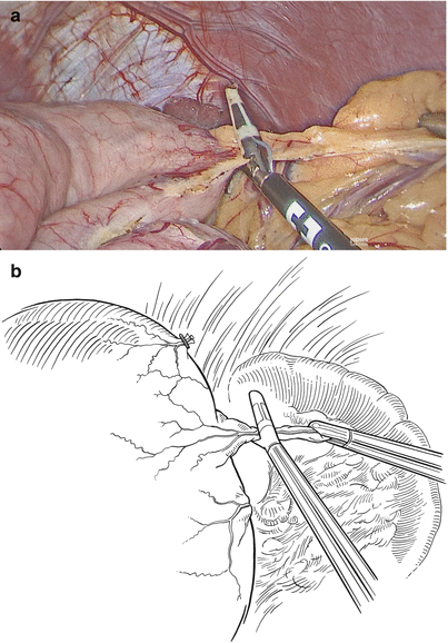
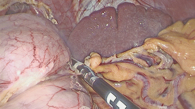
Fig. 6.11
(a) and (b), Taking down the short gastric vessels
Fig. 6.12
Taking down the short gastric vessels
Step 4
A Babcock clamp is applied at the level of the esophagogastric junction and the esophagus is retracted upward.
Related posts:


Stay updated, free articles. Join our Telegram channel

Full access? Get Clinical Tree


