Fig. 49.1
Holy plane of TME dissection
Partial Mesorectal Excision (High Anterior Resection and Mesorectal Transection)
Upper rectal tumors (12–15 cm from the anal verge) may not require TME and mesorectal transection 5 cm distal to the lower edge of the tumor is oncologically acceptable and decreases the risk of autonomic nerve injury and allows a higher anastomosis which is less likely to leak than the much lower anastomosis after TME. Generally a neoplasm within 12 cm of the anal verge requires a TME.
Extended Resections
In order to achieve an R0 resection at times it is necessary to excise en bloc any adherent or involved viscera such as a loop of small bowel, uterus, ovary or the ureter.
Anastomosis
Moran Triple Stapling Technique
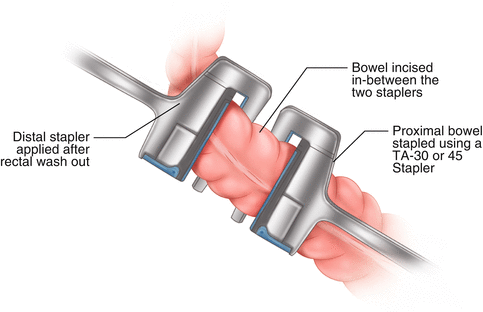
Once dissection has been completed well beyond the tumor a linear stapler (TA 30 or 45) is applied well below the tumor and fired but left in place to seal the muscle tube. The ano-rectal tube is then washed with a cytocidal solution such as water and Proflavine or Povidone-Iodine. A second TA 30 or 45 stapler is then placed distal to the first across the washed bowel and fired (see Fig. 49.2). The bowel is then divided with a scalpel between the two staplers. This reduces the risk of incorporating any spilled intraluminal neoplastic cells into the anastomotic staple line [5, 6]. Downward spread of rectal cancer along the muscle tube is usually not an issue and 2 cm distal clear margin beyond the lower edge of palpable cancer is adequate, though a 1 cm margin and the doughnut is acceptable in order to perform a sphincter saving operation. The TME specimen should be assessed for quality and should be a bilobed fatty pedicle package with no tears with a clear naked eye CRM. The cut end within the proximal stapler is inspected for clearance and if there is any doubt the staples should be opened and mucosa inspected. The distal stapler should only be taken off after clearance has been confirmed as this allows for possible application of a third stapler below the in situ distal one if the margin is not clear. The pelvic cavity should be washed and inspected for any bleeding and hemostasis secured.


Fig. 49.2
Moran’s triple stapling technique
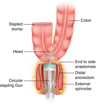
When TME has been performed and a coloanal anastomosis is envisaged a neorectal reservoir has a better functional outcome then a straight end to end coloanal anastomosis. We favor an end to side technique with the side of the distal colon anastomosed to the end of the ano rectum using a circular stapler (28–31 mm head) (see Fig. 49.3). The head is detached and inserted into the lumen of the colon, spike first, after excising part of the staple line on the distal colon. The spike is pushed through the wall on the anti-mesenteric border, midway between the taenia coli 4–5 cm from the distal colonic end. The staple line defect is closed and the staple line is inverted with interrupted sutures.


Fig. 49.3
Reconstruction end to side stapled anastomosis
The anorectal remnant is gently palpated. The circular staple gun is then gently inserted transanally taking care not to disrupt the transverse staple line. On occasions bimanual placement by the abdominal surgeon is required. Adequate visualization and retraction with a St Marks retractor is vital at this stage. Once happy with this position the spike on the gun is opened ideally just behind the transverse staple line. The head of the gun is brought down and engaged with the gun, the colon is checked for orientation to ensure no twists that would compromise its blood supply. The gun is closed slowly and completely until the green marker is visualized. The closed position is maintained for a minute before firing. The gun is fired and then opened again until a click is heard. The gun is slowly withdrawn without twisting and the doughnuts are checked for integrity and the distal doughnut should be sent for histology if there are concerns about clearance in a low rectal cancer. The anastomosis is tested for air-leaks by filling the pelvis with water, occluding the distal colon with a soft clamp or fingers and insufflating air via the anus. A gross leak may be sutured and this may be possible transanally in very low anastomosis. Two abdominal drains (low suction closed type) are placed in the pelvis and a defunctioning stoma is created.
Defunctioning Stoma
Our practice has been to perform a defunctioning loop ileostomy for anterior resection with TME and coloanal anastomosis. A recent randomized trial reported 28 % leak rate in patients without a defunctioning stoma compared with 10 % in those with a stoma [7]. Loop stoma reduces the consequences of an anastomotic leak and the need for emergency surgery. The defunctioning stoma is reversed at 8–12 weeks postop after a contrast enema confirms no leaks from the anastomotic site.
Abdomino Perineal Excision (APE)
Historically APE was the first attempted major curative rectal resectional operation as popularized by Ernest Miles and Charles Mayo [8]. An APE is necessary if it is not possible to preserve the sphincter complex or the sphincters are of poor functional quality. Traditionally APE has been performed in the Lithotomy-Trendelenburg position with synchronous abdominal and perineal dissection. Recent reports suggest worse outcome in patients having APE compared with anterior resection [9]. This is primarily because of specimen perforation, ‘coning’ or ‘waisting’ at the level of the levators. Availability of high resolution MRI has helped to determine preoperatively if the levators or sphincters are involved [10] and it is now possible to tailor the operation accordingly to obtain an R0 resection. Hence instead of a standard APE (SAPE) an intersphincteric APE (ISAPE) or extra levator APE (ELAPE) may be more appropriate [11].
Stay updated, free articles. Join our Telegram channel

Full access? Get Clinical Tree


