Open Lateral Internal Sphincterotomy
S. Alva
Bertram Chinn
Indications
Chronic fissures that fail to respond to nonoperative therapy
Acute fissures with severe pain
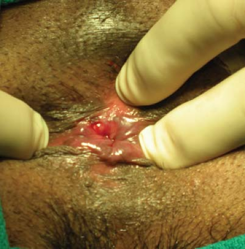
Anal fissure is a common disorder that results in bleeding and painful defecation. It is frequently related to the passage of a hard or a constipated bowel movement. A linear tear is noted in the anoderm and is frequently seen with gentle retraction of the buttocks (Fig. 17.1). Approximately 80–90% of fissures are posterior in location with the remainder in the anterior quadrant (1). Occasionally, fissures may be seen in both regions.
Many fissures heal by increasing dietary fiber to soften and bulk the stool, using an emollient suppository and warm sitz baths (2). Over the last decade, topical nifedipine or nitroglycerin ointments and the injection of Botulinum toxin A into the internal sphincter have improved fissure healing by reducing sphincter spasms (3,4,5).
In chronic conditions, irritation, itching, mucous, and discomfort may be more evident than pain or bleeding. Scarring at the base of the fissure, rolled and indurated edges, a sentinel skin tag, and/or a hypertrophic papilla suggest that the fissure will not heal without surgery (6). A posterior sphincterotomy was once recommended but due to a resultant “keyhole” deformity, Eisenhammer advocated a lateral internal sphincterotomy (LIS) for surgical treatment of fissures (2).
An LIS may also be necessary when pain from an acute fissure is overwhelming.
Contraindications
Diminished sphincter integrity
Inflammatory bowel disease
Infections (tuberculosis and syphilis)
Leukemia and HIV
When considering an LIS, individuals with diminished sphincter tone or incontinence should be evaluated for their candidacy for alternative therapies. An atypical fissure may suggest the presence of other diseases. Fissures in the lateral quadrants should raise the concern of inflammatory bowel disease, specifically Crohn’s disease, tuberculosis, syphilis, leukemia, or HIV. In these situations, treating the underlying disease is recommended instead of performing a sphincterotomy.
Routine preoperative evaluation and planning that include a history and physical examination with meticulous attention to the anorectal region should be performed. Although a phosphate enema is recommended prior to surgery, discomfort frequently precludes its use. If diminished sphincter tone is suspected or if the patient has had prior anorectal surgery, preoperative anorectal physiology testing may be helpful.
Positioning
Prone jackknife position
The patient is placed in a prone jackknife position. This position allows the surgical team full access to the operative field. Retracting 3-in. silk tape that has been placed on the buttocks and securing it to the sides of the operating table provides exposure. Lithotomy and a left lateral or modified Sims’ positions can also be used.
Anesthesia
Monitored anesthesia care (MAC)
A 0.25% bupivacaine and 1:200,000 epinephrine
Although LIS can be performed under general or regional anesthesia, our preference is MAC and a local block. After initially attaining adequate sedation and comfort under MAC, a local block with bupivacaine and epinephrine is used. This block provides analgesia and allows relaxation of the sphincter to facilitate surgery. The vasoconstrictive effects of epinephrine will decrease vascularity during the surgery and increase the period of postoperative analgesia.
Initially, 10 ml of the local anesthetic is injected circumferentially into the perianal skin and the subcutaneous tissue with a 1.5 in. × 25 gauge needle. A circumferential deeper injection into the sphincter is then performed. Typically, a total of 20–30 ml of the local anesthetic is needed to complete the operation.
Whatever anesthesia is selected, patients may benefit from the use of a local block that contains epinephrine in addition to the analgesic/anesthetic. Procedures performed under general anesthesia still benefit from the additional sphincter relaxation and hemostasis attained with the bupivacaine and epinephrine. The vasoconstrictive effects of epinephrine are also helpful in offsetting the vasodilatory effects of a spinal anesthetic.
Technique
Confirmation of a fissure and hypertonic/spastic sphincter
Insertion of a 35-mm Hill-Ferguson retractor with identification of the internal sphincter and intersphincteric groove
Incision of the perianal skin overlying the intersphincteric groove
Isolation of the internal sphincter and division under direct vision
Fissure debridement and excision of a sentinel tag and hypertrophic papilla
Closure of the sphincterotomy site with interrupted absorbable sutures
After appropriate positioning and anesthesia are attained, the presence of a fissure is confirmed with circumferential examination of the anal canal using a Hirschmann anoscope. A 35-mm Hill-Ferguson retractor provides a consistent measure of the diameter of the anal canal. Resistance during insertion of this retractor confirms the presence of a hypertonic/spastic sphincter and a taught, bank-like internal sphincter is seen (Fig. 17.2). Failure to identify a hypertonic sphincter should prompt further evaluation before a sphincterotomy is performed.
Selection of either the left or the right lateral quadrant for the sphincterotomy is contingent upon where the internal sphincter is best noted and whether hemorrhoidal tissue would interfere with the operative field. An incision is made at the intersphincteric
groove and extended 1.5–2.0 cm distally to the perianal skin. A fine, curved hemostat is used to mobilize the anoderm off the internal sphincter up to the level of the dentate line (Fig. 17.3). Caution is used to prevent violation of the anoderm since nonhealing of this may result in a fistula. The intersphincteric plane is accessed and the internal sphincter is isolated with the hemostat up to the dentate (Fig. 17.4). Electrocautery may be used to control small points of bleeding.
groove and extended 1.5–2.0 cm distally to the perianal skin. A fine, curved hemostat is used to mobilize the anoderm off the internal sphincter up to the level of the dentate line (Fig. 17.3). Caution is used to prevent violation of the anoderm since nonhealing of this may result in a fistula. The intersphincteric plane is accessed and the internal sphincter is isolated with the hemostat up to the dentate (Fig. 17.4). Electrocautery may be used to control small points of bleeding.
Stay updated, free articles. Join our Telegram channel

Full access? Get Clinical Tree


