This article presents an overview of the techniques and indications for office-based ultrasound for the clinical urologist. Discussion includes renal, bladder, scrotal, penile Doppler, and prostate ultrasonography and a review of the pertinent literature and images for each anatomic location.
Key points
- •
Renal ultrasonography allows assessment of a dilated upper urinary tract particularly in pediatric patients, assessment of flank pain during pregnancy, and evaluation of hematuria in patients who are not candidates for intravenous pyelography, contrast computed tomography, or magnetic resonance imaging.
- •
Bladder ultrasonography allows assessment of postvoid residual in male patients with benign prostatic hyperplasia, particularly during the initial workup.
- •
Scrotal ultrasonography allows assessment of a scrotal or testicular mass or swelling, assessment of acute scrotal pain, and assessment of male infertility.
- •
Penile Doppler ultrasonography allows assessment of the cavernosal arteries and their spectral waveform evolution following intracavernosal injection of a pharmacostimulant in patients with erectile dysfunction.
- •
Transrectal ultrasound of the prostate is the most common modality for imaging the prostate during biopsy; new modalities include color Dopper prostate ultrasonography, three-dimensional ultrasonography, and elastography of the prostate.
Introduction
Ultrasonography provides the busy office urologist with a minimally invasive, low-risk imaging modality that is easily accessible in the clinic setting. The basic concepts behind ultrasound imaging involve using a frequency (number of sound waves per second, measured in hertz [Hz]) too high for the human ear to hear. Ultrasound waves are generated by a transducer, which is housed in an ultrasound probe that is shaped for the desired application. These waves are then transmitted to the tissue of interest and waves that reflect (or echo) after bouncing off the tissue of interest are incorporated by a receiving element in the transducer. Through a process called acoustic-electric conversion, the transducer transforms the sound energy into electrical energy, which is processed by the ultrasound console computer to generate white pixels corresponding to returning signals displayed on a black background. This article reviews the basic applications of ultrasound imaging in the office setting, including renal, bladder, scrotal, penile Doppler, and prostate ultrasonography.
Renal ultrasonography
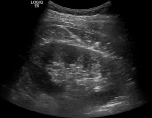
Given a urologist’s knowledge of the anatomy of the kidney and retroperitoneum, performing a focused retroperitoneal ultrasound in the office setting can be useful for specific clinical indications. The ultrasound probe and transducer for renal ultrasonography is a 3.5- to 5.0-MHz curved probe; a 6- to 10-MHz transducer may be used for pediatric patients. The patient is placed in the supine position and scanning begins in the midclavicular line for the kidney of interest. In the sagittal plane, the probe is moved laterally until a midsagittal view of the kidney is obtained; when analyzing the image, the upper pole of the kidney is located on the left side of the monitor ( Fig. 1 ). Imaging of the kidney in the transverse plane is possible by rotating the probe 90° counterclockwise and the kidney is scanned from upper to lower pole. Important office-based indications for renal ultrasound include follow-up of hydronephrosis on prenatal ultrasound, assessment of a dilated upper urinary tract particularly in pediatric patients, assessment of flank pain and monitoring ureteral stent position during pregnancy, and evaluation of hematuria in patients who are not candidates for intravenous pyelography (IVP), contrast computed tomography (CT), or magnetic resonance imaging (MRI).
Pediatric patients requiring renal imaging represent an important subset of patients for which renal ultrasonography is used. Postoperative follow-up of pediatric patients following ureteroscopic treatment of lithiasis is effective with ultrasound. Resorlu and colleagues found negative and positive predictive values of 97.7% and 100%, respectively, for detecting hydronephrosis at 3 months postoperatively for ureteroscopic manipulation of lithiasis. Similar to postureteroscopy upper tract surveillance, ultrasound is important for surveillance of postpyeloplasty patients and can be used to identify patients who may require a mercaptoacetyltriglycine-3 scan in the setting of postoperative deteriorating renal function. Recently, 3-dimensional (3D) ultrasonography has been reported when evaluating pediatric patients. 3D ultrasonography improves visualization of complex anatomy and pathologic condition in any plane and allows evaluation of a dilated collecting system with similar specificity to IVP and MR urography.
When assessing a pregnant patient with renal colic, determining whether this is secondary to physiologic hydronephrosis or lithiasis may be challenging. In experienced hands, ultrasonography has a sensitivity of greater than 95% for diagnosis of nephrolithiasis. A review of 300 pregnant patients presenting with renal colic by Andreoiu and MacMahon found that the accuracy of ultrasonography for predicting a calculus improved from 56.2% to 71.9% when features of obstruction were present, such as the absence of a ureteric jet and an elevated resistive index. Although recent studies have suggested that low-dose CT scan may be safe and improve the efficacy of lithiasis diagnosis in pregnant patients, ultrasound remains an important and safe modality for diagnosing nephrolithiasis and monitoring progression of stone passage.
As part of the evaluation for hematuria, the upper urinary tract has historically been evaluated with an IVP and more recently a contrast CT or MRI of the abdomen and pelvis. In patients with an elevated creatinine, a contrasted study risks further worsening of kidney function providing an opportunity for ultrasonography to evaluate the upper urinary tract. In this subset of patients, ultrasonography offers the ability to detect renal masses and cysts ( Fig. 2 ). Mucksavage and colleagues analyzed 116 patients who underwent ultrasound prior to imaging before definitive therapy. Patients also received an MRI or CT scan and they found that the size differences between CT and MRI compared with ultrasound was less than 3.5% and ultrasound correlated well with both MRI and CT (both P <.001). However, diagnosis of upper tract transitional cell carcinoma with ultrasonography is difficult to differentiate from other causes of filling defects of the renal collecting system, such as fungus balls or sloughed papillae. Ultrasound should not replace an IVP or contrasted CT or MRI for the evaluation of the upper urinary tract in the setting of hematuria; however, in select patients with poor kidney function it may be considered a safe alternative.
Bladder ultrasonography
Bladder ultrasonography is an important tool for the office urologist, allowing evaluation of the lower urinary tract and prostate in men and the bladder in women. A curved probe set at 3.5 to 5 MHz is used with the patient in the supine position. The bladder should be viewed in the transverse and sagittal planes, angling the probe beneath the pubic bone for optimal bladder assessment ( Fig. 3 ). Arguably the most common utilization of bladder ultrasonography in the office setting is assessment of postvoid residual (PVR) in male patients with benign prostatic hyperplasia (BPH). Although the utility of PVR as an objective measure for BPH treatment efficacy and as an indicator for surgical treatment have been inconclusive, it is generally accepted that PVR volume be included in the initial assessment of a patient with BPH and during monitoring of patients undergoing conservative treatment regimens. Additional indications for bladder ultrasonography in the office setting may include evaluation of bladder wall configuration and thickness, detection of ureteroceles, assessment for ureteral obstruction, detection of perivesical fluid collections, evaluation of clot retention, confirmation of catheter position, and guidance of suprapubic tube placement.
Scrotal ultrasonography
Scrotal ultrasonography provides the urologist with high-quality images when evaluating patients with acute scrotal pain, a palpable scrotal mass, or an enlarged scrotum on physical examination. Ultrasonography is typically performed with high frequency (6–12 MHz) and a 4- or 7.5-cm linear array transducer. Patients should be in the supine position with the scrotum supported by a towel. The penis should be out of the way and adequate conducting gel should be used to circumvent artifact that may result secondary to scrotal hair. Transverse and sagittal images should be obtained paying particular attention to each testis, epididymis, and spermatic cord ( Fig. 4 A). The 2 testes should be compared for echogenicity because certain infiltrative processes (eg, leukemic testicular involvement) may result in subtle changes that are only noticeable when a bilateral comparison is used. Furthermore, Doppler ultrasonography should be used to determine blood flow to each testis particularly when clinical suspicion is concerning for testicular torsion. Indications for in-office scrotal ultrasonography include assessment of a scrotal or testicular mass or swelling, assessment of acute scrotal pain, and assessment of male infertility.
Ultrasonography of the scrotum in the setting of a swollen scrotum or a palpable testis mass allows for delineation between extratesticular (eg, hydrocele) (see Fig. 4 B) and intratesticular (eg, tumor, infection) causes (see Fig. 4 C). For medicolegal reasons, evaluation for malignancy should also include a review of the images by a radiologist. Micallef and colleagues retrospectively analyzed 256 patients for scrotal swelling and reported that 75% of cases involved extratesticular causes (most commonly hydrocele) and 25% involved intratesticular causes, most commonly secondary to infection (50%) and tumor (21%). Color Doppler ultrasonography of a testicular nodule is characterized by hypervascularity with irregular branching patterns. Leydig cell tumors often have a unique ultrasound finding of hypoechoic nodules with peripheral hypervascularity and no internal flow. When a hydrocele is suspected on physical examination (no nodules, nonpalpable testis, scrotal swelling), ultrasonography may be used to confirm the diagnosis, demonstrating an increased fluid volume around the testis and in increased pulsatility index.
The assessment of the acute scrotum relies on an accurate history and physical examination and, if needed, ultrasonography. Clinical suspicion for testicular torsion obviating scrotal exploration should not be delayed because of ultrasonographic confirmation. The hallmark of testicular torsion on ultrasonography is the absence of intratesticular blood flow (see Fig. 4 D); paratesticular blood flow secondary to collateral circulation may appear within hours of testicular torsion. Although the paradigm at many tertiary centers is to explore an acute scrotum regardless of ultrasonographic findings for fear of overlooking testicular torsion, a recent report by Altinkilic and colleagues suggests that normal intratesticular perfusion using color-coded duplex sonography (CCDS) obviates scrotal exploration. The authors assessed the diagnostic value of CCDS in 236 patients (median age 13 years) with clinical suspicion of testicular torsion who subsequently underwent exploration whereby the surgeon was blinded to the ultrasound findings. Testicular torsion was the most common cause of acute scrotum (50.4%), followed by torsion of the testicular appendage (34.8%) and epididymo-orchitis (7.6%). The reported sensitivity, specificity, and positive and negative predictive values of detecting testicular torsion with CCDS were 100%, 75%, 80%, and 100%, respectively.
Scrotal ultrasonography may elucidate pathologic conditions affecting male fertility, including a varicocele ( Fig. 5 ), which is the most common abnormality in infertile men with abnormal semen analysis. The degree of testicular size difference that may warrant surgical repair in infertile men has not been defined; however, a 20% size discrepancy has been proposed as a threshold for intervention. Although varicocele is often diagnosed on physical examination of the testis and spermatic cord, accuracy of palpation may be subjective. Pierik and colleagues analyzed 1372 infertile men who were assessed with color Doppler ultrasonography and found a scrotal abnormality in 38% of men. Varicocele (29.7%) was the most common abnormality, followed by epididymal cyst (7.6%), hydrocele (3.2%), testicular microlithiasis (0.9%), testicular cyst (0.7%), and testicular tumor (0.5%). Interestingly, 67% and 60% of overall sonographic findings and varicoceles, respectively, were not evident on physical examination.
Penile Doppler ultrasonography
Penile Doppler ultrasonography allows assessment of the cavernosal arteries and their spectral waveform evolution following intracavernosal injection of a pharmacostimulant in patients with erectile dysfunction (ED). Depending on the institution, this may be performed in the office setting or by an ultrasound technician in a radiology suite, with subsequent interpretation of results by an urologist. In the authors’ experience, the patient is brought to the radiology suite and a 1 mL injection of an alprostadil-papaverine-phentolamine mix using a 25-gauge needle is performed. The injection is made in either corpora cavernosa at the 3- or 9-o’clock position at the base of the penis. Following injection, a linear transducer set at 7 MHz or higher is used; scanning is typically performed on the ventral surface of the penis; however, the dorsal and lateral surfaces may be used if necessary. Images and waveforms are obtained in both the longitudinal and the transverse planes from the base of the penis to the tip of the glans. Attention to the cavernosal arteries, integrity of the corpus cavernosa, and identification of plaques, tumors, and calcifications is important. Color Doppler ultrasonography is necessary for measuring the systolic velocity in the cavernosal arteries and for determining the preinjection ( Fig. 6 A) and postinjection diameters (see Fig. 6 B) of the arteries. The thresholds for normal systolic and diastolic velocities of blood flow relating to the cavernosal arteries following injection of a pharmacostimulant are generally regarded as a peak systolic velocity (PSV) of greater than 25 to 35 cm/s and an end diastolic velocity (EDV) of less than 5 to 7 cm/s (see Fig. 6 C).


