Nutritional Disorders
Severe nutritional deficiency can produce marked small intestinal abnormalities. The classic example is kwashiorkor. Malnutrition of this severity seldom occurs in developed countries, but less severe forms may be recognized after gastric surgery or in patients with chronic debilitating diseases. Malnutrition results from a lack of a suitable diet, faulty metabolism, and inadequate absorption of dietary constituents.
Kwashiorkor
Kwashiorkor is one of the most common pediatric illnesses in underdeveloped countries. It affects many African tribes,
particularly in eastern and southern Africa, causing high morbidity and mortality. Nutrient malabsorption exacerbates the protein–caloric malnutrition, further damaging the gut, impairing immune competence, and increasing the risk of infections (Fig. 6.211). Diarrheal illnesses are common.
particularly in eastern and southern Africa, causing high morbidity and mortality. Nutrient malabsorption exacerbates the protein–caloric malnutrition, further damaging the gut, impairing immune competence, and increasing the risk of infections (Fig. 6.211). Diarrheal illnesses are common.
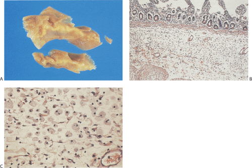 FIG. 6.210. Small intestinal xanthoma. A: Gross photograph showing the prominent submucosal expansion by a clear gelatinouslike fluid collection. B: Histologic features demonstrating the presence of a diffuse histiocytic infiltrate loosely arranged in the submucosa. C: Higher magnification demonstrating the histiocytic cells widely separated from one another. |
The small intestinal abnormalities may be indistinguishable from untreated celiac disease. Histologically, the mucosa appears flattened. The crypts appear coiled with a relative increase in length and a lower than normal mitotic index. The crypts are not uniformly altered. They may be atrophic in some areas, whereas in others they appear normal or hyperplastic (581). Epithelial cell height decreases and the nuclei are arranged irregularly. The lamina propria becomes infiltrated with mononuclear cells and the basement membrane thickens (582). The presence of neutrophilic infiltrates suggests a coexisting infection. The patients respond to a
normal diet with concomitant improvement in the appearance of the intestinal mucosa.
normal diet with concomitant improvement in the appearance of the intestinal mucosa.
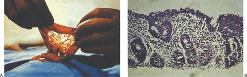 FIG. 6.211. Kwashiorkor. A: Hypoplastic bowel in kwashiorkor visualized at the time of surgery. B: Severe villous atrophy. |
Vitamin E Deficiency and Brown Bowel Syndrome
Patients with vitamin E deficiency develop eosinophilic enteritis and brown bowel syndrome. Vitamin E deficiency occurs alone or it complicates other diseases (583,584). Brown bowel syndrome is characterized by lipofuscin deposits in the muscularis propria. Patients range in age from the 20s to the late 70s, with an average age of 51 years. They present with epigastric pain, mild diarrhea, and chronic malabsorption.
Related posts:


Stay updated, free articles. Join our Telegram channel

Full access? Get Clinical Tree


