and Christopher Isles2
(1)
Institute of Cardiovascular and Medical Sciences, University of Glasgow, Glasgow, UK
(2)
Dumfries and Galloway Royal Infirmary, Dumfries, UK
Disorders of mineral metabolism (aka chronic kidney disease mineral bone disorder (CKD-MBD) or renal bone disease) occur frequently in patients with CKD. Understanding what happens to calcium and phosphate when the kidneys fail is key.
Q1 List the disorders of mineral metabolism that may occur in chronic kidney disease
There are five. Secondary hyperparathyroidism is the classic form of renal bone disease. If left untreated this may progress to tertiary hyperparathyroidism (see later). More than one disorder of mineral metabolism may be present at any one time (Fig. 28.1).
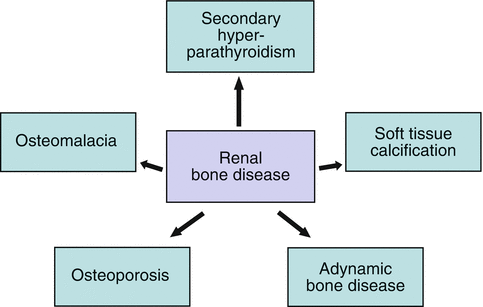

Fig. 28.1
Disorders of mineral metabolism occurring in renal disease
Q2 Why do patients with CKD develop disorders of mineral metabolism?
Two things happen when GFR falls below 30 ml/min. Serum phosphate rises – simply because the kidneys cannot excrete it – and serum calcium begins to fall because the failing kidney cannot activate vitamin D. Vitamin D is normally responsible for increasing the absorption of calcium from the gut. Failure to activate vitamin D therefore leads to hypocalcaemia. Hyperphosphataemia also leads to hypocalcaemia, at least partly because of failure to activate vitamin D (Fig. 28.2).
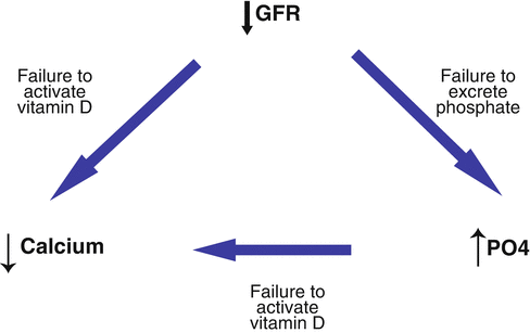

Fig. 28.2
Mechanism through which renal failure causes hypocalcaemia and hyperphosphataemia
Q3 What do you understand by the term secondary hyperparathyroidism?
Secondary hyperparathyroidism occurs when the parathyroid gland is stimulated to secrete PTH by low serum calcium and high serum phosphate. PTH does its best to correct hyperphosphataemia by reducing phosphate reabsorption in the kidney. Its main action, however, is to raise serum calcium. It does this in three ways:
1.
By direct action on the kidney increasing calcium reabsorption.
2.
Indirectly, by activating vitamin D which then increases absorption of calcium from the gut.
3.
By stimulating the osteoclasts in bone. Osteoclasts break down bone releasing calcium into the blood stream.
The ability of the failing kidney to regulate calcium and phosphate reabsorption and to activate vitamin D may then become exhausted, leaving osteoclastic reabsorption of bone as the only way left of normalising serum calcium (Fig. 28.3). As a consequence of increased osteoclastic activity, there is secondary stimulation of new bone formation by osteoblasts, the activity of which is reflected by the level of the enzyme alkaline phosphatase in the blood stream. When all these features are present, secondary hyperparathyroidism is characterised by a low (may sometimes be normal) serum calcium, high serum phosphate, high serum PTH ± high serum alkaline phosphatase.
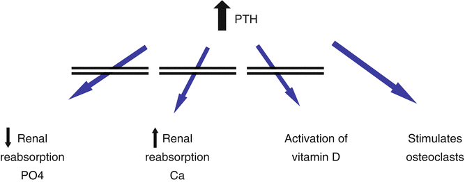

Fig. 28.3
Actions of PTH in renal disease
Q4 What are the clinical features of secondary hyperparathyroidism?
The condition may be completely asymptomatic but may also be characterised by bone pain or fractures. The risk of fracture is increased by the other forms of bone disease, particularly osteoporosis.
Q5 Why do renal patients sometimes develop osteomalacia?
Osteomalacia means failure to mineralise bone adequately and in renal osteomalacia accumulation of aluminium may be responsible. Historically renal osteomalacia occurred in haemodialysis patients before we began removing aluminium from the dialysis water by reverse osmosis. Nowadays renal osteomalacia is seen occasionally in patients who are taking aluminium as a phosphate binder, which is one of the reasons why Alucaps are used less than before.
Q6 Why does soft tissue calcification occur?
Soft tissue calcification, also known as metastatic calcification, describes the deposition of calcium and phosphate in soft tissue such as muscles, joints and arteries. This occurs in all kidney patients but particularly in those in whom the calcium phosphate product (serum calcium × serum phosphate) exceeds 4.5 mmol/L. Arterial calcification may contribute to the high rates of vascular disease seen in renal patients, and is one of the reasons for the move away from calcium containing phosphate binders such as Calcichew and Phosex.
Calcium & Phosphate Product = Serum Calcium × Serum Phosphate.
If this exceeds 4.5, there is significant risk of soft tissue or “metastatic” calcification. Reducing levels of phosphate by binders while maintaining a normal serum calcium is key to treatment.
A flow chart summarising the pathogenesis of secondary hyperparathyroidism, osteomalacia and soft tissue calcification is shown overleaf (Fig. 28.4).
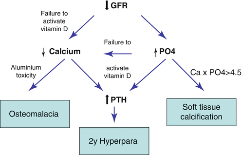

Fig. 28.4
Pathogenesis of renal bone disease
Q7 What is adynamic bone disease?
In health, bone is continuously broken down & rebuilt. Adynamic bone disease is a condition in which this process of “turn-over” is absent or depressed. Uraemic patients develop skeletal resistance to PTH so even a normal PTH level may represent reduced bone turnover. A low serum level of parathyroid hormone may also be a consequence of over-suppression of PTH by alfacalcidol. Patients whose bones are not constantly being remodelled are at greater risk of fracture.
Q8 Why do renal patients develop osteoporosis?
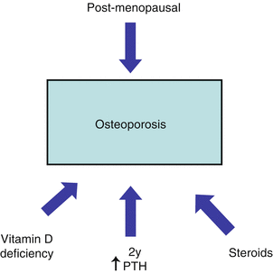
The fifth main disorder of mineral metabolism is osteoporosis, which literally means thin bones. This is an important cause of fracture in renal patients. Renal patients are prone to all the usual causes of osteoporosis eg post-menopausal osteoporosis and have other reasons for developing thin bones which include immobility, vitamin D deficiency, secondary hyperparathyroidism and the use of steroids, particularly after transplantation or the treatment of certain glomerulopathies (Fig. 28.5).
 < div class='tao-gold-member'>


Only gold members can continue reading. Log In or Register to continue
Stay updated, free articles. Join our Telegram channel

Full access? Get Clinical Tree


