Fig. 1
Image showing both testes (single screen)
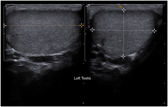
Fig. 2
Left testicular measurements with the split screen: longitudinal view on left and transverse view on right
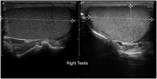
Fig. 3
Right testicular measurements with the split screen: longitudinal view on left and transverse view on the right of the screen
Split Screen (laterality is when looking at screen)
7.
EPIDIDYMIS #1: epididymal head, right and left side, split screen: right testis on left of screen and left testis on right of screen (Fig. 4)
8.
EPIDIDYMIS #2: Epididymal body, right and left side, split screen: right epididymal body on left of screen and left epididymal body on right of screen (Fig. 5)
9.
EPIDIDYMIS #3: Epididymal tail, right and left side, split screen: right epididymal body on left of screen and left epididymal body on right of screen (Fig. 6)
10.
Lateral view of the left and right testis: right testis on left of screen and left testis on right of screen (Fig. 7)
11.
Medial view of the left and right testis: right testis on left of screen and left testis on right of screen (Fig. 8)
12.
Upper (superior pole) view of the left and right testis: right testis on left of screen and left testis on right of screen (Fig. 9)
13.
Lower (inferior pole) view of the left and right testis: right testis on left of screen and left testis on right of screen (Fig. 10)
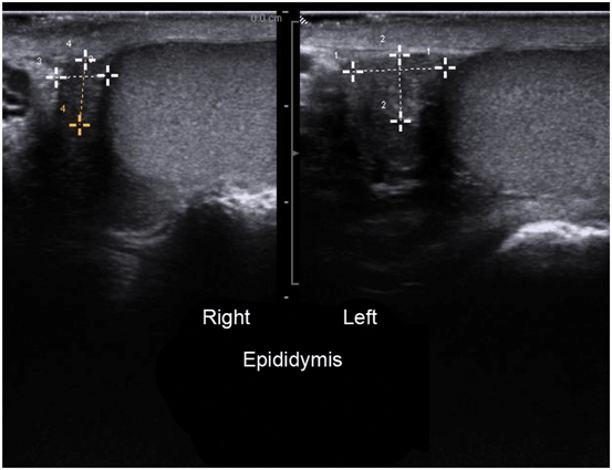
Fig. 4
Epididymis #1: epididymal head, right and left side, split screen: right testis on left of screen and left testis on right of screen
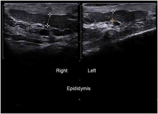
Fig. 5
Epididymis #2: epididymal body, right and left side, split screen: right epididymal body on left of screen and left epididymal body on right of screen
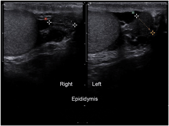
Fig. 6
Epididymis #3: epididymal tail, right and left side, split screen: right epididymal body on left of screen and left epididymal body on right of screen
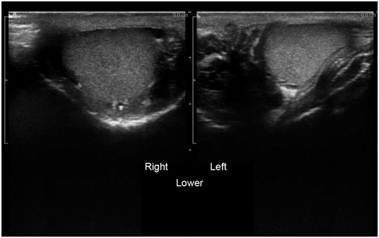
Fig. 7
Lateral view of the left and right testis: right testis on left of screen and left testis on right of screen
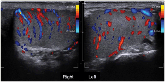
Fig. 8
Medial view of the left and right testis: right testis on left of screen and left testis on right of screen
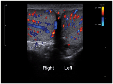
Fig. 9
Upper (superior pole) view of the left and right testis: right testis on left of screen and left testis on right of screen
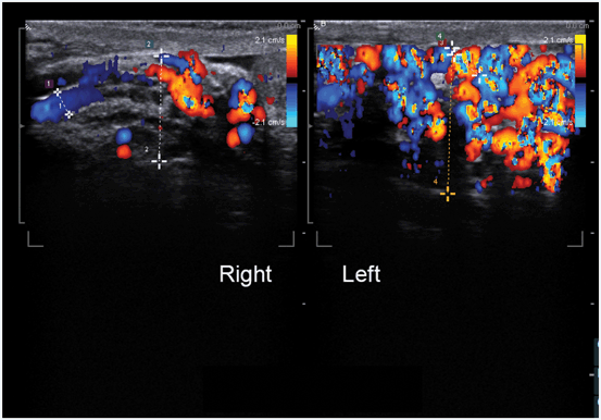
Fig. 10
Lower (inferior pole) view of the left and right testis: right testis on left of screen and left testis on right of screen
Color Doppler
14.
Intratesticular blood flow pattern. Split screen of longitudinal view of both testes with the right testis on left of screen and left testis on right of screen (Fig. 11).
15.
A single screen transverse view of both testes for comparative intratesticular blood flow (Fig. 12).
16.
Varicocele evaluation #1. Longitudinal split screen of spermatochord superior to the testis with the right spermatochord on left of screen and left spermatochord on right of screen. Measurement of inner diameter of largest vein and width of entire complex (Fig. 13).
17.
Varicocele evaluation #2. Longitudinal split screen of spermatochord posterior to the testis with the right spermatochord on left of screen and left spermatochord on right of screen. Measurement of inner diameter of largest vein and width of entire complex (Fig. 14).
18.


Optional: Varicocele evaluation #3. Transverse split screen of spermatochord posterior to the testis with the right spermatochord on left of screen and left spermatochord on right of screen. Measurement of inner diameter of largest vein and width of entire complex (Fig. 15).
Stay updated, free articles. Join our Telegram channel

Full access? Get Clinical Tree


