Liver disorders


Liver disorders


CHOLESTASIS

Piotr Socha MD

Joanna Pawłowska MD

Anil Dhawan MD, FRCPCH


Jaundice and cholestasis

Jaundice results from the deposition of unconjugated or conjugated bilirubin pigment in the skin and mucous membranes. Cholestasis is defined as conjugated hyperbilirubinaemia. Liver disease may present as cholestasis alone, cholestasis that accompanies liver cirrhosis or severe liver disease with liver insufficiency. Ascites, peripheral oedema, bruises and so on can be present which indicate severe liver damage. A jaundiced child with liver insufficiency requires immediate hospitalization with prompt diagnosis to find the underlying liver disease. Prothrombin time can be solely regarded as an indicator of acute liver failure.


Infancy

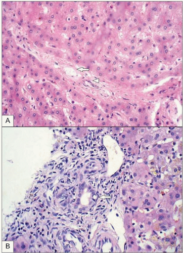
Cholestasis should be diagnosed quickly in infancy as various diseases require immediate therapy. Numerous aetiologies make diagnosis difficult. The American Academy of Pediatrics has elaborated practical guidelines for the management of hyperbilirubinaemia in newborn infants, mainly concentrating on indirect hyperbilirubinemia1. The guidelines emphasise the importance of measuring direct bilirubin in sick infants and in those who are jaundiced at or beyond 3 weeks. If the direct or conjugated bilirubin level is elevated, additional assessment for the causes of cholestasis is recommended. For instance, hypothyroidism, galactosaemia, and tyrosinaemia should be taken into account. Infectious diseases may also cause cholestasis, thus urinary tract infections as well as cytomegalovirus (CMV) and toxoplasmosis should be excluded. The role of CMV as the causative factor of cholestasis is still under debate, as congenital 
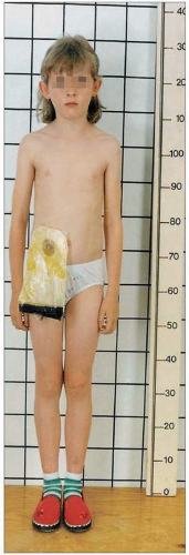
CMV infections may present with liver damage and cholestasis. It is very important to assess stool colour to make a timely diagnosis of biliary atresia (BA). 
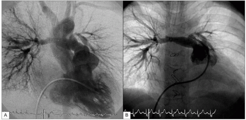
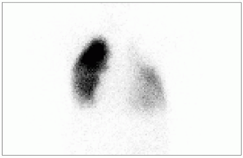
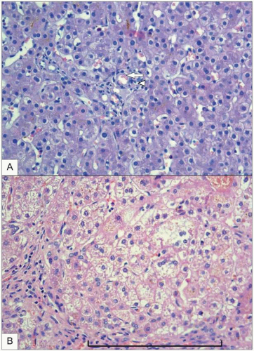
BA is a relatively uncommon disease characterized by a biliary obstruction of unknown origin that presents in the neonatal period2. However, it is the most important surgically correctable cause of cholestatic jaundice in this age group and is the most common indication for liver transplantation in children. Untreated, BA leads to cirrhosis and death within the first years of life. Surgical treatment usually involves an initial attempt to restore bile flow: the Kasai portoenterostomy3 which is performed as soon after diagnosis as possible. Prognosis of the disease seems to be better in infants operated on before 90 days of age4. Other causes of cholestasis with pale stools are: Alagille syndrome, progressive familial intrahepatic cholestasis (PFIC), alpha-1-antitrypsin deficiency (α1-ATD), and cystic fibrosis. Neonatal haemochromatosis, Zellweger syndrome, Niemann-Pick disease type C, and glycogen storage disease type IV should also be considered in the differential diagnosis of infantile cholestasis (Tables 9.1, 9.2).


Table 9.1 Differential diagnosis of cholestatic liver disease of infancy


Extrahepatic cholestasis


Biliary atresia


Choledochal cyst


Choledocholithiasis


Intrahepatic cholestasis


Infectious diseases


TORCH (Toxoplasmosis, Others, Rubeolla, Cytomegaloviral infection, Herpes)


Other viral infections


Reo


Echo


Parvovirus


Listeria


Sepsis


Urinary tract infection


Tuberculosis


HIV


Metabolic diseases


α1-antitrypsin deficiency


Carbohydrate metabolism disturbances


Galactosaemia


Fructosaemia


Glycogenosis: type IV


Amino acid metabolism disturbances


Tyrosinaemia


Lipid metabolism disturbances


Niemann-Pick disease


Cholesterol ester storage disease


Wolman disease


Cystic fibrosis


Bile acid metabolism disturbances


Peroxisomal disorders


Zellweger syndrome


Congenital defects of glycosylation


Familial cholestatic diseases


Idiopathic neonatal hepatitis


Progressive familial intrahepatic cholestasis


Alagille syndrome


Nonsyndromatic paucity of bile ducts


Recurrent intrahepatic cholestasis


Benign-familiar


With lymphatic oedema (Aageneas syndrome)


Anatomical changes


Caroli disease


Multicystic liver and kidney disease


Chromosomal diseases


Down syndrome


Other causes


Hypothyroidism


Histiocytosis X


Necrotizing enterocolitis


Parenteral nutrition-associated cholestasis


Drug-induced cholestasis


Table 9.2 Diagnostic approach to cholestasis of infancy


Urgent diagnosis indicated


Elective diagnosis performed


Biliary atresia


PFIC


Galactosaemia


Alagille syndrome


Hypothyroidism


Cystic fibrosis


Tyrosinaemia


Choledochal cyst (if not accompanied by cholangitis)


Neonatal haemochromatosis


α1-ATD


Zellweger syndrome


Niemann-Pick disease type C


Infections (sepsis, urinary tract infection, CMV, Toxo)


Glycogen storage disease type IV


Childhood

Jaundice is uncommon in children. However, indirect hyperbilirubinaemia can be the presentation of a benign, inherited condition, Gilbert syndrome, which affects about 6% of the adult population. Mutations in hepatic enzyme isoform (1A1) belonging to the urine diphosphoglucuronate glucuronosyltransferase (UGT) family of enzymes are responsible for this condition5. Gilbert syndrome can be confirmed by molecular diagnosis but it should be suspected in an otherwise asymptomatic child whose other liver tests are normal and there are no signs of haemolysis.

Several liver diseases may present with jaundice such as infection with hepatitis B, hepatitis A, and hepatitis C virus, autoimmune hepatitis, Wilson disease, and α1-ATD. Cholecystitis is a rare condition in children and acute clinical symptoms with jaundice and fever are very unusual. Choledocholithiasis is also infrequent in childhood.

Familial cholestatic diseases (Alagille syndrome, PFIC) and postoperative biliary atresia may present with episodes of increased hyperbilirubinaemia or symptoms of liver insufficiency. That is why it is important to follow these patients carefully and the primary care physicians have to be aware of possible exacerbation of symptoms. Cholangitis episodes in postoperative biliary atresia require rapid diagnosis and treatment. Septic and hypoxic damage to the liver can cause cholestasis, and septicaemia seems to be a common reason for referral of a jaundiced child to liver clinics.


9.1 Typical facial appearance in a child with Alagille syndrome.


Clinical characteristic and laboratory findings


Alagille syndrome

Alagille syndrome can present with very early cholestasis of infancy but it may also be asymptomatic for a very long time. Liver damage may progress quickly in some patients with portal hypertension, recurrent cholangitis, and malnutrition6. Diagnosis of Alagille syndrome is aided by the presence of syndromic features, including bile duct paucity on liver histology, chronic cholestasis, cardiac murmur, vertebral abnormalities, peculiar facies, eye findings, renal disease, and xantomas7. Usually differential diagnosis is performed in early infancy when biliary atresia must be excluded.

Typical facial features are a prominent forehead, pointed chin, deep set eyes, moderate hypertelorism, and a saddle or straight nose with a bulbous tip (9.1), but these features are not very typical in early infancy. The parents may also have the typical appearance (9.2) which may indicate the need to diagnose this disease. Pulmonary artery stenosis is a typical finding which may also cause a serious heart disease (9.3, 9.4). Embryotoxon posterior observed in a slit lamp is one of the most common features of the Alagille syndrome. Butterfly vertebrae observed in late infancy are also a characteristic feature (9.5). Liver biopsy is helpful to establish the diagnosis (9.6). Liver disease may slowly progress to liver insufficiency and liver transplantation may be needed. However, in most of the patients liver disease remains stable for a long time.


9.2 The parent of the child usually presents also with very typical facial appearance.


9.3 A, B: Fallot tetralogy and pulmonary artery hypoplasia in a patient with Alagille syndrome (right ventricle angiography in a sitting-up projection of 30°). (Courtesy of the Cardiac Catheterization Laboratory, The Children’s Memorial Health Institute.)


9.4 Scintigraphy may show peripheral stenosis of pulmonary arteries with hypoperfusion of the left lung. (Courtesy of the Radioimmunology Department, The Children’s Memorial Health Institute.)


9.5 Butterfly vertebrae in Alagille syndrome (arrows).


9.6 A: Presentation of bile duct paucity in Alagille syndrome (vein, arrow; artery, arrowhead); B: bile duct (arrow) visible in the portal space. (Courtesy of the Pathology Department, Children’s Memorial Health Institute.)


9.7 Pruritus as a presenting symptom of PFIC.


Table 9.3 Genetic background of cholestastic diseases


PFIC 1


PFIC 2


PFIC 3


Alagille syndrome


Genetics


Autosomal recessive


Autosomal recessive


Autosomal recessive


Autosomal dominant with highly variable expression


Gene


FIC 1


BSEP


MDR 3


JAG 1


Mutation on


18q 21-22


2q 24


7q 21


Short arm of chromosome chromosome 20


Gene product


P-type ATPase


Bile salt pump


Phospholipid transporter


Ligand of Notch1


Cell localization


Gut, bile duct cells, canalicular membrane


Canalicular membrane


Canalicular membrane


Liver, heart, skeletal, eye, kidney


Progressive familial intrahepatic cholestasis

PFIC is an inherited disorder where bile acid excretion is impaired (Table 9.3). This diagnosis should always be suspected in patients with high serum bile acid concentration, low/normal or elevated gamma glutamyl transpeptidase (GGT), and pruritus.

Pruritus is usually the predominant symptom and skin excoriations due to itching may be observed (9.7). Short stature is a typical feature in later age (9.8). Liver histology may reveal giant cell transformation, ductular proliferation, and fibrosis (9.9). The disease may progress to end-stage liver disease. Some experience from centres in the USA and Poland points to a successful therapy with partial external biliary diversion (9.10) when performed early in the course of the disease8. PFIC also seems to be a risk factor for hepatocellular carcinoma9. In general, diagnosis of PFIC is not very urgent but doctors should be aware of the potential health effects.


Benign recurrent intrahepatic cholestasis

Benign recurrent intrahepatic cholestasis (BRIC) is characterized by intermittent episodes of cholestasis without progression to cirrhosis. The onset of the disease usually occurs before the second decade of life. The attacks of cholestasis vary in severity and duration (from 1 to 18 months) and may be preceded by a preicteric phase with malaise, anorexia, and pruritus. Biochemically it is characterized by increased concentrations of bilirubin, serum bile acid, and alkaline phosphatase; GGT remains low. There are no pathological changes in the liver on biopsy besides some hepatocellular cholestasis. In between episodes patients are totally asymptomatic both clinically and biochemically. Treatment is symptomatic.


9.8 Short stature in a 14-year-old girl with PFIC.


9.9 Liver histology in PFIC. A: Missing bile duct in a portal space (arrow, artery; arrowhead, portal tract); B: enlargement of the portal tract with septal fibrosis (brace).


9.10 Partial biliary diversion in a child with PFIC. Catch up growth is observed since the procedure was performed.


Biliary atresia

BA presents with cholestasis in early infancy (1-2 months of age) in an otherwise healthy child, thus diagnosis may be delayed. The most common clinical features are: jaundice with conjugated bilirubin beyond 2 weeks of life (9.11), white stools and dark urine as well as hepatomegaly, which may not always be evident. Usually a complete work-up of cholestatic diseases is performed to confirm the diagnosis and to exclude other liver diseases with early presentation. The gall bladder may not be visible on ultrasound in many patients with biliary atresia, dynamic scintigraphy (HIDA) does not show any bile passage to the gut (9.12), and liver histology may present ductal plate malformation with bile duct proliferation and fibrosis (9.13), which is regarded as a typical feature that confirms the diagnosis. It is recommended that infants with direct hyperbilirubinaemia exceeding 2 mg/dl (34 μmol/l) over 2 weeks of age be referred to liver centres for further work-up to avoid wasting valuable time in making the diagnosis. BA may progress quickly to cirrhosis with ascites (9.14) and oesophageal varices as a consequence of portal hypertension.


9.11 A child with biliary atresia with moderate jaundice and normal nutritional status (A), and pale stools (B).

Only gold members can continue reading. Log In or Register to continue

Stay updated, free articles. Join our Telegram channel

Jun 19, 2016 | Posted by in GASTROENTEROLOGY | Comments Off on Liver disorders

Full access? Get Clinical Tree

Get Clinical Tree app for offline access