Fig. 4.1
Port placement and operating room setup for the laparoscopic transverse colectomy
4.3 Surgical Procedures
After the omentum and transverse colon are moved toward the upper abdomen, tenting the transverse mesocolon creates the optimal operative field: the middle colic pedicle, duodenum, and pancreas can be well visualized from the ventral side if severe adhesion does not occur (Fig. 4.2). In a thin patient, the middle colic pedicle is readily obvious, whereas the pedicle is subtle in an obese patient. The ileocolic pedicle needs to be identified by retracting the right mesocolon. Before starting dissection, it is useful to assume the line of the dissection range according to the location of the tumor and the extent of lymph node dissection (Fig. 4.2, white dotted arrows).
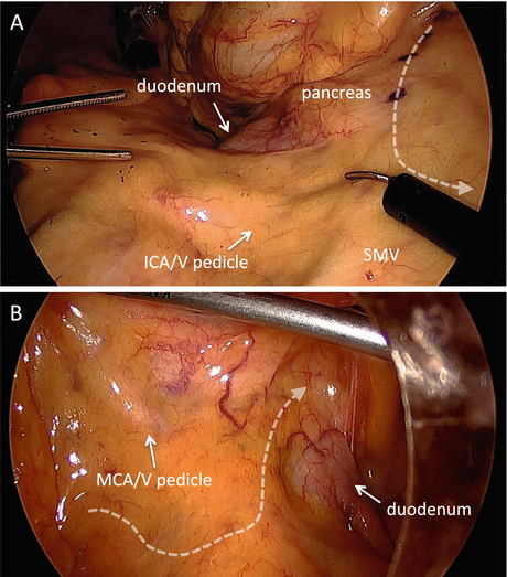

Fig. 4.2
Tenting the transverse mesocolon makes the optimal operative field. Right-sided (a) and left-sided (b) views. White dotted arrows represent the planned line of the dissection range according to the location of the tumor and the extent of lymph node dissection. ICA/V ileocolic artery/vein, MCA/V middle colic artery/vein, SMV superior mesenteric vein
For the right-sided dissection, the ventral peritoneum of the cranial portion of the ileocolic pedicle is incised, followed by medial-to-lateral mobilization of the right mesocolon. Although various approaches such as lateral-to-medial (lateral approach), medial-to-lateral (medial approach), and retroperitoneal approaches have been reported in laparoscopic colon surgery, we believe that the medial approach is the most useful to facilitate exposure of the mesentery, with the assistance of peritoneal fixation of the right colon [6]. The second portion of the duodenum is an important landmark to identify (Fig. 4.3a). The right mesocolon is carefully separated from the retroperitoneal tissues, duodenum, and pancreatic caudal portion up to the hepatocolic ligament (Fig. 4.3b–d). The assistant medially retracts the right colon with both atraumatic graspers to maintain the peritoneal window (Fig. 4.4). This enables the surgeon to provide his own countertraction with forceps of both hands (Fig. 4.5).
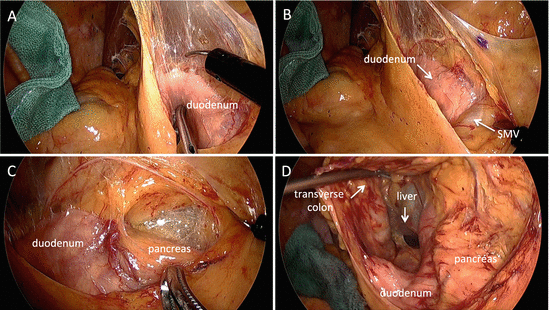
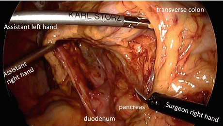
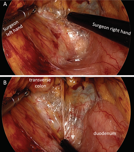

Fig. 4.3
(a) The retroperitoneal portion of the duodenum is an important landmark for medial approach. (b–d) The right mesocolon is separated from the retroperitoneal tissues, duodenum, and pancreatic caudal portion up to the hepatocolic ligament

Fig. 4.4
The assistant retracts the right colon medially with both atraumatic graspers to maintain the right-sided peritoneal window

Fig. 4.5
In the right-sided peritoneal window maintained by the assistant, the surgeon provides his own countertraction with forceps of both hands. Operative fields are shown before dissection (a) and after dissection (b). White dotted arrow represents the motion of the surgeon’s right hand
Next, the peritoneum over the superior mesenteric vessels is incised (Fig. 4.6a), and then ventral surfaces of superior mesenteric vessels are exposed (Fig. 4.6b). The vascular sheath of the superior mesenteric vein (SMV) can be directly exposed, whereas SMA is exposed with being covered by the nerve sheath (Fig. 4.7). When proceeding to dissect the ventral surface of SMV and SMA in a caudal to cranial direction, MCA, MCV, and Henle’s trunk (gastrocolic trunk) are gradually identified (Fig. 4.8). MCA is skeletonized below the pancreas and then divided at its origin (Fig. 4.9a, b). The independent right colic artery, if present, is located around the upper border of the duodenum, although the majority of patients do not have the independent right colic artery. MCV is usually identified just below the inferior border of the pancreas, which can become an important landmark (Fig. 4.10a, b). Understanding anatomical relationship of MCA and MCV is necessary for appropriate lymph nodes dissection in laparoscopic transverse colectomy (Fig. 4.11). Careful dissection onto the duodenum and the pancreatic caudal portion must be exercised to expose the right and middle colic vessels. When the root of the accessory right colic vein (aRCV) is clearly identified, aRCV is divided just before it drains into the Henle’s trunk (Fig. 4.12a, b). However, if aRCV is difficult to confirm in this situation, this vein may be easily detected later at the takedown of the hepatic flexure. It is important to understand that the venous anatomy between the hepatic flexure and the middle colic vessels has many variations, including Henle’s trunk, the fusion between the right gastroepiploic vein (RGEV), and a branch of RCV or MCV. After division of aRCV, RGEV is clearly exposed and then cranially dissected from the transverse mesocolon, which can lead to wide separation between the pancreatic head and transverse mesocolon (Fig. 4.16a).
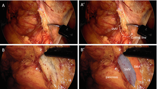

Fig. 4.6


(a) The peritoneum over the superior mesenteric vessels is incised. (a′) Cutting line of the peritoneum is shown. (b) Ventral surfaces of superior mesenteric vessels are exposed. (b′) Overlaid view with colored shadow. SMA superior mesenteric artery
Stay updated, free articles. Join our Telegram channel

Full access? Get Clinical Tree


