Fig. 6.1
Positions of the cannulae and the surgical team. The camera port is at the umbilicus. Two ports on the right side are for a surgeon, and two ports on the left are for the first assistant. The specimen is extracted through an extended wound at the umbilicus
6.3 Procedures
6.3.1 Basic Principles for Dissection
All tissues and organs are covered with connective fibers, the so-called “fascia.” Dissection means the dividing of these fibrous tissues between the other tissues or organs, creating new fasciae on both sides (Fig. 6.2a). Your dissection between the rectum and the neural system, such as the hypogastric nerves, the pelvic nerve plexus, and the neurovascular bundles, creates the so-called “prehypogastric nerve fascia” (fibrous tissues covering the nerves) and the “rectal fascia propria” (fibrous tissues covering the rectum and its proper adipose tissue). A good counter-traction is a prerequisite to identify the fibrous tissues. You may divide the adipose tissue intentionally only at the time when you divide vascular or neural branches to the rectum, because these branches are accompanied with the adipose tissues.
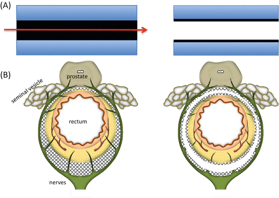

Fig. 6.2
Basic principles of dissection. (A) Dissection means cutting the connective fibers (fascia) between the tissues or organs, creating the new fasciae on both sides. (B) The connective fibers (fascia) between the rectum and the nerves are dissected, creating two new fasciae: the prehypogastric nerve fascia and rectal fascia propria
6.3.2 Medial to Lateral Dissection
The greater omentum and the transverse colon are placed in the left upper quadrant, and the small bowel loops are placed in the upper right quadrant to expose the terminal portion of duodenum and the inferior mesenteric vein (IMV) (Fig. 6.3).
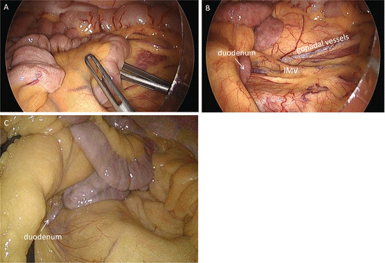

Fig. 6.3
The small intestinal loops are placed in the right upper quadrant to expose the duodenum and IMV (A, B). In an obese patient, the IMV is not clearly seen (C)
Sigmoid colon and rectum are retracted out from the pelvis to identify the tumor location. If the tumor location is unclear, proctoscopy or colonoscopy is recommended to exactly determine the distal cutting line of the rectum. Preoperative tattooing is not recommended because the ink often exudates extramurally to make dissection layer obscure.
The anterior wall of the rectum and the pedicle of superior rectal vessels are grasped gently and retracted ventrally by the assistant to extend the right-side peritoneum of rectum widely (Fig. 6.4). If the grasping of the anterior wall of the rectum is difficult, the assistant can push the rectum ventrally and laterally with an opened intestinal grasper (Fig. 6.5). An incision is made on the peritoneum at the level of promontory, going toward the peritoneal reflection (Fig. 6.6). Picking up the cutting edge of peritoneum facilitates the recognition of the subperitoneal connective fibers (Fig. 6.7). Cutting these fibers, not the adipose tissues, prevents the injury on the hypogastric nerves. Even if you are dissecting a wrong plane with dividing the fibers, it is able to easily return to a correct plane without any oncological as well as physiological complications.

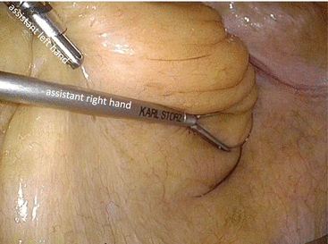
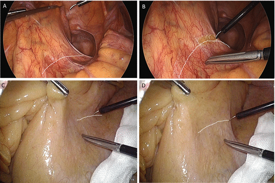


Fig. 6.4
The assistant grasps the superior rectal pedicle by the left hand (A) and the anterior wall of the rectum by the right hand, in order to extend the right surface of mesorectum (B)

Fig. 6.5
The assistant can push the rectum ventrally and laterally with an opened grasper

Fig. 6.6
When the assistant relaxes his retraction, the incisional lines are imagined as the dotted lines along the concave of peritoneum (A, C). Those lines are moved ventrally with full retraction by the assistant (B, D)

Fig. 6.7
Picking up the edge of peritoneum extends the subperitoneal fibrous tissues widely and facilitates easy dissection
Cutting of these fibrous tissues creates fibrous membranes on both sides: the so-called rectal fascia propria (proper rectal fascia) and prehypogastric nerve fascia (Fig. 6.8). You can advance the dissection as deep as possible toward the pelvic floor. If it is difficult, you should move to cephalad dissection. Incision of peritoneum is continued up to the origin of IMA. The assistant should now grasp the IMA pedicle proximally to extend subperitoneal connective tissues. This maneuver facilitates the recognition of fine structure of connective fibers and prevents the injuries to the lumbar splanchnic nerves, the ureter, and the gonadal vessels (Fig. 6.9). The borderline between the lymph nodes along the IMA and the lymph nodes along the aorta is unclear so that you should make the border yourself dividing the adipose tissues between them. I recommend an ultrasonic or a bipolar energy device to divide the adipose tissues. It is safe and easy to dissect between adipose tissues containing lymph nodes and neural tissues surrounding the IMA (Fig. 6.10). You should divide the sigmoidal branches of the lumbar splanchnic nerves, especially the branches from the right lumbar splanchnic nerves, to skeletonize the IMA. Following the skeletonization and division of the IMA, the sigmoidal branches from the left lumbar splanchnic nerves are identified and divided (Fig. 6.11). Or you can divide the IMA distally to avoid the injury to the left splanchnic nerves (Fig. 6.12).
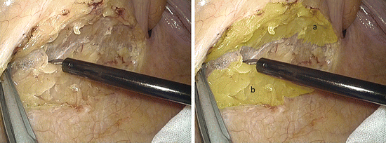
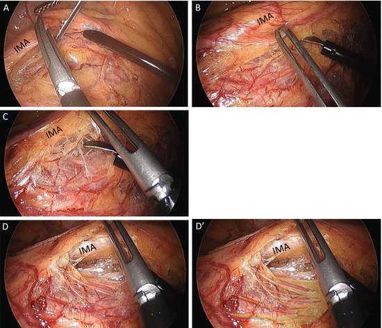

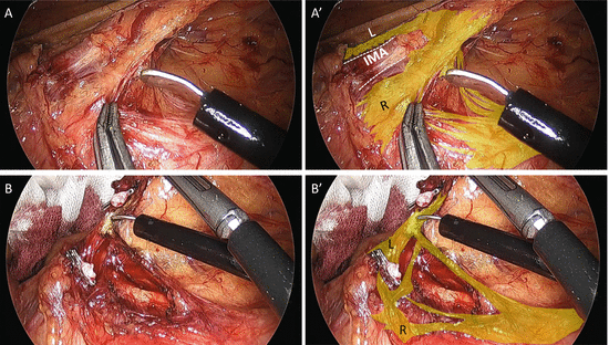
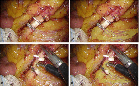

Fig. 6.8
The rectal fascia propria (a) and the prehypogastric nerve fascia (b) are separated

Fig. 6.9
Following the initial peritoneal incision and dissection, the assistant now grasps the IMA pedicle proximally (A). The additional ventral traction of the IMA pedicle by the surgeon extends the fibrous tissues behind the pedicle (B, C) and facilitates the following dissection between the sigmoidal mesentery and retroperitoneum (D). Yellow lines are left lumbar splanchnic nerves (D′)

Fig. 6.10
The adipose tissues (yellow area) containing the lymph nodes around the IMA (dotted white line) are dissected from the IMA, which is covered with the sigmoidal branches of splanchnic nerves

Fig. 6.11
(A) The sigmoidal branches from the right splanchnic nerves are divided prior to the division of the IMA. The skeletonization of the IMA is necessary to preserve the left splanchnic nerves. (B) Following the division of the IMA, the sigmoidal branches from the left splanchnic nerves are divided. Yellow area lumbar splanchnic nerves, L left branch, R right branch

Fig. 6.12
Comparing the case in Fig. 6.11, the IMA is divided more distally so that the left lumbar splanchnic nerves are easily preserved. (A) The IMA is completely skeletonized. (B) The sigmoidal branches from the left lumbar splanchnic nerves are divided. Yellow area lumbar splanchnic nerves, L left branch, R right branch
The assistant now grasps the distal edge of IMA and retracts it ventrally to extend the dissection plane between the sigmoidal mesentery and retroperitoneum. The inferior mesenteric vein (IMV) and the left colic artery (LCA) are divided at the same level of the IMA division. In some case the LCA is running far away from the IMV [1]. The dissection between the mesocolon and the retroperitoneum is advanced laterally and cephalad. Again the dissection means cutting of the connective fibers. In order to recognize these connective fibers, counter-traction is very important. Your own ventral retraction often extends the fibrous tissues between the sigmoidal mesocolon and the retroperitoneum (Fig. 6.13). Dividing the connective fibers creates a fibrous membrane covering the left kidney, the so-called the Gerota’s fascia. You can recognize the lower border of pancreas.
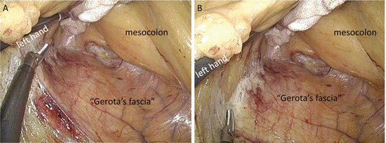

Fig. 6.13
The ventral retraction by the surgeon’s left hand extends the fibrous tissues between the dissected planes: mesocolon and Gerota’s fascia (A). Continuous division of the fibrous tissues creates membranes clearly on both sides (B)
Following the cephalad dissection, you come back to the posterior dissection of the rectum. The assistant grasps the distal edge of the IMA by the left hand and the right edge of the incised peritoneum of the mesorectum by the right hand (Fig. 6.14A, B). The following ventral and cephalad traction by the assistant extends the fibrous tissues between the mesorectum and the retroperitoneum (Fig. 6.14C, C′). The dividing these fibers creates the rectal fascia propria and the prehypogastric nerve fascia on both sides.
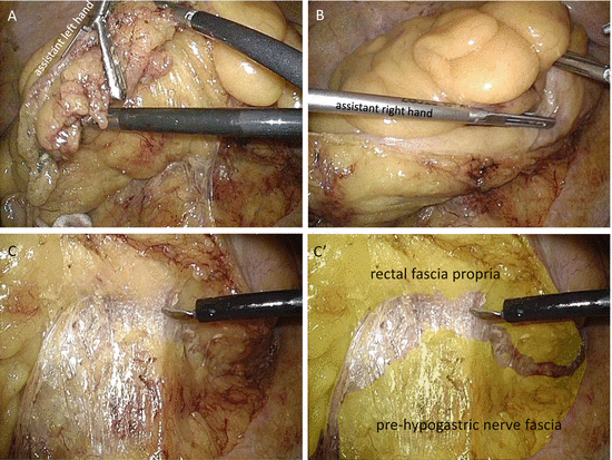

Fig. 6.14
The assistant grasps the distal edge of the IMA pedicle (A) and the right edge of incised peritoneum of the mesorectum (B). The sufficient ventral traction extends the fibrous tissues for the following dissection (C, C′)
6.3.3 Lateral Mobilization and Takedown of Splenic Flexure (Refer to Another Chapter)
Chapter 5 Laparoscopic left sided colectomy (mobilization of splenic flexure and sigmoidectomy)
Stay updated, free articles. Join our Telegram channel

Full access? Get Clinical Tree


