Fig. 3.1
Position of the equipment and the surgical team for the laparoscopic right-sided colectomy. The second assistant (camera operator) stands between the patient’s legs or on the left side depending on situations.
3.2 Instruments
Specific instruments recommended for laparoscopic right hemicolectomy are 5 cannulas (1 × 12 mm, 4 × 5 mm), 1 dissecting device (ultrasonic shears and electrocautery), 1 laparoscopic dissector, and 4 laparoscopic bowel graspers.
3.3 Cannula Positioning
We routinely use the “open Hasson” technique to safely insert the first cannula through the umbilicus. Pneumoperitoneum is established and maintained at 8 mmHg. Then, cannulas are placed in the following order (all 5 mm): left lower abdominal region, left middle, right lower, and right middle abdominal regions. The surgeon uses the left-sided cannulas and the assistant uses the right-sided cannulas [1] (Fig. 3.1).
3.4 Technique
The procedure is divided into four parts: (1) mobilization of the right colon and its mesentery; (2) division of the vascular pedicles; (3) division of the lateral attachments, hepatic flexure, and omental attachments; and (4) specimen extraction and anastomosis. There are four commonly used approaches to a laparoscopic right-sided colectomy, namely, the medial, inferior, lateral, and superior approaches. We prefer to use the inferior approach to mobilize the right colonic mesentery because it enables faster and wider mobilization of the right colon compared with the medial approach and also makes the subsequent division of the vascular pedicles easier. The operating table is tilted into the Trendelenburg position with the right side slightly down to move the small intestine toward the right upper quadrant. After the omentum and transverse colon are moved toward the upper abdomen and the small intestine toward the upper right quadrant, the assistant grasps the small bowel mesentery near the terminal ileum and near the ligament of Treitz and retracts widely to allow the surgeon to identify the imaginary dissection plane between the small bowel mesentery and the retroperitoneum (Fig. 3.2). This is at a slight reverse angle to the scope and takes time to master, so the surgeon should help the assistant to grasp the mesentery properly. The surgeon needs to confirm the correct orientation of the field; if the imaginary dissection plane is not identified, another attempt is made in cooperation with the assistant. After creating the proper field, the surgeon incises the mesentery to identify the dissection plane, which lies between the shiny surface of the small bowel mesentery and meshwork tissue of the retroperitoneum (Fig. 3.3). The plane is developed underneath the small bowel mesentery until the duodenum is identified cranially (Fig. 3.4). The plane is developed laterally to identify the right ureter and the gonadal vessels underneath the retroperitoneum. Using the left hand, the surgeon lifts up the small bowel mesentery and, using the right hand, sweeps down the right ureter and the gonadal vessels underneath the retroperitoneum (Fig. 3.5). Dissection to the caudal portion of the pancreas is needed to facilitate the medial approach. After completing the lateral dissection to the base of the ileal mesentery, the peritoneum is incised, and the lateral attachment of the ascending colon is divided around the cecum. Surgical gauze could be placed on the dissection plane as a landmark for the medial approach.
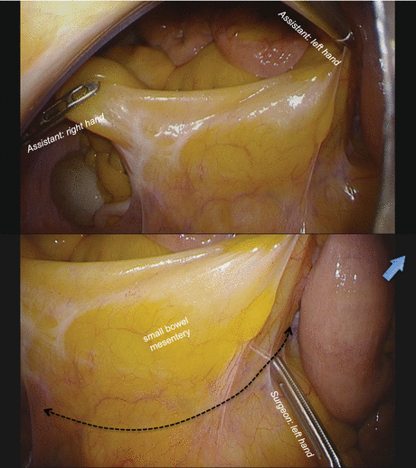
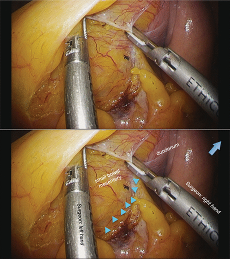
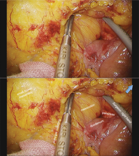
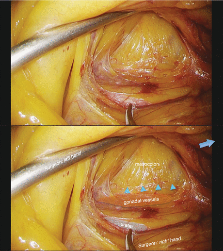
Fig. 3.2
The assistant grasps the small bowel mesentery near the terminal ileum and near the ligament of Treitz and retracts widely to allow the surgeon to identify the imaginary dissection line (dotted line) between the small bowel mesentery and the retroperitoneum. The blue arrow indicates the caudal direction
Fig. 3.3
The dissection plane lies between the shiny surface of the small bowel mesentery and meshwork tissue of the retroperitoneum (blue arrowheads). The blue arrow indicates the caudal direction
Fig. 3.4
The plane is developed underneath the small bowel mesentery until the duodenum is identified cranially. The blue arrow indicates the caudal direction
Fig. 3.5
The right ureter and the gonadal vessels were swept down from ascending mesocolon underneath the meshwork tissue of the retroperitoneum (blue arrowheads). The blue arrow indicates the caudal direction
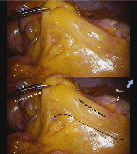
Next, the medial approach is commenced. The operating table is reversed and tilted slightly into the Trendelenburg position with the left side down. The small intestine is moved to the left side of the abdomen, the omentum and transverse colon are moved toward the upper abdomen, and the ventral side of the right colon is visualized. The surgeon lifts up the mesocolon near the ileocecal junction to identify the “ridge” of ileocolic vessels, which is grasped by the assistant’s right hand (Fig. 3.6). The peritoneum is incised between the ridge and the periphery of the superior mesenteric vein to identify the previously dissected plane (surgical gauze) easily. The assistant retracts the ileocolic pedicle ventrally using the right hand and raises cephalad the transverse mesocolon slightly using the left hand in order to identify the ileocolic vessels and the superior mesenteric vessels. The peritoneum is incised over the superior mesenteric vessels from the previously incised peritoneum below the ileocolic pedicle. The surgeon should grasp and retract the cut edge of the peritoneum with adequate traction of the ileocolic pedicles in order to confirm the root of the ileocolic vein and the surface of the superior mesenteric vein (Fig. 3.7). The mesocolon is dissected to expose the second portion of the duodenum and the pancreas head widely (Fig. 3.8). Care must be taken not to dissect between the duodenum and the pancreatic tissue. The root of the ileocolic vessels is usually located at the lower border of the duodenum. The ileocolic artery runs either in front of or behind the superior mesenteric vein, with approximately the same frequency. In the former case, the ileocolic artery is cut at the root; in the latter case, the artery is cut at the right border of the superior mesenteric vein, although the boundary for radical lymph node dissection along the surgical trunk for advanced right colon cancer is always on the left border of the superior mesenteric vein (Figs. 3.9 and 3.10). The dissection of the cellular adipose tissue at the ventral side of the superior mesenteric vessels is continued to expose the origin of the ileocolic vessels, and the ileocolic vessels are cut at their root. After dividing the ileocolic vessels, the ascending mesocolon is separated from the retroperitoneal tissues, duodenum, and pancreas head, up to the hepatocolic ligament cranially. The surgeon lifts up the ascending mesocolon, and the assistant holds it with two bowel graspers placed through the dissection window. The right mesocolon is dissected away from the duodenum and the retroperitoneal structures from medial to lateral, and the hepatocolic ligament could be ruptured with continued dissection (Fig. 3.11). The most important points are to separate the pancreas head and mesocolon widely and maintain the elevation of the ascending and transverse mesocolon. This maneuver is accomplished by the assistant grasping the pedicle of the ileocolic vessels with a grasper in the left hand and holding the ascending mesocolon with another grasper in the right hand (Fig. 3.12). This part of the procedure reveals the course of right colic artery and vein (if present) and the accessory right colic vein running on the dorsal surface of the transverse mesocolon. The dissection along the superior mesenteric vessels is continued cephalad to expose the root of the gastrocolic trunk, and then the imaginary line connecting the gastrocolic trunk and the accessory right colic vein is identified (Fig. 3.13). The right colic artery or the middle colic artery is occasionally identified to the right of the gastrocolic trunk, and in such cases, these arteries could be cut at their roots prior to the dissection around the gastrocolic trunk.
 Evidence of Laparoscopic Surgery for Colorectal Cancer
 Laparoscopic Transverse Colectomy
 Robotic Total Mesorectal Excision
 Laparoscopic Right Lateral Pelvic Lymph Node Dissection (LPLND) with Pelvic Autonomic Nerve Preservation
 Laparoscopic Left-Sided Colectomy (Mobilization of Splenic Flexure and Sigmoidectomy)
 Transanal Minimally Invasive Surgery for Rectal Cancer


Related posts:
Evidence of Laparoscopic Surgery for Colorectal Cancer
Laparoscopic Transverse Colectomy
Robotic Total Mesorectal Excision
Laparoscopic Right Lateral Pelvic Lymph Node Dissection (LPLND) with Pelvic Autonomic Nerve Preservation
Laparoscopic Left-Sided Colectomy (Mobilization of Splenic Flexure and Sigmoidectomy)
Transanal Minimally Invasive Surgery for Rectal Cancer
Stay updated, free articles. Join our Telegram channel
Full access? Get Clinical Tree
