(1)
Department of Gastrointestinal Surgery, Kameda Medical Center, Kamogawa, Japan
Keywords
Laparoscopic low anterior resection of the rectumFascial compositionThe fascia propria of the rectumDenonvilliers’ fascia4.1 Introduction
Outcomes of rectal cancer treatment depend on the operative technique adopted. Complications vary, and can occur during mobilisation of the rectum, with damage to the ureter, the autonomic nerves, and the rectum itself. Complication rates can be reduced by careful dissection of the correct tissue plane in the pelvic space. To date, fascial composition of the pelvic space has been studied based on clinical anatomy and histological examination of cadaveric specimens. However, clarification of fascial composition is clearly limited, to a certain extent, in histological examinations compared with clinical anatomy. Some degree of dissociation must exist between the histological examination and clinical anatomy. Surgeons should not consider fascia encountered intraoperatively as an artefact. To address these difficult issues, considerations should be made purely from the perspective of clinical anatomy. Originally, the trunk was embryologically regarded as a multi-layered structure (in analogy like an onion) [1]. Understanding the fascial composition of the abdomen is comparatively easy when approached from this perspective. If this theory is adapted to the pelvic space in order to avoid antilogy, an understanding of the fascial composition of the pelvic space should also be possible (See the Chap. 1).
4.2 Resection Range and Degree of the Lymph Node Dissection
Laparoscopic low anterior resection (LapLAR) compared with LAR with laparotomy is similar in terms of the resection range and the degree of lymph node dissection, except for their respective approach to the abdominal cavity. Lymph node dissection of the rectum is performed based on lymph node mapping information obtained by computed tomography (CT) imaging studies that have improved to sufficiently high quality to detect lymph nodes. There are only a few patients indicated for high tie operation performed with division the root of the inferior mesenteric artery (IMA) for lymph node dissection at the root of IMA. Low tie operation, which is reserved for inferior mesenteric root nodes and the left colic artery, is a suitable dissection procedure for most cases of rectal cancer.
4.3 Fascial Composition and Fusion Fascia of the Rectum
In order to match the surgical procedure with knowledge of the clinical anatomy, it is essential to have a basic knowledge of gastrointestinal embryology. In other words, an understanding of the embryonic peritoneal placement and the relation to the body wall, as well as the resulting clinical anatomy obtained from the knowledge of the fusion concepts are essential (See the Chap. 1).
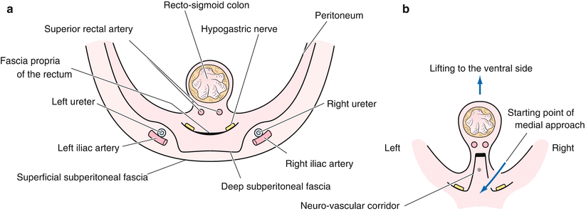
The pelvic fascial composition will be discussed below with a cross-sectional view of the pelvis from the cranial side to the caudal side (Fig. 4.1). However, it should be noted that operative procedure begins at the promontory and is often in the order (2)(1)(3)(4)(5).
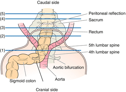
Fig. 4.1
Cross-sectional view of the pelvic space. The pelvic fascial composition will be discussed with a cross-sectional view of the pelvis from the cranial side to the caudal side. However, it should be noted that the operative procedure begins at the promontory and is often in the order (2)(1)(3)(4)(5) [2, with permission]
4.3.1 Level of Aortic Bifurcation (Fig. 4.1 (1))
The superior hypogastric plexus divides to form the right and left hypogastric nerves in the caudal centre of the aorta. A fascia is present at the ventral (deeper) side of the deep subperitoneal fascia that covers the ureter and the aortic bifurcation (Fig. 4.2). This fascia fuses to the deep subperitoneal fascia in the most cranial part, and is called the fascia propria of the rectum. The deepest fascia is thought to be the fascia propria of the rectum according to Dr. Takahashi, based on his concept derived from embryological considerations of organ dominance of the autonomic nerve fibres [3–7]. The deep subperitoneal fascia turns back to the cranial side as the recto-sacral ligament and forms the fascia propria of the rectum (Fig. 4.3). The fascia propria of the rectum then converges towards and fuses with the deep subperitoneal fascia at the superior hypogastric plexus (Fig. 4.4). However, as this part is located on the dorsal side of the sigmoid mesocolon, there may be notable resistance for acceptance of the term if given the name the fascia propria of the rectum. The reason for this will be explained later.
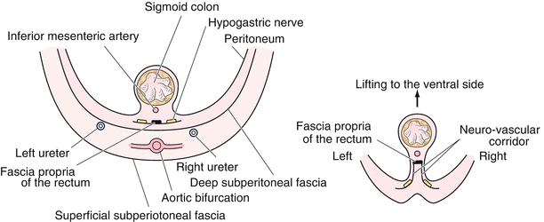
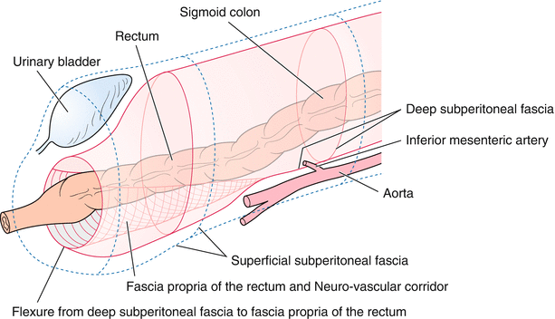
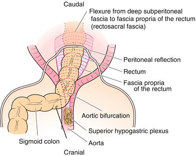
Fig. 4.2
Level of the aortic bifurcation (Fig. 4.1 (1)). The superior hypogastric plexus divides to form the right and left hypogastric nerves in the caudal centre of the aorta. A fascia is present at the ventral (deeper) side of the deep subperitoneal fascia that covers the ureter and the aortic bifurcation. This fascia fuses to the deep subperitoneal fascia in the most cranial part and is called the fascia propria of the rectum [2, with permission]
Fig. 4.3
The fascia propria of the rectum in accordance with the concept of Dr. Takahashi. The deep subperitoneal fascia turns back to the cranial side as the rectosacral ligament and forms the fascia propria of the rectum. The fascia propria of the rectum is the route for the autonomic nerve fibres [2, with permission]
Fig. 4.4
The fascia propria of the rectum from the ventral view. The fascia propria of the rectum converges towards and fuses with the deep subperitoneal fascia at the superior hypogastric plexus
4.3.2 Level of the Promontorium (Fig. 4.1 (2))
This represents the starting point for the medial approach in LapLAR (Fig. 4.5b, arrow). The space between the deep subperitoneal fascia and the fascia propria of the rectum broadens (Fig. 4.5a). At this level the recto-sigmoidal mesenterium is markedly shortened (Fig. 4.5a). This section originates from the fascial propria of the rectum and the neuro-vascular corridor. In other words, this is the portion that lines the mesorectum and the corridor of the neural branches from the hypogastric nerve (Fig. 4.5b).
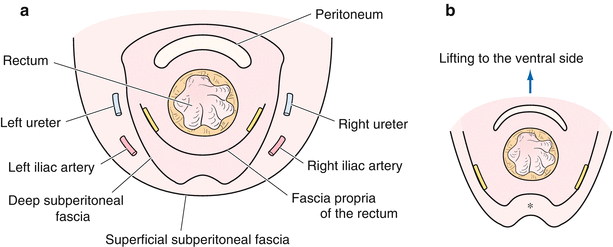
4.3.3 Level of the Rectovesical Fossa (Fig. 4.1 (3))
Part of the rectum is covered with peritoneum only in the rectovesical fossa. The annulus of the deep subperitoneal fascia shrinks with respect to the cranial side and covers the internal and the external iliac arteries and the ureter on the lateral sides, and then continues towards the ventral side of the rectovesical peritoneum that shifts into Denonvilliers’ fascia. The fascia propria of the rectum, as a fascia of the deepest part, is spread caudally. The fascia then closes at the reflection of the deep subperitoneal fascia, which has become the rectosacral ligament (Fig. 4.6a). The retrorectal space is difficult to visualise if the ventral side of the rectum is elevated (Fig. 4.6b*).
4.3.4 Level of the Lateral Ligament (Fig. 4.1 (4))
The space between the fascia propria of the rectum and the deep subperitoneal fascia in the dorsal rectum is closed at this level, and becomes the position for mutual reflection (rectosacral ligament). The two fasciae are fused on the lateral sides of the rectum. The pelvic plexus is then formed, with connective tissue binding the middle rectal vessel, lymph ducts and branches of the rectal nerve, thus forming the lateral ligament (Fig. 4.7).
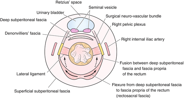
Fig. 4.7
Level of the lateral ligament (Fig. 4.1 (4)). The space between the fascia propria of the rectum and the deep subperitoneal fascia in the posterior rectum is closed at this level, and becomes the position of mutual reflection (rectosacral ligament). On the ventral side of the rectum, the deep subperitoneal fascia lies just ventrally to Denonvilliers’ fascia. The surgical neuro-vascular bundle can be confirmed if the dissection on the medial side is advanced to the right or left. A supralevator space is apparent if the rectosacral ligament is incised on the posterior side of the rectum (solid arrow). This space has been confirmed to lead to the bladder-side space and to the bladder front cavity (Retzius’ space) from the lateral side of the rectum [2, with permission]
On the ventral side of the rectum, the deep subperitoneal fascia exists just ventrally to Denonvilliers’ fascia. This represents the deep subperitoneal fascia that forms the boundary between the urogenital organs and the rectum, as well as the abdomen. The neurovascular bundle can be confirmed if the dissection on the medial side is advanced to the right or left (Fig. 4.7). However, since neurovascular bundles are covered by prostate fascia by definition [8], the neural branch-sprouting out from the pelvic plexus and towards the prostate along the rectum is referred to as the “surgical neurovascular bundle” for convenience (Fig. 4.7).
4.3.5 Level of the Caudal Portion of the Terminal End of the Deep Subperitoneal Fascia (Fig. 4.1 (5))
The ventral side is the final line of the deep subperitoneal fascia. The end of the caudal side is the perineal body. There should be a slight remnant of the deep subperitoneal fascia at this point (Fig. 4.8).
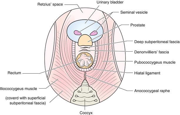
Fig. 4.8
Level of the caudal portion of terminal end of the deep subperitoneal fascia (Fig. 4.1 (5)). The ventral side is the final line of the deep subperitoneal fascia. The end of the caudal side is the perineal body. There should be a slight remnant of the deep subperitoneal fascia at this point. The pubococcygeus muscle and the iliococcygeus muscle form the pelvic floor. They are covered with the superficial subperitoneal fascia. The final portion of the rectum is surrounded by the hiatal ligament. The anococcygeal raphe is located between the hiatal ligament and the coccyx. The puborectal muscle on the caudal side of the hiatal ligament supports the dorsal side of the anal canal
The rectum migrates to the anal canal. The pubococcygeus muscle and the iliococcygeal muscle together form the pelvic floor. They are covered with a superficial subperitoneal fascia. The final portion of the rectum is surrounded by the hiatal ligament that is responsible for the synchronisation of the rectum, bladder neck (upper portion of the vagina), and motion of the pubococcygeal muscle. The anococcygeal raphe is located between the hiatal ligament and the coccyx. The puborectal muscle on the caudal side of the hiatal ligament supports the dorsal side of the anal canal [9] (Fig. 4.8).
4.4 Operative Procedures (in Men)
4.4.1 Intraoperative Positions, Trocar Site and the Elimination of the Small Intestine
(See the Chap. 2).
4.4.2 Operative Procedures (Figure Shows the Case of Men)
For details relative to the dissection of the sigmoid colon and dissection of central lymph nodes, see the section on Laparoscopic sigmoidectomy (See the Chap. 3).
4.4.2.1 Dissection in the Lesser Pelvis (Especially in the Retrorectal Space)
Scope: umbilicus.
The operator’s right hand: an electrosurgical knife with spatula-type blade.
The operator’s left hand: performs traction of the fascia propria of the rectum to the ventral-caudal side.
The assistant’s right hand: assists in the fine manoeuvres of the operator.
Assistant’s left hand: adequately raises the fascia propria of the rectum to the ventral side.
The bilateral parietal peritoneum along the rectum is excised towards the rectovesical fossa. It is important that the assistant maintains the spread of operative field. The vascular stump is retracted ventrally by the assistant’s right hand, and the assistant’s left hand catches the fascia propria of the rectum and retracts it to the ventral side sufficiently enough to provide an adequate surgical plane. Next, given that the assistant’s right hand is free, the assistant eliminates what has now become an obstacle to the operator by his right hand.
In the field of view of the retrorectal space, the fascia propria of the rectum is dissected from the dorsal side midway between the branches of the hypogastric nerves (Fig. 4.9). Furthermore, it is conducted towards the pelvic floor as far as possible without moving to the lateral sides of the rectum (Figs. 4.5b*, 4.6b*, and 4.9). After dissecting the retrorectal space, the rectal branches stemming from the hypogastric nerves are extended and divided (Fig. 4.9).
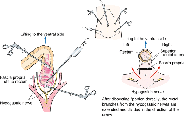
Fig. 4.9
Field of view and the dissection of the retro-rectal space. It is important that the assistant maintains the extent of the operative field. In the field of view of the retro-rectal space, the fascia propria of the rectum is dissected from the dorsal side midway between the branches of the hypogastric nerves. After dissecting the retro-rectal space, the rectal branches stemming from the hypogastric nerves are extended and divided
The operator dissects and cuts along the midline towards the caudal-ventral side using his right hand while holding the ventral rectum caudal-ventrally with forceps in his left hand. At the midline, the retrorectal space is dissected as far as possible, and the rectosacral ligament, which is the fold of the deep subperitoneal fascia leading towards the ventral-cranial side, is identified (Figs. 4.3, 4.4, and 4.7). Because the neural branches from the hypogastric nerves are close to the rectum, it is important to proceed without removing the midline. By cutting the rectosacral ligament (Fig. 4.7, solid arrow line), the pelvic floor can be reached. At this point, the supralevator space can be dissected off like a comma-shaped jewel on both sides, and the dissection along the inside of the pelvic plexus can be performed.
At this point, the rectal branches from the pelvic plexus are raised to the ventral side, and the space between the rectum and the pelvic plexus can be visualised (Fig. 4.7, dotted arrow). It is possible to cut off the rectal branches from the pelvic plexus near the mesorectum (Fig. 4.7, dotted arrow). Furthermore, following the exposure of the caudal edge of the S4 pelvic splanchnic nerve and the division of the branch to the rectum, the levator ani muscle is sufficiently visible. By dissecting the rectum along the caudal-ventral direction on the bilateral side, the supralevator space forms a shape similar to that of a comma-shaped jewel (Fig. 4.10b).
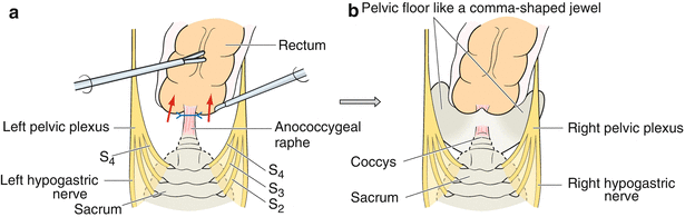
Fig. 4.10
Disconnection of the anococcygeal raphe (a). After performing the dissection of the rectum in the caudal-ventral direction on the bilateral side on the pelvic floor, the supralevator space assumes a comma-shaped jewel form (b). In addition, the anococcygeal raphe is removed from the midline of the rectum and is divided at the hiatal ligament (a)
After the supralevator space is dissected like a comma-shaped jewel, and the rectum is dissected to the caudal-ventral side, the anococcygeal raphe that runs from the coccyx to the rectum can be seen. Furthermore, if the anococcygeal raphe is dissected along the slightly bilateral side, it is possible to expose the anococcygeal raphe even more clearly (Fig. 4.10a). The rectum is dissected further to the caudal side and the anococcygeal raphe is then divided at the hiatal ligament (Fig. 4.10a). When the hiatal ligament is incised along the rectum at the bilateral side, the puborectal muscle, which is located behind the hiatal ligament, can be seen as having a V-shape at the dorsal side because the U-shaped becomes V-shaped when raised ventrally (Fig. 4.8). Having dissected the inside of the puborectal muscle, it is possible to enter the anal canal.
In the presence of bleeding within the area of the pelvic floor, it is difficult to continue surgery using this surgical plane. Thus, it is important to proceed with adequate haemostasis.
4.4.2.2 Incision and Dissection of the Ventral Side of the Rectum (Dissection of the Ventral Side of Denonvilliers’ Fascia)
Scope: umbilicus.
The operator’s right hand: an electrosurgical knife with spatula-type blade.
The operator’s left hand: develops the ventral side of the rectum.
The assistant’s right hand: ensures traction of the serosa at the urinary bladder ventrally (or traction of the serosa at the urinary bladder to the lateral-ventral side).
The assistant’s left hand: ensures traction of the rectum cranially (or traction of the serosa at the urinary bladder to the lateral-ventral side).
For the dissection of the retrorectal side, if the field of view is felt to be insufficient for the procedure, dissection of the ventral side of the rectum should be performed.
At approximately 1 cm from the transition of the serosa to the rectovesical fossa, the serosa incision should be performed and bilateral peritoneal divisions continued (Fig. 4.11). If the rectum falls within the operative field, the assistant’s left hand bowel forceps grips the rectum and can lead it to the cranial side. The assistant’s right hand bowel forceps lift the serosa ventrally at the urinary bladder to form a counter-traction with the bowel forceps in the operator’s left hand (Fig. 4.11a). The dissection is easily visualised; the visualisation of the surface plane for dissection is established by the assistant’s hands and the operator’s left hand. It is also possible for the assistant to control the side of the urinary bladder and for the operator to control the rectal side (Fig. 4.11b).
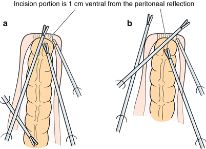
Fig. 4.11
Dissecting and dividing of the ventral side of the rectum. At about 1 cm from the transition of the serosa in the rectovesical fossa, the serosa incision should be performed and bilateral peritoneal divisions are continued. A plane for dissection is established by both hands of the assistant and by the surgeon’s left hand. (a) If the rectum falls within the operative field. (b) If the separating portion is likely to expand
At the medial part, the procedure continues with the dissection of the caudal tissue from the ventral side of Denonvilliers’ fascia. In other words, this is achieved by entering between the dorsal side of the deep subperitoneal fascia and the Denonvilliers’ fascia. Without continuing to dissect on both sides, it is important to dissect the median in order to reveal the inner sides of the bilateral seminal vesicles (Fig. 4.12). The dissection continues between the dorsal side of the seminal vesicle and Denonvilliers’ fascia. If the seminal vesicle is confirmed to be covered with the deep subperitoneal fascia, it is not to be mistaken for the dissecting layer. Following careful haemostasis of the small veins from the cranial and lateral side of the seminal vesicles, the dissection can continue towards the caudal side. It is better to dissect the Denonvilliers’ fascia caudally as far as possible. There are visible dents indicating the passage to the supralevator space along the bilateral side of the rectum.
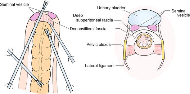
Fig. 4.12
Dissection between Denonvilliers’ fascia and the deep subperitoneal fascia. At the medial part, the procedure requires to continue the dissection caudally along the dorsal side of the deep subperitoneal fascia from the ventral side of Denonvilliers’ fascia. If the seminal vesicle is confirmed covered by the deep subperitoneal fascia, it is not to be mistaken for the dissecting layer
With regards to the dissection of the dorsal side and the ventral side of the rectum, the optimal order has not yet been determined. Therefore, if the field of view can be secured and the traction for the view is effective, it is better to continue to advance the field of the operation. Repeating the dorsal and the ventral dissection is appropriate.
4.4.2.3 Dissection of the Right Lateral Ligament
Scope: umbilicus.
The operator’s right hand: an electrosurgical knife with spatula-type blade or an ultrasonically activated device (USAD).
The operator’s left hand: maintains traction of the dorsal side of the rectum to the left.
The assistant’s right’s hand: maintains traction of the rectum ventrally.
The assistant’s left hand: maintains traction of the rectum to the cranial and ventral side.
In order to divide the right rectal branches from the pelvic plexus, the method involves the separation of a sheet of the branches using an USAD in the dorsal view of the rectum. Subsequently, a sheet of branches can be divided from the ventral side of the rectum without causing any further damage to the pelvic plexus (Fig. 4.13). Advancing the dissection along the ventral side along inner surface of the pelvic plexus, the operator ultimately reaches the seminal vesicle that was previously dissected on the ventral side. In this way, a sheet of rectal branches from the pelvic plexus is guided like a tent towards the rectum. If the apex of the tent can be treated sharply by an electrosurgical knife and dissected bluntly, the so-called lateral ligament can be completely excised (Fig. 4.14).
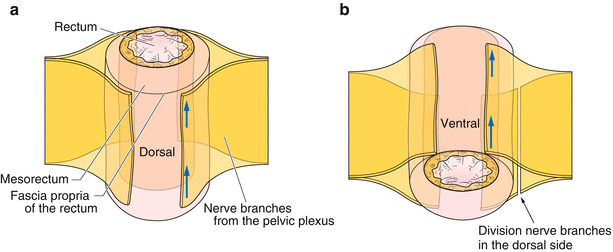
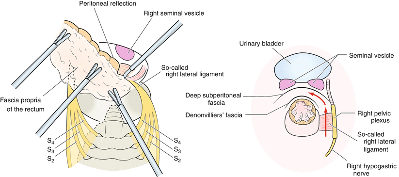
Fig. 4.13
Imaginary view of the division of the rectal branches from the pelvic plexus. To divide the rectal branches from the pelvic plexus, the procedure involves the separation of a sheet of branches in the dorsal view of the rectum (a). Subsequently, a sheet of branches can be divided from the ventral side of the rectum without causing any further damage to the pelvic plexus (b)
Fig. 4.14
Dissection and division of the right lateral ligament. The pelvic plexus is guided like a tent towards the rectum. If the apex of the tent can be treated sharply by electrocautery and dissected bluntly, the so-called lateral ligament can be completely excised
4.4.2.4 Dissection of the Left Lateral Ligament
Scope: umbilicus.
Related posts:


Stay updated, free articles. Join our Telegram channel

Full access? Get Clinical Tree


