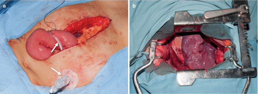
Fig. 16.1
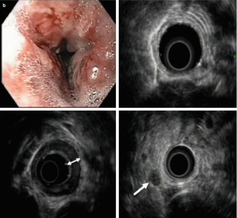
(a) Conventional chest x-ray demonstrating a history of sternotomy for coronary bypass surgery, as well as previous placement of a cardiac pacemaker. (b) Endoscopy (upper left) showing ulcerated mass in the distal esophagus, and endoscopic ultrasound showing transmural extension of the tumor into the adventitia in the area of the ulcerated mass (cT3) (two-headed arrow) and multiple enlarged paraesophageal lymph nodes (cN1) (arrow)
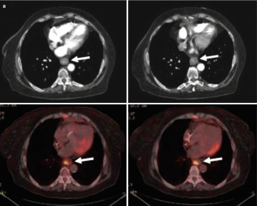
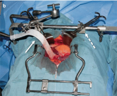
Fig. 16.2
(a) CT scans at initial staging (top) showing the distal esophageal tumor (arrows); PET/CT scans (bottom) with increased standardized uptake value (SUV 14) (arrows). (b) Limited midline incision from the xiphoid to 3–4 cm above the umbilicus. Mobilize liver by taking down the triangular ligament, and retract the liver to the patient’s right with a fixed retractor (solid arrow). Ideally, a fixed retraction system (dashed arrow) lifts the costal margin to verticalize the diaphragm and provide an unobstructed view of the proximal stomach and esophagogastric (EG) junction
16.2 Procedure
Make a limited midline incision from the xiphoid to 3–4 cm above the umbilicus (Fig. 16.2b). Mobilize the liver by taking down the triangular ligament, and retract the liver to the patient’s right with a fixed retractor. Ideally, the fixed retraction system will provide an unobstructed view of the proximal stomach and esophagogastric (EG) junction.
Figure 16.3a shows the completed dissection of the EG junction, encircled by a Penrose drain. Transhiatal dissection mobilizes the esophagus, mediastinal fat, and level 8 lymph nodes. Dissection should be completed over 8–10 cm. Any attachment or invasion of crura or diaphragm should be resected en bloc. Following Kocherization of the duodenum, assess for pyloric stenosis (Fig. 16.3b). Whether a pyloric procedure is done remains controversial. We do not do a pyloroplasty unless pyloric stenosis is noted at preoperative esophagogastroduodenoscopy (EGD) or intraoperatively. Ideally, the duodenum should be mobilized sufficiently to place the pylorus 3–4 cm below the hiatus.
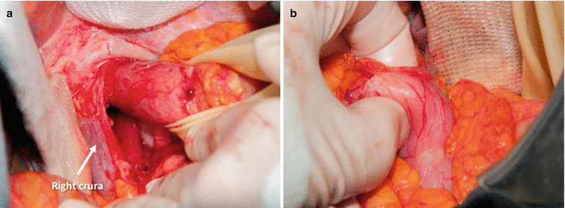

Fig. 16.3
(a) Completed dissection of the EG junction, encircled by a Penrose drain. (b) Assessing for pyloric stenosis following Kocherization of the duodenum
Figure 16.4a shows the dissection of the greater curvature, ensuring preservation of the right gastroepiploic arcade. Omental attachments are taken down, starting at the watershed area between the left and right gastroepiploic arcades. The right gastroepiploic arcade is preserved (Fig. 16.4b). When the greater curve is completely mobilized (Fig. 16.4c), ideally the omentum is preserved along the upper body and cardia of the stomach to cover the anastomosis (see Fig. 16.16c). The mobilized stomach shown in Fig. 16.4d demonstrates posterior access to the left gastric pedicle. Lymph node dissection includes supraceliac, suprapancreatic, and paragastric nodes. Figure 16.5a shows dissection of the left gastric vein. The left gastric artery is ligated immediately above the celiac axis (Fig. 16.5b).
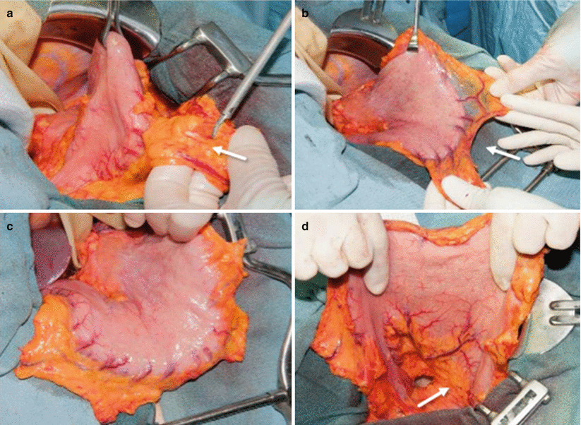
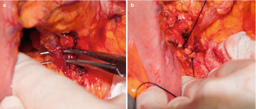

Fig. 16.4
(a) Dissection of greater curvature, ensuring preservation of the right gastroepiploic arcade. Starting at the watershed area (arrow) between the left and right gastroepiploic arcades, omental attachments are taken down. (b) Right gastroepiploic arcade is preserved (arrow). (c) Greater curve completely mobilized. (d) Mobilized stomach demonstrating posterior access to left gastric pedicle (arrow). Lymph node dissection includes supraceliac, suprapancreatic, and paragastric nodes

Fig. 16.5
(a) The entire suprapancreatic and supraceliac lymph node fields are dissected. This image shows dissection of the left gastric vein. (b) The left gastric artery is ligated immediately above the celiac axis
The lesser curvature is dissected 7–10 cm distal to the EG junction (Fig. 16.6a), at a location guided by pretreatment endoscopy and endoscopic ultrasound to provide a minimum of 5–7 cm of distal resection margin. The conduit is then fashioned with sequential firing of a linear stapler (Fig. 16.6b–d). It is important to keep the stapled margin oriented and parallel to the greater curve.
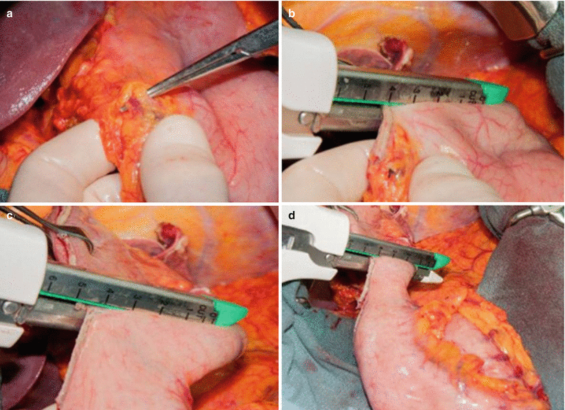

Fig. 16.6
(a) The lesser curvature is dissected 7–10 cm distal to the EG junction, providing a minimum of 5–7 cm of distal resection margin. (b–d) The conduit is then fashioned with sequential firing of a linear stapler
The extent of gastric resection can vary. It should be based on findings at endoscopy and endoscopic ultrasound. The resection in Fig. 16.7a provides a margin of 10–12 cm from the EG junction. The width of the conduit remains controversial; we aim to fashion a gastric conduit 3–4 cm in width (Fig. 16.7b), preserving the right gastroepiploic arcade and the proximal right gastric arcade.
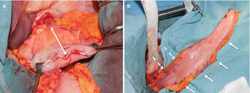

Fig. 16.7
(a) This gastric resection provided a 10- to 12-cm resection margin from the esophagogastric junction (arrow). (b) We aim to fashion a gastric conduit 3–4 cm wide, with preservation of the right gastroepiploic arcade (arrows) and proximal right gastric arcade (dashed arrows)
To decrease the risk of bleeding, we recommend oversewing the staple line with interrupted 3.0 silk sutures (Fig. 16.8a). The last imbricating stitches at the tip of the conduit are left long, with the needle attached (Fig. 16.8b). These sutures are used to attach the tip of the conduit to the gastric portion of the specimen (Fig. 16.8c) so it can be drawn up into the chest following esophageal mobilization.
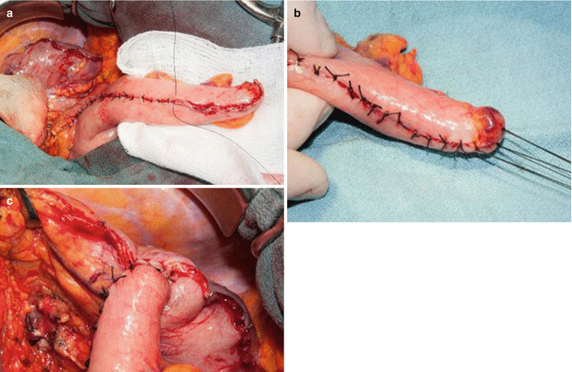

Fig. 16.8
(a) The staple line is oversewn with interrupted 3.0 silk sutures. (b) The last imbricating stitches at the tip of the conduit are left long, with the needle attached. (c) These sutures are used to attach the tip of conduit to the gastric portion of the specimen
A 14 Fr feeding jejunostomy is placed 60–80 cm from the ligament of Treitz (Fig. 16.9a). The tube is imbricated into the antimesenteric jejunum over a 2-cm distance and suspended to the peritoneal surface circumferentially. To avoid torsion, it is then tacked proximally and distally to the peritoneal surface over 3–4 cm.
The second stage of the procedure is initiated with a limited thoracotomy, typically performed at the fourth or fifth interspace (Fig. 16.9b). The visceral pleura is incised and the azygous vein is ligated (Fig. 16.10a). The esophagus is mobilized just distal to the azygous vein (Fig. 16.10b). Esophageal mobilization proceeds distally, mobilizing all paraesophageal nodes en bloc. Subcarinal nodes can be mobilized en bloc or separately.
