15 Inflammatory bowel disease
Case
A 28-year-old female presents with a 3-month history of diarrhoea, weight loss, fatigue, and right lower quadrant abdominal pain. She complains of six to eight non-bloody bowel movements daily with at least one at night that awakens her from sleep. She has lost 10 kg since her symptom onset. She has fevers to 38.5°C without chills. She also complains of perianal pain with purulent drainage. She has generalised joint aches with intermittent swelling of the knees and mouth sores. Her past medical history is otherwise unremarkable and she does not take any medications. She denies taking any non-steroidal anti-inflammatory drugs. She is an active smoker of one pack a day for the last 5 years. She has no family history of inflammatory bowel disease.
On examination, she is a normally developed, thin-appearing female in no distress. Her heart rate is 110 beats per minute, blood pressure is 90/40 mmHg and temperature 38°C. There are aphthous-type ulcerations in the buccal mucosa of the mouth. There is mild right knee effusion without erythema. Abdominal examination reveals soft diffuse tenderness with localisation to the right lower quadrant and a possible mass. There are no guarding and no rebound. Rectal exam finds a draining perianal fistula without fluctuance, and a mild anal stricture.
Pertinent admission studies of blood haematology and chemistry show the following: haemoglobin 9 g/dL, haematocrit 27, white blood cell count 24,000, blood urea nitrogen 30, creatinine 1.0, anion gap 12 and albumin 2.8 g/dL.
Abdominal and chest plain films are negative. CT enterography shows terminal ileal thickening with narrowing. There is mesenteric stranding with borderline enlarged lymph nodes.
Stool studies show Clostridium difficile toxin, ova and parasites; culture, Escherichia coli O157:H7 pending.
The patient was admitted. Intravenous hydration and antibiotics were administered. Intravenous methylprednisolone 50 mg daily was started for a presumptive diagnosis of Crohn’s disease. A tuberculosis skin test (PPD) was placed. On day 2 the patient was afebrile and underwent a colonoscopy, which showed ulceration of the terminal ileum and right colon and also in the lower rectum (biopsies subsequently showed findings consistent with Crohn’s disease). Patient shows some signs of clinical improvement on day 3 with better volume status and some decrease in diarrhoea. Stool studies and PPD were negative. Methylprednisolone was switched to oral prednisone. She was infused with infliximab and continued to show clinical improvement and was discharged on hospital day 5. Prednisone was tapered off over 4 weeks. She was seen after her second infliximab infusion 9 days later. Her diarrhoea had cleared, her energy improved and her mouth sores and joint symptoms resolved. Her perianal drainage, while still present, had decreased. Infliximab was continued for remission maintenance. At last follow-up 6 months later she was completely asymptomatic and her perianal fistula had closed.
Introduction
Inflammatory bowel disease (IBD) is traditionally divided into ulcerative colitis (UC) and Crohn’s disease (CD). Unlike infectious colitis that is typically self-limited, IBD is a chronic illness that is punctuated by disease exacerbations and remissions. While some similarities exist, UC and CD are very different diseases with regard to their clinical presentation, pattern of bowel involvement, response to therapy and prognosis (Table 15.1 and Box 15.1). With current methods, proper diagnosis of CD or UC can be made, although 10–15% of patients are labelled indeterminate colitis. Incidence and prevalence rates for UC and CD vary throughout the world but are highest in developed countries of northern Europe and North America.
Table 15.1 Pathological features in inflammatory bowel disease
Ulcerative colitis | Crohn’s disease |
---|---|
Distribution | |
Macroscopic appearance | |
Microscopic appearance | |
∗ Preservation of crypt architecture suggests acute self-limited colitis.
† Granulomas may occur in tuberculosis, schistosomiasis and syphilis.
Ulcerative Colitis
Aetiology
The aetiology of ulcerative colitis is unknown. Current research focuses on genetic associations and interactions of bowel flora with the colonic mucosa but no definitive cause has been found. While genetic predisposition is the strongest risk factor for the development of UC its contribution is minor with only about 10% having a positive family history. Cigarette smoking appears to be protective, either by reducing the risk of UC development or delaying its onset. Diet appears not to influence disease onset or course while emotional stress may lead to disease flares.
Clinical presentation
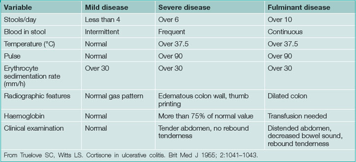
UC typically presents in the third or fourth decade of life. Clinical presentation is based on disease location and severity. Severity is classified as mild, severe or fulminant with moderate disease between mild and severe (Table 15.2). Diarrhoea, usually bloody with cramping typically awakening the patient from sleep, signifies mucosal inflammation. Mild disease may be accompanied by frequent diarrhoea but is not associated with significant life impairment. Moderate disease, between mild and severe, typically leads to significant symptoms including profound diarrhoea (usually bloody), dehydration, fatigue and weight loss. This may progress to severe disease, which may require hospitalisation. Fulminant disease is most severe often requiring intensive care or surgery.
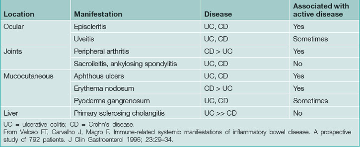
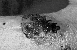
UC may be associated with extraintestinal manifestations in some patients (Table 15.3) which may be associated with disease activity (Fig 15.1). Those not associated with disease activity may run a clinical course independent of the disease and persist after colectomy.
Disease extent and location
UC can be divided into three groups based on the disease location: proctitis or proctosigmoiditis (continuous involvement, from the anus, of the rectum or rectosigmoid); left-sided colitis (continuous involvement up to the splenic flexure); and pancolitis (continuous involvement extending proximal to the splenic flexure). Patients with more extensive disease have a worse prognosis. Proximal progression is common. Approximately half the patients with proctitis and two-thirds of patients with left-sided colitis will progress to a more proximal involvement aover time.
Frequent low-volume bowel movements, urgency, rectal bleeding and tenesmus alone suggest proctitis. However, patients with or without these symptoms may also present with prostration, fever, tachycardia, dehydration and complications of blood loss, suggesting more severe disease or more extensive bowel involvement. Advanced cases may present with massive abdominal distention because of megacolon. In fulminant colitis, severe diarrhoea and high C-reactive protein despite intensive treatment predict eventual colectomy. Fortunately, the presentation of fulminant colitis is relatively uncommon.
Differential diagnosis
The presenting clinical features of ulcerative colitis are non-specific, especially among patients without a prior history. Acute colitis secondary to infection and ischaemia are important considerations depending on the patient and are of particular importance in elderly patients. A history of previous episodes is important but distinguishing a flare of UC from an infectious colitis is not possible on clinical grounds alone. Crohn’s disease that is confined to the colon may be difficult to distinguish from UC, but may present with rectal sparing and skip lesions.
Diagnosis
Among patients without a prior history of ulcerative colitis, presenting features are non-specific so that additional diagnostic testing is required. Diagnosis is confirmed by clinical features, endoscopic and histological findings as well as stool examination to exclude acute colitis. Infection, ischaemia, non-steroidal anti-inflammatory medication-induced colitis, diverticular colitis and Crohn’s disease are important considerations.
Stool studies
Infectious colitis may present independently or result in a flare of existing UC. Exclusion of enteric infection is paramount since these infections can mimic clinical and endoscopic features of active UC. Specific stool studies to exclude common enteric pathogens such as Shigella spp., Campylobacter spp., Yersinia spp., Entamoeba spp. and enterohaemorrhagic Escherichia coli are essential. Stool studies may be negative even in the presence of infection and it is very important that the clinician understand what is routinely tested for in his or her facility when stool culture, ova and parasite analysis are ordered. Exclusion of C. difficile infection among patients with UC is very important. While commonly associated with prior antibiotic use, hospitalisation or residence at a long-term care facility, patients with IBD are at a higher risk for this infection even in the absence of these usual risk factors. Stool C. difficile toxin should be requested on all patients with IBD presenting with symptoms of a flare.
Stool biomarkers
Reliable stool biomarkers to predict disease activity, recurrence or likelihood of relapse in IBD would avoid invasive endoscopy and/or radiation exposure. Calprotectin, a biomarker that can easily be detected in stool, has been shown to be a sensitive and specific marker of relapse in UC but not CD. Faecal lactoferrin is elevated in active IBD and may increase prior to IBD relapse. Unfortunately, while results are encouraging, data on biomarkers are limited. More information is needed before they can be reliably used in clinical practice.
Haematology and biochemistry
Haematological measurements may vary based on the severity and duration of the colitis. Patients with mild colitis may have minimal abnormalities whereas patients with more severe disease may have a leukocytosis related to inflammation and anaemia due to blood loss. Anaemia may be more pronounced and associated with frank iron deficiency with longer duration of illness. Thrombocytosis may also be present due to inflammation or iron deficiency. Albumin may be low due to severe disease or malnutrition. C-reactive protein or sedimentation rate may be elevated. Electrolyte imbalance and prerenal azotemia may occur secondary to dehydration. Alkaline phosphatase may be elevated if primary sclerosing cholangitis is present or as a result of drug toxicity.
Radiology
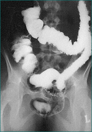
Plain abdominal x-rays should be performed to exclude toxic megacolon and perforation among severely ill patients. Computed tomography (CT) scanning may be considered if there is concern of other aetiologies such as Crohn’s disease. CT of the abdomen may reveal colonic thickening and provide additional information as to the extent of the disease. Barium enema is rarely performed because of the widespread use of sigmoidoscopy and colonoscopy (Fig 15.2).
Sigmoidoscopy, colonoscopy and biopsy
Endoscopic visualisation with biopsy is essential for all patients with suspected UC and is often necessary to document disease activity for a suspected flare. Disease activity, if present, should be visible in the rectum and may extend more proximally. Rectal sparing or segmental colitis raises the possibility of infection, ischaemia, Crohn’s disease or other aetiology. Biopsies confirm the presence of chronic colitis and exclude acute colitis as seen with infection. Among patients with colitis refractory to therapy, especially if they are receiving corticosteroid or immunosuppressive therapy, cytomegalovirus should be excluded by biopsy with special studies. Severely ill patients may undergo an unprepped flexible sigmoidoscopy whereas patients with mild to moderate disease are often prepped for colonoscopy. There is usually little value in performing a complete colonoscopy on patients with active UC and it may be dangerous. However, prepping for colonoscopy, if possible, will allow for better visualisation above the sigmoid colon, especially if findings are not typical for UC or it is important to determine extent of disease for therapy. Flexible sigmoidoscopy with biopsy is often sufficient for those with established disease to confirm disease activity. Among patients with long-standing quiescent disease proximal to the rectum, colonoscopy with biopsies is routinely performed for dysplasia surveillance.
Pathology
Ulcerative colitis results from chronic inflammation confined to the mucosal layer of the colon. Inflammation is continuous and may involve only the rectum or may spread more proximally. Confusion with CD occurs when patients with UC have a discrete area of inflammation in the caecum (a ‘caecal patch’), or those with a flare of pancolitis have an inflamed terminal ileum (‘backwash ileitis’). Both of these presentations are consistent with ulcerative colitis.
Treatment
Treatment goals for UC are induction and maintenance of remission and are based on disease extent, clinical severity and previous medication history.
Mild to moderate disease
Oral mesalamine
The best option for patients with mild to moderate UC is mesalamine (5-aminosalicylate), orally or rectally. Response rates vary because of differences in dose, disease activity and definitions of outcomes but about two-thirds will respond. Sulfasalazine, the first of this class, is both an inexpensive and effective medication. It remains essentially unchanged until it reaches the colon where it is cleaved into the active moiety 5-aminosalicylate and sulfapyridine by bacteria. Sulfapyridine (a sulfa antibiotic) is responsible for most of the observed toxicity that includes allergic reaction or renal toxicity (Box 15.2). The remaining mesalamine formulations differ on their mechanism and site of drug delivery. Newer preparations release in the colon, require fewer tablets and can be dosed once or twice daily to improve compliance. These medications are well tolerated. Headache is the most common adverse effect and, rarely, renal toxicity occurs. A baseline serum creatinine should be determined prior to starting therapy. Pancreatitis has also been described with sulfasalazine due to its sulfa moiety but also with mesalamine, although the likelihood with mesalamine is extremely low.
Box 15.2 Side effects of sulfasalazine∗
∗ Start at one 500 mg table, twice daily on the first day after meals, then increase by two tablets every second day until the patient is taking the full amount (typically, 1 g four times a day in acute disease; and 1 g twice a day for prophylaxis). Usually efficacious within 2–4 weeks (in 40–80% of patients) A blood count and urinalysis should be performed before starting therapy and then regularly during the first 3 months of treatment, and thereafter every 6 months. Supplement with folate, 1 mg/day orally. The drug may also be of specific benefit for the arthropathy in ankylosis spondylitis associated with inflammatory bowel disease.
Mesalamine is not useful in moderate to severe UC and in mild to moderate disease is often administered at too low a dose. In appropriate doses all are probably equally effective. Sulfasalazine is relatively inexpensive and once or twice daily preparations will enhance compliance. Olsalazine should be avoided if possible, because it can cause secretory diarrhoea. Common preparations with the usual doses to induce and maintain remission are outlined in Table 15.4.
Table 15.4 Oral mesalamine preparations for induction and maintenance of remission in mild to moderate ulcerative colitis
Optimal daily dose for remission (g) | ||
---|---|---|
Drug | Induction | Maintenance |
Oral mesalamine | ||
Sulfasalazine | 4–6 | 2–4 |
Eudragit S coated | 3.6–4.8 | 2.4–3.6 |
Ethylcellulose coated | 3–4 | 2–3 |
Balsalazide | 6.75 | 3–4.5 |
Topical mesalamine | ||
Mesalamine suppository | 0.5–1 twice daily | 1 daily |
Mesalamine enema | 4 nightly | 4 nightly or every other night |
From Kornbluth A, Sachar DB. Ulcerative colitis practice guidelines in adults. Am J Gastroenterol 2010; 105:501–523.
Topical preparations
The most underutilised group of mesalamine medications are the topical rectal preparations. These are effective for symptoms of urgency, pain and tenesmus. In distal colitis they are more effective than oral preparations and a combination of oral and topical mesalamine enema improves overall efficacy. Patients with proctitis (disease limited to the last 15 cm of the colon) benefit from mesalamine suppositories, 1000 mg once or twice daily, while mesalamine enema therapy (4 g at bedtime) is useful for treatment of disease up to the splenic flexure. For patients who cannot hold a whole enema, start with half of the preparation, which will also be beneficial, and work up to a full enema daily. Oral corticosteroids are rarely required for disease confined to the rectosigmoid region.
Hydrocortisone enema may be added in the morning if the patient has not responded to rectal mesalamine after a period of 2–3 weeks. Rectal corticosteroids are helpful in resistant cases but mesalamine enemas are more effective as first-line therapy. After response, wean the topical steroids and maintain rectal and topical mesalamine. If after several weeks response is maintained, try to taper topical mesalamine but it may be required indefinitely. Oral mesalamine should be continued as maintenance therapy to prevent relapse.
Moderate to severe colitis
In cases of moderate to severe UC systemic corticosteroids or infliximab are the best options.
Systemic corticosteroids
The most common errors made with corticosteroids are an inadequate or excessive starting dose, tapering too quickly and use for remission maintenance. Although various preparations have been tested, parenteral methylprednisolone and oral prednisone are most commonly used. Once-daily dosing of 50 mg methylprednisolone parenterally in hospitalised patients and 40–60 mg of prednisone daily in outpatients are adequate doses for efficacy. Parenteral hydrocortisone (300 mg) is equally effective and may be substituted for methylprednisolone (60 mg).

Stay updated, free articles. Join our Telegram channel

Full access? Get Clinical Tree

