Fig. 4.1
Colpocystodefecography in the resting phase. The anorectal junction, the vaginal fornices, and the bladder neck lie over the pubococcygeal line (dashed line). The anorectal angle measures 93°
During voluntary contraction of the pelvic floor, the ARA decreases to about 75°, and the ARJ migrates cranially. The puborectal impression becomes more evident because of the contraction of the levator ani, pulling the ARJ toward its insertion at the symphysis pubis.
When the patient is asked to strain, the converse is seen: the ARA increases with partial to complete loss of puborectal impression, and the pelvic floor descends. The degree of caudal migration, as measured in relation to the bony landmarks, is considered normal when it is less than 3.5 cm relative to the resting position.
During evacuation, the anal canal and the rectum migrate caudally. The ARA increases in relation to the relaxation of external and internal sphincters and puborectal muscle, respectively. Puborectalis sling impression on the rectum posterior wall disappears almost completely, and the anal canal reaches the widest diameter (it should open to a mean anteroposterior diameter of 1.5 cm).
During the late phases of evacuation, the rectal bulb funnels and its walls progressively collapse. A small amount of infolding of redundant anterior and posterior rectal walls is considered normal. The entire process lasts less than 30 s in physiologic conditions.
At the end of evacuation, the resting condition is reached when the anal sphincters close and levator ani restores its tone, pulling the ARJ cranially. The rectum is completely empty, and only minimal barium dye can be found [2].
4.3 Magnetic Resonance Defecography Technique
Pelvic floor anatomy is complex and DCP does not show the anatomical details that pelvic magnetic resonance imaging (MRI) provides. Technical advances, allowing acquisition of dynamic rapid MRI sequences, have been applied to pelvic floor imaging [6]. MRD, although still an experimental technique, may be a useful tool in preoperative planning of these disorders and may lead to a change in surgical therapy, reducing postoperative relapses especially in patients with multicompartmental disorders [7–9].
Concerns about the risks related to radiation, especially in women of childbearing age, and the spreading of pelvic floor MRI in addition to questions relating to the clinical significance of DCP findings have added to these controversies. Furthermore MRD allows the detection of incidental pathologic conditions, such as urethral and bladder diverticula, endometrial polyps, malignant lesions, fibroids, and adnexal lesions [10–12].
A potential disadvantage of MRD is its less physiologic nature if performed with the patient in the supine position, but sitting dedicated systems have been developed [13, 14]. In some studies, MRD has been performed in the orthostatic position also [15].
4.3.1 Preparation
The patient performs a rectal cleaning enema at home. The MRI protocol requires no oral or intravenous contrast agents, since the small bowel is inherently delineated. In some protocols, contrast material is not used to opacify the bladder and patients are requested not to void for 1–2 h prior to their examination.
4.3.2 Technique
Protocols vary by institution, with MRI studies performed with the patient in both supine and upright positions, either with closed or open magnets. Studies have been performed without contrast material, with vaginal and rectal markers, and with rectal, vaginal, urethral, and bladder contrast material.
In the majority of cases, a 1.5 T system is used and T2-weighted turbo spin echo and balanced fast field echo sequences are acquired, with the patient in the supine position. Pelvic or torso phased-array coils are centered low on pelvis to ensure visualization of prolapsed organs.
At the beginning of examination, the patient is positioned on the left side with flexed knees and the rectum is filled with 200 mL echographic gel; the vagina is also filled with an echographic gel suspension of 100 mL. Finally, the bladder is filled with 180 mL physiological solution via a 16 F double-way Foley catheter, which is removed soon after filling; otherwise the patient is asked not to void for 1–2 h before the examination.
With the patient at rest, static images of the entire pelvis are acquired in the axial, sagittal, and coronal planes using rapid half-Fourier T2-weighted sequences (HASTE, half-Fourier acquisition single-shot turbo spin echo) (recommended parameters: TR 3,000 ms; TE 90 ms; flip angle 90°; slice thickness 5 mm; FOV 250 × 250, matrix 448 × 345).
After the midline between the pubic symphysis and the coccyx is localized, dynamic images are acquired with a balanced sequence in the sagittal plane; a single stack of images is acquired continuously at rest, and during maximal contraction, straining, and defecation (recommended parameters: TR 2.7 ms; TE 1.35 ms; flip angle 45°; slice thickness 10 mm; FOV 300 × 300, matrix 160 × 160; dynamic scan time 0.432 s, dynamic images 100).
At our institution, MRD is also performed on a permanent open magnet with changeable position and static 0.25 T field. The magnet table is provided with a tilting mechanism going from 0° to 90° and pitched at 80° to allow a semi-orthostatic acquisition [15].
A surface lumbar spine DPA coil is used as the receiving coil, composed of a stiff base and a flexible anterior band with variable dimensions.
Before the examination, the rectum is filled with 200 mL mashed potato mixed with 1 mL paramagnetic contrast medium. The bladder is also filled with 180 mL physiological solution mixed with 3 mL contrast medium via a catheter, left in place during the entire study. Finally vagina is filled with an echographic gel suspension mixed with 0.5 mL paramagnetic contrast medium.
The three orthogonal image planes are acquired at rest using three-dimensional HYCE (hybrid contrast enhanced), a type of gradient echo (GE) balanced sequence (TR 10 ms; TE 5 ms; flip angle 90°; sections 20; section thickness 2.5 mm; FOV 280 × 280, matrix 200 ×160).
Static images are acquired in the sagittal plane at rest, during sphincter contraction and straining using a GE T1-weighted sequence (TR 35 ms; TE 10 ms; flip angle 90°; section 1; section thickness 5.5 mm; FOV 300 × 300, matrix 192×128).
Finally, the dynamic phase is performed during defecation using a GE T1-weighted sequence in the midsagittal plane (TR 30 ms; TE 6 ms; flip angle 90°; section 1; section thickness 5.5 mm; FOV 300 × 300, matrix 192×128; acquisition time 3 s/image).
The urinary study is performed after removing the bladder catheter.
4.3.3 Parameters
The interpretation of the images should begin in the sagittal plane by drawing the PCL, defining pelvic floor base, extending from the inferior border of pubic symphysis to the last visible coccygeal joint [13]. The distance between the PCL and the lowest recognizable part of pelvic organs (the bladder neck, vaginal fornices, and ARJ) should be measured at rest, during maximal sphyncterial contraction, and during straining and evacuation (Fig. 4.2).
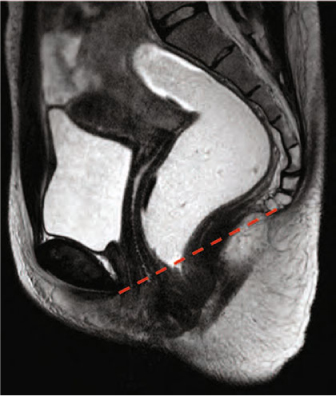
Fig. 4.2
Magnetic resonance defecography performed on a 1.5 T magnet. T2-weighted turbo spin echo sequence acquired at rest in the sagittal plane. The anorectal junction, the vaginal fornices, and the bladder neck are located above the pubococcygeal line (dashed line). The anorectal angle measures 90°
Two other lines should be measured according to the HMO system: the H line, which represents the anteroposterior width of the levator hiatus and is drawn from the inferior aspect of the pubic symphysis to the ARJ; the M line, representing the vertical descent of the levator hiatus and is drawn perpendicular from the PCL to the ARJ; and the O component, which represents the organ prolapse [8].
The ARA and the posterior urethrovescical angle are measured similarly to conventional defecography examinations.
On the axial and coronal images, the puborectal and iliococcygeal muscles should be examined for thinning and tears. The pelvic sling appears relatively symmetric and is hypointense on T2-weighted images. Paravaginal fascial tears can be inferred from posterior displacement of the vaginal fornix. Lateral pubovesical ligaments can also be seen.
4.3.4 Normal Findings
In the upright position, the support to pelvic organs is primarily provided by the bony pelvis, while the pelvic floor muscles and the endopelvic fascia counteract intermittent increases in abdominal pressure. On axial images, the entire the levator ani should be of similar thickness and homogeneous low signal intensity. No tears should be identified. On coronal images, the iliococcygeal muscle should be intact and upwardly convex.
On axial images, the urethral cuff is located anteriorly with a bull’s eye configuration; the vagina should have an H-shaped configuration, which indicates adequate lateral fascial support; the perineum is a diamond-shape structure, with the anorectum lying behind the transverse perineum (Fig. 4.3).
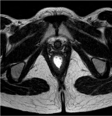
Fig. 4.3
Magnetic resonance defecography performed on a 1.5 T magnet. T2-weighted turbo spin echo sequence acquired at rest in the axial plane. The pelvic sling appears symmetric and hypointense
In healthy, continent patients, even with maximal downward pelvic strain, MR images demonstrate minimal descent of the pelvic organs (M line < 2 cm).
The ARA and the posterior urethrovescical angle can be measured in the saggital plane; normal values are similar to those cited for conventional defecography (see Section 4.2.4).
4.4 Pathological Findings
4.4.1 Intussusception and Rectal Prolapse
Rectal prolapse can be categorized as intrarectal, rectoanal, or external, depending on extension inside the viscus; and as simple or complete, depending on the involved wall layers.
The pathological condition called simple prolapse or procidentia occurs when the mucosal layer protrudes into the lumen. Complete prolapse or intussusception can be observed when all layers of the wall are involved and there is an invagination of the rectal wall into the rectal lumen or the anal canal.
Clinical manifestations frequently associated with rectal prolapse are outlet obstruction and persistent desire to defecate by blocking the rectal ampulla during evacuation, tenesmus, hematochezia, and incontinence. Symptoms are caused by the obstructive effect of the prolapsed wall on the propulsion of rectal contents and on sphincter irritation or weakness. This condition is frequently found in association with solitary ulcer syndrome [16].
4.4.1.1 Conventional Defecography
At the end of defecation, small infoldings of less than 3 mm thickness can be frequently observed without any clinical significance. Larger protrusions have also been observed in asymptomatic patients. Intussusception usually originates 6–8 cm above the anal canal at the level of the main rectal fold and can be either anterior, circumferential, or posterior in location.
Related posts:


Stay updated, free articles. Join our Telegram channel

Full access? Get Clinical Tree


