and Hubert Lepidi1
(1)
UER Médecine, Aix-Marseille Université, Marseille, France
Abstract
Significant knowledge of the bulbo-clitoral organ has been acquired thanks to the enormous progress made in relation to new medical imagery techniques, which have been used to explore the pelvis and especially the female pelvis. X-Ray computed tomography and then NMR (nuclear magnetic resonance) are at the forefront of these new exploration techniques and are mutually complementary. At the same time, great progress has been achieved in medical echography, which has not only become a routine and essential process for pregnancy monitoring but also a sophisticated method for studying pelvic organs and more particularly female genitalia.
14.1 General
Significant knowledge of the bulbo-clitoral organ has been acquired thanks to the enormous progress made in relation to new medical imagery techniques, which have been used to explore the pelvis and especially the female pelvis. X-Ray computed tomography and then NMR (nuclear magnetic resonance) are at the forefront of these new exploration techniques and are mutually complementary. At the same time, great progress has been achieved in medical echography, which has not only become a routine and essential process for pregnancy monitoring but also a sophisticated method for studying pelvic organs and more particularly female genitalia.
14.1.1 Computed Tomography (CT Scan Imagery)
The images obtained by X-Ray CT appeared, as soon as this process was developed, to be extremely interesting as they could be compared to (and almost superimposed on) the cadaveric sections prepared by anatomists. The progress made since then, in only 20 years, is considerable and at present the 64-slice scanners, due to their speed of acquisition and to the multiplicity of the cross-sections, can be used to reconstruct (CT image reconstruction) the organs under study in 3D and to generate three-dimensional images.
The bulbo-clitoral organ appears to be extremely complex and complete on the most superficial sections of the pelvis, especially on the sections made in the planes of the anterior perineum. On such sections, which are strictly comparable to anatomical sections (compare Figs. 14.1b and 13.1), all the parts forming the bulbo-clitoral organ are clearly observed. However, this observation varies according to the levels of the sections:
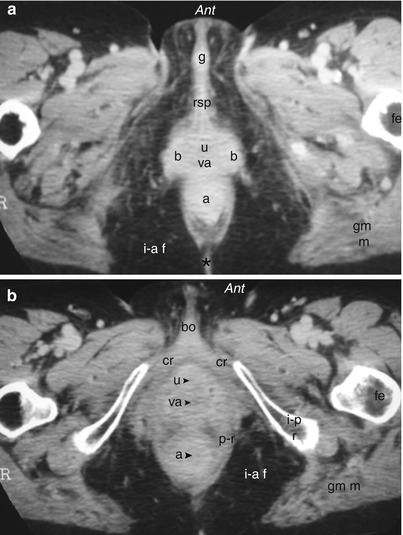
Fig. 14.1
CT scan sections of the bulbo-clitoral organ (scanner of female perineum). (a) Section concerning the spongy parts. (b) Section concerning the cavernous parts. a anus, a (with arrowhead) anal canal, b bulb, bo clitoral body, cr crus clitoridis, fe femur, g glans clitoridis, gm m gluteus maximus muscle, i–a f ischio-anal fossa, i–p r ischio-pubic ramus, p–r pubo-rectalis muscle (levator ani muscle), rsp residual spongy part, u external urethral orifice, u (with arrowhead) urethra, va vaginal orifice, va (with arrowhead) vagina, *ano-coccygeal ligament
On sections tangent to the pelvic outlet but not concerning this bony part (Fig. 14.1a), it is especially possible to observe 2 spongy bulbs bordering the urethral and vaginal ducts and then further ahead, in the extension of the commissure of the bulbs, the entire IC RSP (infra-clitoral residual spongy part) up to the glans.Related posts:

Stay updated, free articles. Join our Telegram channel

Full access? Get Clinical Tree


