Histologic Pattern Approach for Differential Diagnosis of Renal Tumors
Satish K. Tickoo
As is also true for the resected specimens, the initial approach to arriving at the diagnosis of the tumor type requires consideration of both the architecture and cytologic features in renal needle core biopsies. One may start either with the architectural pattern recognition or with the cytologic features, but ultimately, a combination of both will be needed to arrive at the final diagnosis. At times, these features may not be sufficiently diagnostic, particularly for pathologists with limited experience in renal core biopsy interpretation. In such situations, use of a limited panel of immunohistochemical stains will help in arriving at the diagnosis in the overwhelming majority of cases (1,2). Because of the limited material that may sometimes make the architecture difficult to discern at first look, it may be easier to first focus on the cytologic features of the tumor in biopsy specimens. The following represents a practical approach of arriving at the final diagnosis on renal needle core biopsy specimens.
TUMORS WITH CLEAR CELL CYTOLOGY
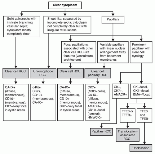
There can be multiple diagnostic possibilities for renal tumors that show cells with clear cytoplasm (Fig. 2.1). To arrive at a particular diagnosis, first, the “clear” cytoplasm requires further qualification. For example, is the cytoplasm completely clear (optically transparent) or does it have fibrillary cytoplasmic strands, rarified granular cytoplasm, or somewhat vacuolated clear appearance (3)? Optically transparent cytoplasm is a typical feature in clear cell renal cell carcinoma (RCC), as well as clear cell papillary RCC, and may also be seen in translocation-associated RCC. On the other hand, “clear” cytoplasm in chromophobe RCC often shows fine fibrillary cytoplasmic strands, whereas “clear cell” areas in papillary RCC often show rarified granular cytoplasm frequently associated with intracytoplasmic, finely granular hemosiderin granules. Cells with
clear cytoplasm with a uniform, vacuolated or bubbly appearance may in fact represent ectopic adrenal cortical tissue or adrenal cortical tumors. Such cells closely resemble the cells seen in sebaceous glands. However, occasional clear cell RCCs may also contain cells with such bubbly appearance. Clear cells may also be present in rare cases of angiomyolipoma (AML), particularly the epithelioid variant.
clear cytoplasm with a uniform, vacuolated or bubbly appearance may in fact represent ectopic adrenal cortical tissue or adrenal cortical tumors. Such cells closely resemble the cells seen in sebaceous glands. However, occasional clear cell RCCs may also contain cells with such bubbly appearance. Clear cells may also be present in rare cases of angiomyolipoma (AML), particularly the epithelioid variant.
In addition to the clear cell cytologic features, other crucial morphologic criterion for diagnosis, both in the resection and biopsy specimens, is the architectural pattern. Solid, variably sized nests separated by intricately dividing vascular septae that surround the cell nests are typical of clear cell RCC. Although often present in needle core biopsies, given the limitation of size, such vasculature may not be obvious in higher grade tumors with a large solid alveolar pattern, solid sheet-like architecture, or sarcomatoid
areas. However, even the focal presence of such architecture in a biopsy is sufficiently diagnostic of clear cell RCC. Nested or solid alveolar pattern may also be seen in translocation-associated RCC or even rare cases of epithelioid AML. If such differential diagnostic possibilities arise on the biopsy material, immunohistochemical confirmation of the diagnosis will be required.
areas. However, even the focal presence of such architecture in a biopsy is sufficiently diagnostic of clear cell RCC. Nested or solid alveolar pattern may also be seen in translocation-associated RCC or even rare cases of epithelioid AML. If such differential diagnostic possibilities arise on the biopsy material, immunohistochemical confirmation of the diagnosis will be required.
Tubular, tubulopapillary, or papillary architecture with clear cell cytology raises the differential diagnostic possibilities of clear cell papillary RCC; papillary RCC; translocation-associated RCC; or RCC, unclassified. Clear cell papillary RCC has optically transparent cytoplasm with lowgrade nuclei that are arranged in a linear fashion away from bases of the cells in the mid or apical cytoplasm. Instead of showing optically transparent cytoplasm, type 1 papillary RCC with areas of “clear” cytoplasm will usually show rarified granular cytoplasm in these “clear” cell areas, as discussed earlier. Rare, fine hemosiderin granules may also be observed at high-power evaluation. The clear cells in translocation-associated RCC are often admixed with cells showing eosinophilic cytoplasm. Many cells often appear voluminous or ballooned out. Immunohistochemical stains or FISH testing for TFE3 or TFEB will be required to confirm this diagnosis (4


Stay updated, free articles. Join our Telegram channel

Full access? Get Clinical Tree


