Histologic Features
The mucosa is smooth and without folds except distally in the rectum. There are no villi. The mucosal surface is flat and regular with interconnected territories containing 10 to 100 parallel crypt mouths measuring 25 to 50 μm in diameter, separated by deep clefts (3). The orifices of the crypts appear as regularly arranged and spaced round depressions that have a uniform size and diameter. The clefts are known as the innominate grooves and they appear as small depressions surrounded by elevations of luminal cells. Visualization of the normal surface pattern has become more important with the increasing use of chromoendoscopy and magnifying
endoscopy to detect neoplastic and preneoplastic lesions at the time of colonoscopy.
endoscopy to detect neoplastic and preneoplastic lesions at the time of colonoscopy.
The crypts are straight tubular structures that lie absolutely parallel to one another and measure approximately 0.5 mm in length. They are unbranched, and extend from the lumen to the muscularis mucosae. However, sections through the crypts making up the innominate grooves demonstrate a branching or cloverleaf pattern. The epithelial lining is columnar in shape and contains an abundance of enterocytes and goblet cells and a small number of endocrine cells. Mucin flows from the crypt openings and covers the mucosal surface (Figs. 13.11 and 13.12) (3).
The colonic epithelium contains undifferentiated cells, goblet cells, absorptive cells, tuft cells, and endocrine cells (4). An orderly pattern of differentiation proceeds from the crypt base to the luminal surface (5). Undifferentiated stem cells in the crypt bases resemble those in the small intestine. The crypt sides are lined by mucus-secreting cells, immature absorptive cells, and rare endocrine cells. Most of the cells lining the middle and upper crypt are in the second replicative zone (i.e., the zone in which partially differentiated cells can still undergo mitosis). The surface epithelium contains goblet cells and mature absorptive cells (5). Cells in the upper 25% of the crypt do not divide but continue to differentiate to form mature absorptive or goblet cells. In addition, M cells are present in the epithelium overlying lymphoid follicles, and intraepithelial T lymphocytes lie scattered within the colonic epithelium.
Absorptive Cells
Absorptive cells outnumber goblet cells 3:1 (Fig 13.13) (6). Mature absorptive cells absorb water and electrolytes; have numerous short, regularly spaced microvilli; and are joined by junctional complexes and lateral desmosomes. The intercellular spaces are variably dilated depending on the physiologic activity of the colon (5). A glycocalyx covers the microvilli on the luminal surface of the epithelial cells. Microvilli increase in number and elongate as the cells mature. Hairlike, loosely packed projections extend perpendicularly from the plasma membrane into the lumen and radiate from the microvilli. Small, round, 30- to 100-nm vesicles, called C bodies, are embedded among the filaments (7). Similar bodies are seen in the apical cytoplasm of mature absorptive cells. These are called R bodies and measure 106 to 250 nm in diameter. As the cells mature, there is a concomitant migration of the R bodies to the apex of the cell. The cytoplasmic microfilaments form a loose, stratified network that retains the same basic organization as that found in the small intestine (8). The apical cytoplasm contains a terminal web composed of microfilaments and core rootlets. Mitochondria, lipid droplets, lysosomes, apical vesicles, and round basally located nuclei are present in the cytoplasm (5). These cells secrete secretory component, which participates in the epithelial translocation of IgA as discussed in Chapter 6.
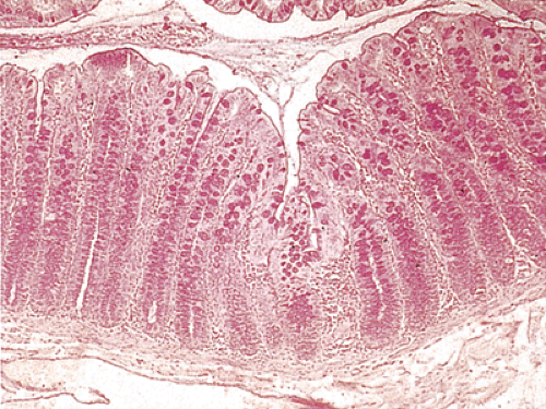 FIG. 13.11. Periodic acid–Schiff–stained section of colon demonstrating mucin secretion. Fine strands of mucoproteins cover the surface. |
Goblet Cells
There are more goblet cells in the large intestine than in the small intestine. They are most abundant in the sigmoid colon and rectum, with approximately one goblet cell for every four columnar cells (Figs. 13.12 and 13.13). Their broad shape creates the false impression that they constitute the majority of the cells. As they differentiate, they migrate toward the mucosal surface and become progressively filled
with mucus granules. The nucleus is compressed into a small dense structure in the basal region of the cell. Mucus secretion occurs throughout the life span of the cell. The membrane-bound mucin granules migrate toward the apical surface of the cell, where they are released by exocytosis. As the cells become more distended with mucin, the microvilli become sparser. The lateral cell surfaces are interlocked by cytoplasmic processes. Junctional complexes are present at the cell apices, and desmosomes are located at various points along the lateral surfaces.
with mucus granules. The nucleus is compressed into a small dense structure in the basal region of the cell. Mucus secretion occurs throughout the life span of the cell. The membrane-bound mucin granules migrate toward the apical surface of the cell, where they are released by exocytosis. As the cells become more distended with mucin, the microvilli become sparser. The lateral cell surfaces are interlocked by cytoplasmic processes. Junctional complexes are present at the cell apices, and desmosomes are located at various points along the lateral surfaces.
Related posts:


Stay updated, free articles. Join our Telegram channel

Full access? Get Clinical Tree


