CHAPTER 77 Hepatitis A
The development of liver biopsy techniques in the 1930s allowed the recognition of hepatic necroinflammation that characterizes all forms of viral hepatitis. Subsequent experimental work in humans led to the clinical recognition that viruses are etiologic agents of hepatitis A (“infectious hepatitis”) and hepatitis B (“serum hepatitis”).1,2 Later, the existence of two hepatitis viruses was demonstrated—hepatitis A virus (HAV) and hepatitis B virus (HBV) (see Chapter 78).3 Additional viral causes of acute and chronic hepatitis were identified subsequently (see Chapters 79 to 81). HAV was first characterized in 1973, when scientists detected the virus in stools from human volunteers who were infected with HAV.4 The ensuing development of sensitive and specific serologic assays for the diagnosis of HAV infection and the isolation of HAV in cell culture5 were important advances that permitted understanding of the epidemiology of HAV infection and, ultimately, control of the disease.
VIROLOGY
In 1982, HAV was classified as an enterovirus type 72 belonging to the Picornaviridae family. Subsequent determination of the sequence of HAV nucleotides and amino acids led to questioning of this classification, and a new genus, Hepatovirus, was created for HAV.6
HAV has an icosahedral shape and lacks an envelope. It measures 27 to 28 nm in diameter, has a buoyant density of 1.33 to 1.34 g/cm3 in cesium chloride, and has a sedimentation coefficient of 156 to 160S on ultracentrifugation. HAV survives exposure to ether and an acid environment at pH 3. It also survives heat exposure of 60°C for 60 minutes but is inactivated at 85°C for 1 minute. HAV is capable of surviving in sea water (4% survival rate), in dried feces at room temperature for 4 weeks (17% survival), and in live oysters for 5 days (12% survival).7
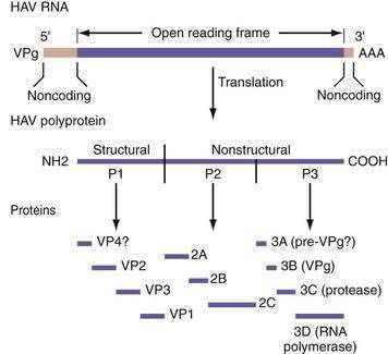
HAV has only one known serotype, and no antigenic cross-reactivity with the hepatitis B, C, D, E, or G agents. The HAV genome consists of a positive-sense RNA that is 7.48 kb long, single-stranded, and linear (Fig. 77-1). HAV RNA has a sedimentation coefficient of 32 to 33S and a molecular weight of 2.8 × 10.4 The HAV RNA has a long open reading frame, consisting of 6681 nucleotides, and is covalently linked to a 5′ terminal protein and a 3′ terminal polyadenosine tract.

Figure 77-1. Genomic organization of hepatitis A virus (HAV). VP, viral protein; VPg, 5′ terminal protein.
(From Levine JE, Bull FG, Millward-Sadler GH, et al. Acute viral hepatitis. In: Millward-Sadler GH, Wright R, Arther MJP, editors. Wright’s Liver and Biliary Disease, 3rd ed. London: WB Saunders, 1992. p 679.)
The onset of HAV replication in cell culture systems takes from weeks to months. Primate cells, including African green monkey kidney cells, primary human fibroblasts, human diploid cells (MRC-5), and fetal rhesus kidney cells, are favored for cultivation of HAV in vitro. The virus is not cytopathic, and persistent infection in the cell cultures is the rule. Two conditions control the outcome of HAV replication in cell culture.8 The first condition is the genetic makeup of the virus; HAV strains mutate in distinct regions of the viral genome as they become adapted to cell culture. The second condition is the metabolic activity of the host cell at the time of infection. Cells in culture, although infected simultaneously, initiate HAV replication in an asynchronous manner. This asynchronicity may be caused by differences in the metabolic activity of individual cells, but definitive evidence of cell-cycle dependence of HAV replication is lacking.9
An initial step in the life cycle of a virus is its attachment to a cell surface receptor. The location and function of these receptors determine tissue tropism. Little is known about the mechanism of entry of HAV into cells. Some work has suggested that HAV could infect cells by a surrogate-receptor binding mechanism (involving a nonspecified serum protein). HAV infectivity in tissue culture has been shown to require calcium and to be inhibited by the treatment of the cells with trypsin, phospholipases, and β-galactosidase.10 A surface glycoprotein, named HAVcr-1, on African green monkey kidney cells has been identified as a receptor for HAV. Blocking of HAVcr-1 with specific monoclonal antibodies prevents infection of otherwise susceptible cells. Experimental data suggest that HAVcr-1 not only serves as an attachment receptor but also may facilitate uncoating of HAV and its entry into hepatocytes.11
Whatever the entry mechanism, once HAV enters a cell, the viral RNA is uncoated, cell host ribosomes bind to viral RNA, and polysomes are formed. HAV is translated into a large polyprotein of 2227 amino acids. This polyprotein is organized into three regions: P1, P2, and P3. The P1 region encodes structural proteins VP1, VP2, VP3, and a putative VP4. The P2 and P3 regions encode nonstructural proteins associated with viral replication (see Fig. 77-1).
The HAV RNA polymerase copies the plus RNA strand. The RNA transcript in turn is used for translation into proteins, which are used for assembly into mature virions. Down-regulation of HAV RNA synthesis appears to occur as defective HAV particles appear.12 In addition, a group of specific RNA-binding proteins has been observed during persistent infection.13 The origin and nature of these proteins is unknown, but they exert activity on the RNA template and are believed to play a regulatory role in the replication of HAV.14
Numerous strains of HAV exist, with considerable nucleotide sequence variability (15% to 25% difference within the P1 region of the genome). Human HAV strains can be grouped into four different genotypes (I, II, III, and VII), whereas simian strains of HAV belong to genotypes IV, V, and VI.15 Despite the nucleotide sequence heterogeneity, the antigenic structure of human HAV is highly conserved among strains.
The HAV VP1/2A and 2C genes are thought to be responsible for viral virulence. This conclusion is based on experiments in which the genotypes and phenotypes of viruses were compared after animals were infected with one of 14 chimeric virus genomes derived from two infectious cDNA clones that encoded a virulent HAV isolate and an attenuated HAV isolate (HM175 strain), respectively.16
Variations in the HAV genome are thought to play a role in the development of fulminant hepatic failure (FHF) during acute HAV infection. The 5′ untranslated region of the HAV genome was sequenced in serum samples from 84 patients with HAV infection, including 12 with FHF.17 The investigators observed fewer nucleotide substitutions in the HAV genome from patients with FHF than in those from patients without FHF (P < .001). The differences were most prominent between nucleotides 200 and 500, suggesting that nucleotide variations in the central portion of the 5′ untranslated region influence the clinical severity of HAV infection.
EPIDEMIOLOGY
Acute hepatitis A is a reportable infectious disease in the United States. The incidence has declined by 90% since 1995. In 2006, 3579 cases of HAV infection were reported, corresponding to a rate of infection of 1.2 per 100,000, compared with 4 per 100,000 in 2001. With the underreporting of cases and the occurrence of asymptomatic infections taken into consideration, the true number of HAV infections in 2006 was calculated to be 32,000, compared with 93,000 in 2001. The greatest rate of decline has been among children from states where routine vaccination of children was recommended in 1999. The highest rate of reported disease historically has been among children ages 5 to 14 years. Because of the rapid rate of decline of disease in children, however, rates are now similar among age groups, with adults ages 20 to 44 having the highest rate of disease in 2006.18
The epidemiologic risk factors for HAV infection reported for the U.S. population in 2006 were as follows: unknown, 65%; international travel, 15%; contact with a patient who has hepatitis, 12%; sexual or household contact with a patient who has hepatitis A, 10%; men having sex with men, 9%; food or waterborne outbreak, 7%; child or employee in a daycare center, 4%; contact with a daycare child or employee, 4%; and injection drug use, 2%.18
HAV infection generally follows one of three epidemiologic patterns.19 In countries where sanitary conditions are poor, most children are infected at an early age. Although earlier seroepidemiologic studies routinely showed that 100% of preschool children in these countries had detectable antibody to HAV (anti-HAV) in serum, presumably reflecting previous subclinical infection, subsequent studies have shown that the average age of infection has risen rapidly to 5 years and older, when symptomatic infection is more likely. For example, 82% of 1393 Bolivian school children were shown to have detectable anti-HAV, but when they were stratified into two groups according to family income, a significant difference was found between the groups: 95% of children from low-income families, but only 56% of children from high-income families, had detectable anti-HAV.20
The second epidemiologic pattern is seen in industrialized countries, where the prevalence of HAV infection is low among children and young adults. In the United States, prior to universal HAV vaccination, the prevalence of anti-HAV was approximately 10% in children but 37% in adults.21
Whatever the epidemiologic pattern, the primary route of transmission of HAV is the fecal-oral route, by either person-to-person contact or ingestion of contaminated food or water. Although rare, transmission of HAV by a parenteral route has been documented after transfusion of blood22,23 or blood products.24 Cyclical outbreaks among users of injection and noninjection illicit drugs and among men who have sex with men (up to 10% may become infected in outbreak years) have been reported.25 Table 77-1 provides information about the detection of HAV and its infectivity in human body fluids.26–33
Table 77-1 Detection of Hepatitis A Virus (HAV) and Infectivity of Human Secretions or Excretions
| SECRETION/EXCRETION | COMMENT | REFERENCES |
|---|---|---|
| Stool | Main source of infection. HAV is detectable during the incubation period and for several weeks after the onset of disease. After the onset of symptoms, HAV is detectable in 45% and 11% of fecal specimens collected during the first and second weeks of illness, respectively, whereas HAV RNA (by polymerase chain reaction assay) is detectable for 4 to 5 months. | 26,27 |
| Blood | Viremia is present during the incubation period. Blood collected 3 and 11 days before the onset of symptoms has caused post-transfusion infection in recipients. Chronic viremia does not occur. | 28,29 |
| Bile | HAV has been detected in the bile of chimpanzees infected with HAV. | 30 |
| Urine | HAV is detected in low titer during the viremic phase. A urine sample was reported to be infectious after oral inoculation. Urine contaminated with blood was also infectious. | 31,32 |
| Nasopharyngeal secretions | Unknown in humans. HAV has been identified in the oropharynx of experimentally infected chimpanzees. | 33 |
| Semen, vaginal fluid | Uncertain. HAV may be detectable during the viremic phase. | — |
From 11% to 22% of patients with acute hepatitis A require hospitalization, with an average length of stay of 4.6 days, costing on average $7926 per patient in 2004. In one outbreak involving 43 persons, the total cost was approximately $800,000. On average, 27 workdays are lost per adult case of hepatitis A. In adolescents and adults, the combined direct and indirect costs associated with HAV infection in the United States totaled approximately $488.8 million in 1997, compared with $93 million in 2006.25,34,35 The decline in costs is a direct result of the dramatic reduction in the number of infections seen since the introduction of the HAV vaccine combined with changes in vaccination policies in the United States (see later).18
PATHOGENESIS
After HAV is ingested and survives gastric acid, it traverses the small intestinal mucosa and reaches the liver via the portal vein. The precise mechanism of hepatic uptake in humans is unknown (see earlier). In an experimental model using African green monkey kidney cells,11 the putative cellular receptor for HAV has been identified as a surface glycoprotein. Once the virus enters the hepatocyte, it starts replicating in the cytoplasm, where it is seen on electron microscopy as a fine granular pattern, but it is not present in the nucleus. HAV is distributed throughout the liver. Although HAV antigen has been detected in other organs (lymph nodes, spleen, kidney), the virus appears to replicate exclusively in hepatocytes. When the virus is mature, it reaches the systemic circulation via the hepatic sinusoids and is released into the biliary tree through bile canaliculi, passed into the small intestine, and eventually excreted in the feces.
CLINICAL FEATURES
Infection with HAV does not result in chronic infection, only in an acute self-limited episode of hepatitis. Rarely, acute hepatitis A can have a prolonged or a relapsing course, and occasionally profound cholestasis can occur.36 The incubation period is commonly 2 to 4 weeks, rarely up to 6 weeks. The mortality rate is low in previously healthy persons. Morbidity can be significant in adults and older children.
The clinical characteristics of cases of hepatitis A reported in 2002 were similar to those in previous years, with a preponderance of cases in men in all age groups. Overall, 72% of patients manifested jaundice, 25% required hospitalization, and 0.5 % died.37 The need for hospitalization rose with age, from 5% among children younger than 5 years to 34% among persons 60 years or older.37
Patients with HAV infection usually present with one of the following five clinical patterns: (1) asymptomatic without jaundice, (2) symptomatic with jaundice and self-limited after approximately 8 weeks, (3) cholestatic, with jaundice lasting 10 weeks or more,36 (4) relapsing, with two or more bouts of acute HAV infection occurring over a 6- to 10-week period, and (5) FHF.
Stay updated, free articles. Join our Telegram channel

Full access? Get Clinical Tree


