Chapter 26 Granulomatous liver disease
1 Granulomas consist of activated macrophages (epithelioid macrophages) accompanied by T lymphocytes and other immune cells, which infiltrate liver tissue as nodular lesions in reaction to foreign or indigestible antigen or as a hypersensitivity reaction.
2 The major causes of hepatic granulomas include infectious agents (especially tuberculosis), sarcoidosis, primary biliary cirrhosis, drugs, systemic diseases (e.g., Crohn’s disease), and neoplasms (e.g., Hodgkin’s lymphoma).
3 An elevated serum alkaline phosphatase level is the most common abnormality in serum liver biochemical test levels.
Pathogenesis of Granulomas
Definition
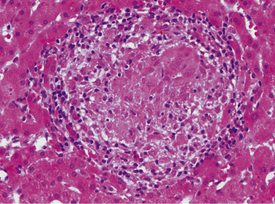
1. Granulomas are rounded, 1- to 2-mm collections of activated macrophages, T lymphocytes, and other immune cells that infiltrate a host of tissues, including the liver, in response to a foreign or indigestible antigen or as a hypersensitivity reaction (Fig. 26.1).
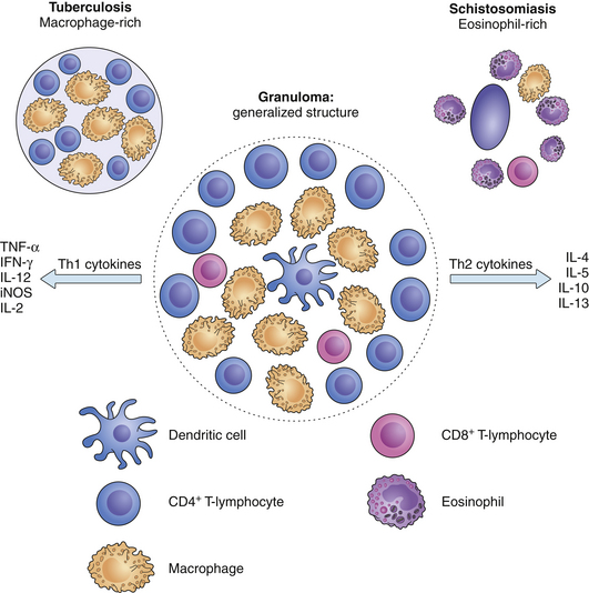
2. The principal immune cells in granulomas are activated macrophages resembling epithelial cells (epithelioid macrophages), CD4+ T cells (T-helper, or Th, lymphocytes), and sometimes multinucleated giant cells that develop from macrophage fusion.
3. The cause of the granuloma influences the constituent immune cells and secreted products: Th type 1 (Th1) cells and cytokines predominate in mycobacterial granulomas, whereas Th2 cells and cytokines predominate in schistosomal granulomas (Fig. 26.2).
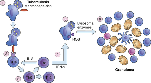
4. Granulomas evolve by the elaboration of secretory products (cytokines and chemokines) by their constituent cells (interferon gamma and interleukin-2 from Th lymphocytes), expansion of macrophage and T-cell pools, and specialization of macrophages for antigen digestion (Fig. 26.3).
Morphologic types
Several types of granulomas are described in liver disease according to their histologic features and constituents (Table 26.1).
| Type | Histologic features | Cause(s) |
|---|---|---|
| Caseating | Peripheral macrophages with or without giant cells; central necrosis | Tuberculosis |
| Noncaseating | Cluster of macrophages with or without giant cells | Sarcoidosis Drugs |
| Lipogranuloma Fibrin-ring (doughnut granuloma) | Lipid vacuole(s) surrounded by macrophages and lymphocytes Central lipid vacuole or empty space Macrophages and lymphocytes Ring of fibrin | Fatty liver Mineral oil Q fever Allopurinol Hodgkin’s lymphoma |
Causes of Hepatic Granulomas
The etiology is multifactorial, but the major causes and examples are shown in Table 26.2.
TABLE 26.2 Causes of hepatic granulomas
| Etiology | Specific cause(s) |
|---|---|
| Infections | |
| Primary biliary cirrhosis | Early stages most commonly |
| Foreign bodies | Suture, talc |
| Systemic disease | Sarcoidosis, Crohn’s disease |
| Drugs | Allopurinol, phenytoin, penicillin |
| Neoplasia | Hodgkin’s lymphoma |


