Fecal Incontinence


Fecal Incontinence


R. John Nicholls


My wind exploded like a thunder-clap Iaso blushed a rosy red And Panacea turned her head Holding her nose: My wind’s not frankincense.

—ARISTOPHANES: Plutus

Fecal incontinence is common, especially in the elderly, and can cause considerable morbidity and diminution of quality of life. It may not be life threatening, but it is traumatizing and often disabling. Many patients feel too embarrassed to discuss the symptom with a physician. Incontinence can result from intestinal disease causing irritability and urgency of defecation due to inflammation or loss of capacity of the rectum or neorectum, or from a defective anal sphincter mechanism. Both factors can be present in the same patient. The management of incontinence associated with intestinal diseases is part of the treatment of those conditions. Most patients suffering from incontinence will, however, fall into the category of having an incompetent sphincter mechanism. This chapter will deal predominantly with these.

There have been several advances in the understanding and management of fecal incontinence in the past 10 years. Previously, the condition had been regarded more as a mechanical disorder, but it has become increasingly apparent that there is a large functional component. Behavioral therapies have been developed, and their value is now established. Importantly, surgical correction is reserved for patients who have a mechanical disruption of the sphincter mechanism. Its use has, therefore, declined having been largely replaced by neuromodulation in many centers, which includes stimulation of the sacral nerve (SNS or SNM) and more recently of the posterior tibial or pudendal nerves or the dorsal nerve of the clitoris. Artificial sphincters either biologic in the form of graciloplasty (stimulated or nonstimulated) or prosthetic in that of the artificial bowel sphincter (ABS [Acticon, AMS]) have been found to be less effective than was originally hoped. Anal procedures including the injection of bulking agents into the anal canal lining and so-called radio frequency energy delivery (SECCA) to promote scarring of the anal canal have been developed, particularly for patients with passive incontinence due to failure of the internal sphincter.

Clinical assessment combined with anal ultrasound to establish the anatomy of the sphincter and physiologic testing by manometry will result in reasonable decision making in most circumstances. Anal ultrasound is now routinely applied for the assessment of continence. The axial view of a normal anal canal shown in Figure 16-1 demonstrates the concentric layers of the submucosa, internal sphincter, longitudinal muscle, and external anal sphincter. Some of the physiologic parameters thought to be important in the past, such as electromyography and others (see Chapter 7), are regarded by many to be no longer useful in clinical practice.

The outcome for the patient is now expressed as much by quality of life as by the frequency of incontinent episodes. The important symptom of urgency, which may be the only manifestation of a continence disturbance, can now be quantified, thus enabling improved assessment. The development of continence scoring systems has also improved our ability to assess the effectiveness of treatment.

As with other areas in colorectal surgery, there has been a general tendency toward specialization. The treatment
options for incontinence are now such that the patient is best served in a unit with the capacity to investigate and maintain continuity of care. Thus, a patient suitable for neuromodulation, for example, should be managed in a formally constituted physiology unit.60


FIGURE 16-1. Normal axial image. Orientation and five-layer pattern.

The techniques used for the evaluation of incontinence overlap with those used for other functional disturbances, including difficulty in defecation and prolapsing disorders. Furthermore, some patients with incontinence may, in reality, have another associated condition—for example, an evacuation disorder such as constipation. It is therefore essential for the physician to be aware of such a possibility.


▶ SCORING SYSTEMS

The development of scoring systems has been an important feature of practice. Their use is now widespread in the reporting of results. Parks introduced a simple system based on the degree of leakage with A, being normal; B, incontinent to flatus; C, incontinent to liquid stool; and D, incontinent to solid stool.350 Pescatori and colleagues358 identified 13 classifications suggested by various authors, including one by Corman. It took into account both the degree and frequency of symptoms in which A, B, and C reflect increasing problems with incontinence for stool and the number system indicates the frequency of the problem (occasional, weekly, and daily).


TABLE 16-1 The Jorge-Wexner Incontinence Scoring System


FREQUENCY


Type of Incontinence


Never


Rarely


Sometimes


Usually


Always


Solid


0


1


2


3


4


Liquid


0


1


2


3


4


Gas


0


1


2


3


4


Wears pad


0


1


2


3


4


Lifestyle alteration


0


1


2


3


4


0 = Perfect


20 = Complete incontinence


Never = 0 (never)


Rarely = <1/month


Sometimes = <1/week, >1/month


Usually = <1/day, >1/week


Always = >1/day


In 1993, Jorge and Wexner proposed a continence grading scale, which is now termed the Cleveland Clinic Incontinence Score (Table 16-1) and has now come to be used by many investigators.216

The score is determined by adding points from this table, which includes the type and frequency of incontinence and the extent to which it alters the patient’s life. This was one of the first attempts by a scoring system to assess quality of
life. Being a benign condition, this is the most important end point for a patient with fecal incontinence, a determination that has been emphasized by several investigators.54,388

Although these scoring systems were useful in comparing the severity of continence before and after treatment, an attempt at standardization was made by a consensus group consisting of representation from five well-recognized academic divisions of colon and rectal surgery in the United States. This resulted in the publication in 1999 of the Fecal Incontinence Severity Index (FICI).216 Four incontinence events were used to determine the FICI score, all calculated based on the frequency of incontinence of flatus, mucus, liquid stool, and solid stool. Each included five frequencies: one to three times per month, once per week, twice per week, once per day, and twice per day. The same group also turned its attention to the development of a health-related quality of life (HRQoL) scale.378 This selfcompleting questionnaire addressed four domains including lifestyle, coping/behavior, depression/self-perception, and embarrassment.

More recently, a scoring system was developed at St. Mark’s Hospital,465 which combined the Pescatori and Wexner scales along with a system developed for the assessment of artificial bowel sphincter by American Medical Systems (AMS). It added further points, including the need for antidiarrheal medication, and improved the assessment of urgency by classifying this symptom to be more or less than 15 minutes. Used in 33 patients, the score that ranged from 0 to 24 (completely incontinent) more accurately coincided with the clinician’s assessment than did the other scoring systems. In a further study of 390 patients, the score agreed moderately well with the patients’ opinions.277

Scoring systems have considerable value, particularly when comparing symptoms before and after treatment. They may also allow some comparison between different units. Being a numerical scale, however, they have the weakness that an idea is being replaced by a figure, and it can be easy to forget what the scale really means in practice. For example, a fall from a score of 14 to 9 sounds impressive, but the patient is still left with troublesome symptoms. A more meaningful statement would include the number of incontinent episodes per unit time—for example, a week or the interval between the arrival of the stimulus and the actual time the patient has in minutes to get to the bathroom. These are also quantifiable in digit integer form and are immediately understandable to patient and physician alike.


▶ PREVALENCE AND ETIOLOGIC FACTORS

Available studies indicate the prevalence of incontinence in the general population to be around 2%, with an increased incidence in the elderly and those in psychiatric, geriatric, and community-based accommodations as high as 50%. It is the second most common cause of institutionalization in the elderly, and in 1993, it accounted for one-half billion dollars per year in the United States for the cost of adult diapers.216 Significant positive associations for fecal incontinence in the nursing home population include urinary incontinence, tube feeding, loss of activity, diarrhea, pressure ulcers, dementia, impaired vision, fecal impaction, constipation, male gender, age, and increased body mass index (BMI).323

In an earlier article, Nelson and colleagues attempted to determine the prevalence and characteristics of anal incontinence in the general community.324 A total of 2,570 households comprising almost 7,000 individuals were surveyed. The overall incidence of anal incontinence was 2.2%. Thirty percent of those affected were older than 65 years, and approximately two-thirds were women. Of those with anal incontinence, 36% were incontinent for formed stool, 54% for liquid stool, and 60% for gas. This figure is similar to that reported by Varma and coworkers who studied a randomly selected group of women (average age, 56 years) from the Reproductive Risks of Incontinence Study at the Kaiser Permanente facility. Although self-reported incontinent episodes in the prior year were recorded by 24% of patients, in only 2.1% was this weekly or more.474

Goode and colleagues studied 1,000 individuals aged 65 to 106 years who were recipients of State Medicare benefits.160 The prevalence of fecal incontinence was 12.0% (12.4% males; 11.6% females). In a subsequent study from the same group, Markland and associates found the incidence of fecal incontinence over a 4-year period in three rural and two urban districts of Alabama to be 17%, with 6% experiencing episodes at least monthly.286

Although age and sex are regarded to be risk factors, definitions are important. This may have accounted for the results of a telephone interview study of 1,153 households in Winnipeg (population, 650,000) in which the overall incidence of an incontinent episode in the previous 12 months was 3.7% (2.0% when physician-diagnosed disease was removed from the analysis) and was not related to age or sex. The average age of the respondents was 47 years; the lower age limit for inclusion in the study was 18 years.203

In a study of 271 identical twin pairs, Abramov and colleagues analyzed the returns of the Colorectal Anal Distress Inventory.5 They found the following variables to be associated with fecal incontinence: age older than 40 years, menopause, parity >2, and urinary stress incontinence. Obesity was very significantly related to incontinence (P < .007). Cesarean delivery was not significantly related to a lower incidence, but no patient having delivery by this means experienced fecal incontinence.


Male Patients

There is a difference in the type of lesion responsible for incontinence between men and women. The published data from 1990 to 2007 were reviewed by Shamliyan and associates.413 They extracted information from 21 observational studies and 4 randomized, controlled trials among community-dwelling elderly men. The pooled prevalence of fecal incontinence was 5%. This was primarily related to age in men older than 85 years, but also to the prevalence of kidney disease.

In a prospective study of 59 males with incontinence, there were 36 with fecal incontinence and 23 with leakage. Overall there were only 5 patients with a sphincter defect (4/36 and 1/23, respectively), 4 of whom had had previous anal surgery. Anal pressures were normal in patients with leakage but were reduced in those with incontinence (resting pressure, 58 vs. 85 [normal]; squeeze, 167 vs. 248 [normal]).453 In another prospective study of 85 consecutive males presenting over 1 year (408 females in the same period), the etiology was determined in 72%. The most important causes were prior anal surgery (23), treatment for cancer of the
prostate (9), and spinal injury (9). Eight patients had soiling of unknown origin. A sphincter defect was present in 35%, compared with 70% in female patients. Treatment resulted in complete resolution of symptoms in 17%, and a further 48% were improved.78


Comorbidity

It is clear that incontinence is not only common in society, especially in the elderly and in those who are institutionalized, but it is also related to comorbid and social factors. In a recent systematic review, Hägglund and colleagues172 evaluated 48 publications on the assessment, management, and prevention of dementia with respect to urinary and fecal incontinence. The prevalence of fecal incontinence among the elderly population aged 75 to 90 years was 17%,429 whereas among demented patients it was 32%47 and 34.8%181 in two studies, a prevalence higher than the available data for the normal elderly population.

In a randomly selected group of women, aged 56 years, gleaned from the Reproductive Risks of Incontinence Study, not surprisingly fecal incontinence reduced the quality of life and increased the “bother.” It was also related to obesity (odds ratio [OR] = 1.2), pulmonary obstructive airways disease (OR = 1.9), irritable bowel syndrome (OR = 2.4), urinary incontinence (OR = 2.1), and colectomy (OR = .9).474 Incontinence was related to chronic diarrhea (OR = 4.55), urinary incontinence (OR = 2.65), hysterectomy (OR = 1.93), poor self-perceived health status (OR = 1.88), geriatric depression score >5 (OR = 2.83), living alone (OR = 2.38), transient ischemic attacks (OR = 3.11), and prostatic symptoms (OR = 2.29).160 In females, risk factors included white race, depression, chronic diarrhea, and urinary incontinence. In males, only urinary incontinence was associated.286 Other risk factors include kidney disease (OR = 1.9) and prostatic surgery or radiotherapy.272 Impaired cognition is also related to incontinence.413


Obesity

There is an extensive literature linking fecal incontinence to obesity. Erekson and colleagues127 measured BMI in 519 new patients attending a clinic for pelvic floor disorders and found a significant correlation with the presence of fecal incontinence (OR = 1.25 [95% CI, 1.09-1.44]) while the relationship with a defecatory disorder was not significant. Looked at from the other perspective, of the 256 women (BMI mean, 49.3 ± kg/m2) attending a bariatric surgery seminar, 61% were found on questionnaire to have experienced incontinence, with a median Wexner score of 7 (1-20) and a score of 10 or more in 34%. Multivariate analysis showed that obstetric injury (OR = 2.4) and urinary infection (OR = 1.2) were significantly related to incontinence, but age, BMI, parity, diabetes, and hypertension were not.485 There is a link between obesity, urinary incontinence, and fecal incontinence. Among 336 women aged 53 ± 10 years with more than 10 or more episodes of urinary incontinence over 7 days, 55 (16%) had fecal incontinence. This was related to low-fiber intake, a high depressive index, and urinary tract infection.287

The connection between urinary and fecal incontinence with obesity was further amplified in a report of the outcome of bariatric surgery in 404 patients whose symptoms were determined by questionnaire. Of the females, 78% had experienced urinary incontinence prior to the operation, of whom 39% felt that they had improved postoperatively. Only 21% of men experienced urinary incontinence preoperatively. The prevalence of fecal incontinence to liquid stool before bariatric surgery was 42% and 48% when comparison is made between the sexes—21% and 30% for solid stool, respectively. The operation resulted in worsening incontinence in 55% of women and in 31% of men. This was attributed to postoperative diarrhea.377 In an age of increasing obesity surgery, this is an important consideration when advising patients and obtaining consent, as is discussed in a Bharucha’s editorial article.30

In summary, fecal incontinence is related to generalized morbidities, including renal, pulmonary, cardiovascular disease, and obesity. It is related to intestinal symptoms, such as diarrhea and the irritable bowel syndrome, and to dementia. Social factors and psychiatric status are also important. The causes associated conditions differ between the sexes.


▶ MECHANISMS OF INCONTINENCE


Normal Continence

Continence is a balance between the ability of the anal sphincter mechanism to withstand the propulsive efforts of the distal intestine, which depends on motility patterns and adequate rectal capacitance. The anal sphincter and pelvic floor form a complex mechanism with motor and sensory nerve components.


Take your hands if you will and cup them. Put in them a mixture of gas, liquid and solid, And try to let only the gas escape—your will fail! The anal sphincter mechanism can do this. Furthermore, the sphincter mechanism can tell Whether you are with friends or alone, Whether you are sitting or standing, or whether you have your pants on or off. There is no muscle that is so prepared to preserve the dignity of mankind Yet so ready to come to one’s relief.

—ROBERT W. BEART, JR, MD

The anal sphincter mechanism includes the anal sphincter itself and the levator ani (see Chapter 1). The latter forms the pelvic floor. It is composed of three parts, including the ischiococcygeus, the pubococcygeus, and the puborectalis (see Figures 1-11 and 
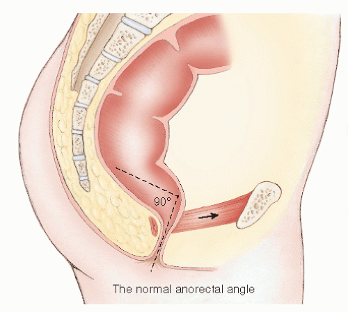
1-12). The first two form a muscular sheet across the pelvic outlet with attachments to the back of the pubis, the obturator fascia, and the coccyx. This is penetrated in the midline by the urethra, vagina, and gut tube in female and by the urethra and gut tube in male. The puborectalis arises on each side from the back of the pubis and passes posteriorly to form a sling around the gut tube. This and the levator ani are in a constant state of tonic activity mediated by muscle spindles located in the levator ani. As a result, the puborectalis draws the gut tube forward to create an angle between the rectum and anal canal. Under normal resting conditions, the long axes of each form an angle, the anorectal angle, which is about 90 to 100 degrees. The puborectalis sling marks the functional junction between the anal canal and the rectum and is easily palpable on digital examination (Figure 16-2).


FIGURE 16-2. Normal anorectal angle.

The anal canal comprises the internal and external sphincters and its epithelial lining, which is richly supplied by sensory nerves. The internal sphincter is responsible for 70% of the resting tone.27 It is maximally contracted at rest and shows reflex relaxation in response to distension of the rectum. This is referred to as the rectoanal inhibitory reflex (RAIR), which is under autonomic control and requires an intact submucous nervous plexus for it to function.267 The pelvic floor musculature relaxes during defecation. Sensory factors include the sampling reflex that is disordered in incontinence.178 It has been estimated that about 15% of anal resting tone is due to the hemorrhoidal complexes.261 The longitudinal length of the anal sphincter complex in female is less than that of male.493


Causes of Incontinence

A classification of the causes of incontinence is shown in Table 16-2.


Increased Intestinal Propulsion

Fecal incontinence can be due to uncontrollable defecation as a result of inflammatory or functional disease of the rectum and more proximal bowel. Diarrheal disorders associated with urgency include any form of proctitis, but in clinical practice ulcerative colitis or Crohn’s disease are the most important. Infective proctocolitides, dysentery, is usually self-limiting or responds to treatment, but it is necessary to consider this in the differential diagnosis of incontinence, particularly because the incidence of infective proctocolitis is increasing in Western society, especially in institutions (see Chapter 33). Organisms responsible include among others Shigella, Clostridium difficile, and Campylobacter. C. difficile and ulcerative colitis can present in an identical manner. There has been an “alarming” increase in C. difficile- associated disease (CDAD) in the United States. This has been dramatic in patients with inflammatory bowel disease (IBD). Rodemann and colleagues reported that between 1998 and 2004 in the United States, CDAD in ulcerative colitis patients rose from 18.4 to 57.6 (more than threefold) per 1,000 admissions.379 In endemic areas, Entamoeba histolytica should be borne in mind.

Urgency may be the predominant feature in some patients with irritable bowel syndrome. It is important to recognize this because surgery has no role to play in the treatment, although neuromodulation may have a place.

Urgency incontinence may also occur as a result of inadequate capacitance of the rectum or “neorectum” in patients who have undergone restorative surgery or radiotherapy. In colorectal practice where many patients will have had a low anterior resection, for example, this form of incontinence is fairly common. Functional symptoms after anterior resection are referred to as the anterior resection syndrome and include the frequent passage of small-volume stool with passive and sometimes urgency incontinence. Those with this disorder may also have a weak sphincter (see next section).


Intrinsic Sphincter Incompetence

The anal sphincter mechanism may be unable to retain stool and flatus if it suffers damage to its nerve supply or as a consequence of direct trauma. Myopathic degeneration can also occur. There is a gradual physiologic weakening with age that can be identified by physiologic testing.205


Age

Attempts have been made to compare functional estimations of anal sphincter capacity in the elderly population with younger individuals. Bannister and coworkers measured anorectal function in 37 elderly patients and compared the results with 48 young, physiologically normal subjects.18 The former had lower anal pressures, required lower rectal volumes to inhibit anal sphincter tone, and had increased rectal pressures as measured by balloon distension, implying lower compliance. Some suggest that internal anal sphincter dysfunction may be the important factor.21 Others believe that, especially in women, the pudendal and somatic pelvic nerves are injured when there is perineal descent on straining such as occurs with aging or during childbirth.244 Many geriatricians believe that fecal incontinence is more likely to be due to a remediable cause, such as fecal
impaction, rather than to dementia, the postmenopausal state, or to old age.283


TABLE 16-2 Causes of Incontinence


Increased intestinal propulsion


Inflammatory bowel disease


Functional bowel disease


Incompetent sphincter mechanism


Age


Neurologic disease


Myopathy


Anorectal disease


Congenital anomaly


Extrarectal fistula


Trauma


Obstetric injury


Anorectal surgery


Accidental trauma


Reduced rectal capacity


Anterior resection


Radiotherapy


Activation of the anorectal inhibitory reflex in fecal impaction


Neurologic Disease

Any neurologic disease may affect bowel control. Upper and lower motor neuron lesions seen in general neurologic diseases, injury to the spinal cord or cauda equina, spina bifida, and neuropathies can all be responsible. In diabetes mellitus,484 there may be the dual factors of diarrhea from autonomic neuropathy and diffuse sphincter weakness due to the neuropathy.

General neurologic diseases including multiple sclerosis206 and Parkinson’s disease can be associated with incontinence. It is clearly necessary to be aware of the possibility of these when assessing the patient. Dementia due to cerebral degeneration in the elderly has already been mentioned. It and presenile dementia are among the most common causes of incontinence. Other conditions leading to cerebral damage include cardiovascular accident, trauma, and tumor.

Spinal cord injury results in weakness of the pelvic floor muscles, but it can also lead to failure of rectal evacuation, impaction, and soiling. Reflex defecation precipitated by sensory cutaneous stimulation above the level of the lesion may be maintained.

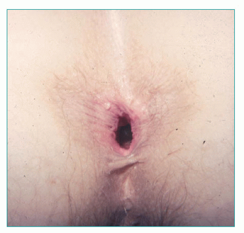
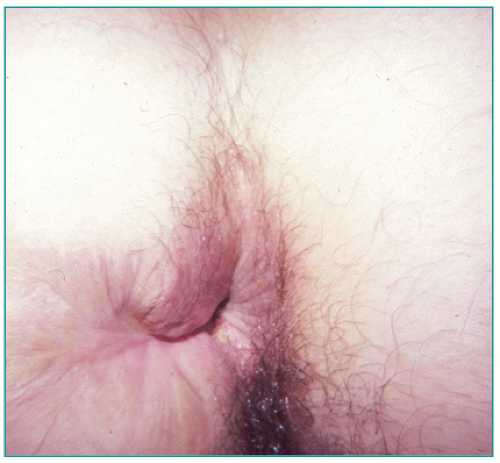
Trauma to the cauda equina, with damage to sacral 4 and 5 segments, causes lower motor paralysis of the pelvic floor and sphincter muscles. Voluntary contraction of the levator ani and external sphincter is reduced or absent depending on the severity of the lesion. Internal sphincter tone is preserved, however, because this is mediated by autonomic activity. There is sensory loss of the perineal skin and of the anorectum and patulousness after digital examination (Figure 16-3).

Spina bifida occurs in association with meningocele or myelomeningocele. Involvement of sensory or motor nerves may produce urinary and fecal incontinence and, with time, rectal prolapse, which will further exacerbate the incontinence. This condition is described in Chapter 21.

Rectal impaction causes internal sphincter relaxation through activation of the RAIR, owing to distension of the rectum by stool. This is a temporary effect that will resolve once the impaction is treated. Schiller and colleagues, in their study of 16 diabetic patients, concluded that incontinence was due to internal anal sphincter dysfunction and that those without diarrhea have no impairment of fecal control.400


FIGURE 16-3. Patulous appearance of the anus immediately following rectal examination. Patient’s incontinence was a consequence of spinal nerve injury.


Idiopathic Incontinence (Pudendal Neuropathy)

Idiopathic incontinence is the term used to describe denervation injury to the sphincter muscle that occurs for no apparent cause. Habitual straining during defecation, nerve entrapment, the preprolapse state, and the descending perineum syndrome have all been implicated. It may be seen after traumatic obstetrical delivery.

The condition was first formally defined by Parks and colleagues352 in a group of patients with incontinence or rectal prolapse associated with diffuse weakness of the anal sphincter muscles. Biopsies taken from the external sphincter showed histologic evidence of atrophy of type I fibers. These patients also had neurophysiologic evidence of denervation as determined by single-fiber electromyographic (EMG) studies. This technique enables an estimate of the ratio of nerve to muscle fibers in the motor neuron unit. When denervation occurs, there is reinnervation by branching of intact nerve axons in an attempt to reach denervated muscle fibers. Consequently, the ratio of nerve to muscle fibers increases, and this can be detected neurophysiologically by direct recording of a needle electrode introduced through the skin into a nerve ending.320 In addition, measurement of the speed of conduction in the pudendal nerve showed the latency to be increased.227 Further studies in patients with idiopathic incontinence identified that most also have degenerative changes in the internal sphincter.245,266 It was postulated that the neuropathic changes might be due to damage to the pudendal nerve through stretching, thereby causing a degree of apraxia as might occur with excessive descent of the pelvic floor, such as in the descending perineum syndrome or during childbirth. It is unlikely that stretching occurs at the level of the ischial spine because at this level the pudendal nerve has not yet branched to supply the sphincter, perineum, and clitoris. Computer modeling has hypothesized that potential stretch during delivery is more likely to damage the inferior rectal nerve.263

Objective evidence of a sensory deficit can be found in patients with idiopathic neuropathic incontinence, as would be expected in the presence of nerve damage. Others have supported the hypothesis that sensory function may be an independent factor contributing to continence.22,31

The normal anal canal is extremely sensitive to temperature. Moreover, there appears to be a temperature gradient between the rectum and the anal canal.308,309 Ambulatory pressure monitoring has shown that the sphincter relaxes several times an hour, allowing equalization of rectal and anal pressures, which permits entry of small amounts of rectal contents into the anal canal.307 This so-called sampling reflex may be lost with neuropathic change. Patients with idiopathic incontinence have reduced thermal sensation at all levels within the anal canal when compared with normal individuals.306 Another report from the same unit involving patients with hemorrhoids and others with incontinence showed reduced sensation in incontinence but also in those with hemorrhoids, albeit to a lesser degree.308 Others have challenged the importance of thermal sensitivity on the basis that the maximum temperature difference was merely 0.13° and was in the region of the anorectal junction.381 Such a
difference was considered too small in their view to have a meaningful role in sampling.


Myopathic Disease


Physiologic

Estrogen and progesterone receptors have been found in the internal sphincter, derived from tissue obtained from hemorrhoidectomy specimens in premenopausal and postmenopausal women. Although none of the differences was considered to be statistically significant, the numbers (17 in each group) were small—a type II error. In premenopausal patients, estrogen receptors were present in 23.5% compared with 11.8% in postmenopausal women. The respective proportions of progesterone receptors were 41.2% and 11.8%.348


Internal Sphincter Degeneration

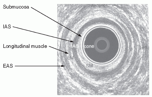
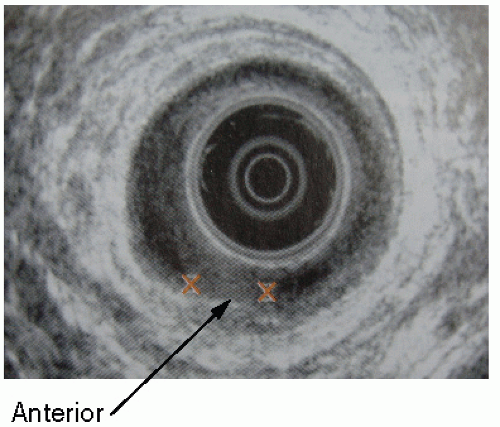
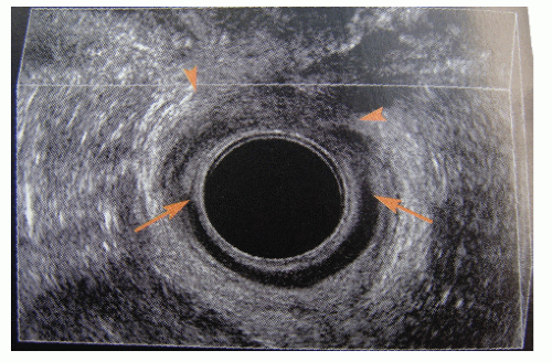
Internal sphincter degeneration was described by Vaizey and colleagues in 1997.466 They reported a series of 45 patients (35 females) who presented with passive incontinence associated with reduced resting anal and normal squeeze pressures. The pudendal nerve terminal motor latencies (PNTMLs) were normal. The internal sphincter on ultrasound was thinned and showed hypoechoic changes (Figure 16-4). The increased thickening seen with normal aging is not seen. Such patients may be suitable for neuromodulation (see later).


Scleroderma

Scleroderma is a chronic multisystem disorder characterized by excess deposition of connective tissue in skin and internal organs, associated with microvascular changes and immunologic abnormalities leading to nerve and muscle degeneration. This results in motility disturbances of the gastrointestinal tract. Thus, gastric emptying is delayed in up to 75% of patients, with small bowel involvement occurring in 17% to 57% of patients. The migrating motor complexes are reduced or absent, predisposing to bacterial overgrowth. Barium enema examination demonstrates pancolonic involvement in 10% to 50% of patients associated with wide-mouthed diverticula. Pseudoobstruction may occur. Complications include pneumatosis cystoides intestinalis, stercoral ulceration, and perforation.


FIGURE 16-4. Anal ultrasound demonstrates internal sphincter degeneration.

Systemic sclerosis may produce incontinence on either a neurologic or a myopathic basis.66,257 The condition affects the internal sphincter, which shows morphologic changes compatible with both neuropathy and myopathy, with atrophy and fibrosis. Resting tone is reduced, and the RAIR may be absent.114

Anal manometry may demonstrate a reduction or loss of the RAIR. Treatment includes biofeedback, sacral nerve stimulation, and surgery.114,232


Anorectal Disease

Soiling may occur in anorectal disease from such conditions as hemorrhoids, fissure, and anal fistula, even without surgical intervention. Prolapse of rectal mucosa or hemorrhoids and true procidentia may interfere with closure of the anal canal. With time, the protruding mass stretches the sphincter and may lead to further weakening. In addition, the rectal prolapse is likely to be associated with internal sphincter atrophy and pudendal neuropathy, both of which are factors responsible for its occurrence in the first place. The incontinence is often ameliorated with definitive treatment of the prolapse (see Chapter 21).395


Congenital Anomaly (See also Chapter 3)

Failure of embryologic development of the hindgut, pelvic floor, and related structures results in varying degrees of anatomical and functional deformity. The condition occurs in approximately 1 in 5,000 births. There are several classifications that mostly divide patients into those with a low, intermediate, or high anomaly. A high anomaly is often associated with a degree of sacral agenesis and in addition to anatomical loss of the lower gut tube; there is also sensory and functional loss of that part of the rectum that is present. The anal sphincter mechanism is not developed and may be too weak to maintain continence even after surgical procedures, such as pull-through operations and posterior sagittal anorectoplasty (PSARP). A megarectum may be present.

The adult colorectal surgeon may encounter patients with a congenital anomaly after leaving pediatric care. Many will have undergone surgery in infancy, either in the form of a pull-through procedure or a PSARP.62,355,356

Those with a low anomaly may have been satisfactorily managed by surgical recanalization when the lumen is absent by dilatation of the stenosed anal segment. The functional results may be disappointing. The condition is described in greater detail in Chapter 3.


Extrarectal Fistula

In this condition, there is an abnormal communication between the intestine above the level of the anal sphincter and the outside, usually the perianal skin. Etiologies and associated conditions include neoplasms, inflammatory conditions (Crohn’s disease, diverticular disease), and anastomotic leakage. A fistula from an intra-abdominal organ to the perineum or vagina will result in fecal incontinence. A pouch-vaginal fistula is one of the most common conditions encountered in colorectal surgical practice.185,412 Management is directed to the cause of the fistulization. This can be located in the intestine, the female genital tract, or to the urologic system. Figure 16-5 demonstrates a fistula to
the perineum from a segment of diverticular disease of the sigmoid colon. The diagnosis and management of extrarectal fistula will be dealt with in Chapter 14.


FIGURE 16-5. Magnetic resonance imaging of a fistula from the sigmoid colon to the perineum as a complication of diverticular disease.


Trauma

The anal sphincter mechanism can be damaged by direct injury in the form of surgery, itself, or by civilian or military trauma. It can also be damaged during childbirth. In the practice of colon and rectal surgery, childbirth injury is now the most common cause of traumatic incontinence.


Obstetrical Injury


Direct Sphincter Trauma and Nerve Damage

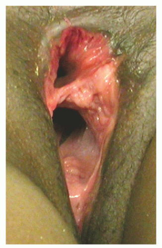
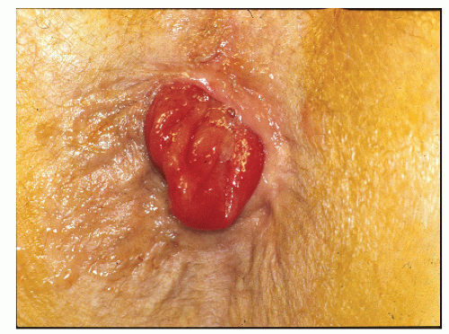
During delivery, the pelvic floor is at risk from trauma due to stretching by the fetal head on the perineum. This may cause a tear in the anterior part of the sphincter. Such an injury may involve only the sphincter ring or it may result in a cloacal deformity if a complete rupture of the anal canal opens into the introitus (Figure 16-6). In recent years, more emphasis has been placed on this form of damage than on denervation injury. Both are, however, important and often occur simultaneously. Subclinical nerve damage during delivery may be a cause of incontinence years later as the natural process of neuromuscular degeneration leads to a situation where the sphincter is no longer competent. This may account for somewhat disappointing results following surgical repair.

Evidence of pudendal nerve damage from childbirth was first reported by Snooks and colleagues in 1984 who measured fiber density and PNTMLs in 79 women 2 months following delivery. Compared with controls, fiber density (1.37 vs. 1.67) and PNTML (1.9 vs. 2.1) were both abnormal in multiparous women, with recovery being evident in those who were primiparous.418,419 Rieger and colleagues375 found a fall in resting and squeeze pressures at 6 weeks after delivery among 53 primiparous women. Although a sphincter defect was found in 41%, this was not associated with lowered anal canal pressures. In a series of 259 women assessed at 6 weeks prior to and 8 weeks following delivery, a sphincter defect occurred in 16.7% and only in those undergoing a vaginal delivery. Multivariate analysis showed forceps delivery (OR = 12), perineal tear (OR = 16), episiotomy (OR = 6.6), and parity (OR = 8.8) to be risk factors for incontinence. Overall, 9% had symptoms of incontinence, but of these only 45% were found to have a defect.


FIGURE 16-6. Cloacal defect following obstetrical injury. There is only a limited separation between the rectum and vagina.

It has become clear that incontinence after childbirth can occur without a sphincter defect.6 When pudendal nerve function was measured in 128 unselected women before and at 6 to 8 weeks after delivery, there was a significant increase in PNTML determinations in both primipara and multiparous women but not in 7 patients who underwent elective cesarean delivery, although not in those having a section after a trial of labor. Twelve women with increased PNTML at 6 weeks were reexamined at 6 months; 8 had returned to normal values.435 In incontinent patients, the PNTML was increased and related to perineal descent, indicating the presence of neurologic damage to the whole pelvic floor in some patients.245 Persistent evidence of pudendal neuropathy was evident among 14 multiparous women who were followed for 5 years.422 Physiologic studies on postpartum women have shown that multiparity, forceps delivery, increased duration of the second stage of labor, and high birth weight may lead to pudendal nerve damage and to sphincter atrophy.
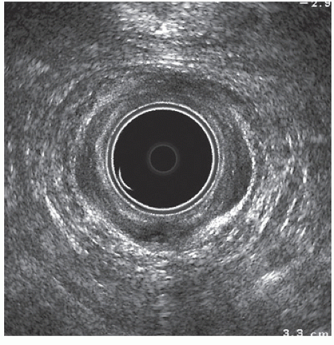
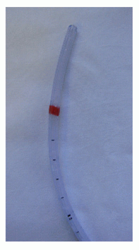
421

Anal ultrasound has demonstrated a high incidence of subclinical sphincter damage following delivery (Figure 16-7). In many cases, the presence of a defect on ultrasound has been interpreted as an indication for surgical repair. Many of these instances, however, are not associated with disruption of the sphincter ring, and repair offers, therefore, no benefit in this circumstance. Poor case selection may be another factor to explain some of the unsatisfactory results following repair.


Incidence

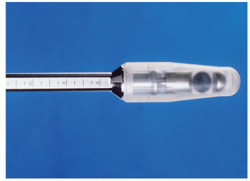
The use of ultrasound (Figure 16-8) has enabled the incidence of sphincter injury during childbirth to be studied objectively. In a group of 202 women who had an anal ultrasound prior to delivery, a repeat examination was undertaken in 150 at 6 weeks and in 32 at 6 months after delivery. Symptoms of anal incontinence were present in 10/79 (13%)
of primiparous individuals and in 11/48 (23%) of those who were multiparous. A sphincter defect was found in 28/79 (35%) and 19/48 (40%) at 6 weeks and in 22/79 (28%) of primiparae at 6 months. None of the 23 patients who had a cesarean delivery were found to have a defect. Eight of 10 patients who had forceps assistance developed a defect compared with none who had vacuum extraction. There was a correlation between a sphincter defect and symptoms of incontinence, including urgency as well as soiling.436


FIGURE 16-7. Three-dimensional ultrasound image of an occult tear of the external anal sphincter in the left anterior position. Note that the internal anal sphincter is intact.

A similar finding was reported by Pollack and colleagues from a questionnaire survey of 349 nulliparous women who then became pregnant.366 Of these, 242 returned the questionnaire at 5 and 9 months and 5 years after delivery. At 5 years, incontinent symptoms were reported in 53% of women who had a sphincter tear and in 32% of those who did not. Age, sphincter tear, and subsequent childbirth were all risk factors for incontinent symptoms at 5 years. It is important to point out that in most of these patients “incontinence” was no more than occasional loss of flatus and that fecal incontinence was rare. de Leeuw and colleagues found that 12 (35%) of their 34 patients who suffered a sphincter injury at the time of delivery experienced difficulty with bowel control.98 Sphincter defects were demonstrated in the majority. The protective effect of cesarean delivery was demonstrated in a study of 1,200 women, half of whom underwent assessment after delivery.503 In those who had a vaginal delivery, there was a fall in anal canal resting pressure, although this did not occur after cesarean delivery. The first vaginal delivery appeared to cause a permanent lowering of resting anal pressure. Frudinger and colleagues found that anal continence deteriorated in 27.6% of women following delivery, 43.2% of whom had sonographic evidence of sphincter trauma.144 Others have shown a high prevalence of anal sphincter defects on ultrasound (up to 62%) in incontinent, parous women without a prior history of anal surgery.91,318

In a study of 3,002 primiparous women who delivered between 1983 and 1986 recorded on the U.K. National Health Service database, 62% responded to a questionnaire sent two decades later. Of the 985 with adequate data, 54% recorded some form of pelvic floor symptom. About 5% had severe impairment of continence to solid stool. Cesarean delivery was protective for both urinary (OR = 0.47) and fecal (OR = 0.32) incontinence, but of 76 who had only a cesarean delivery, 9 (12%) had experienced some degree of fecal continence disturbance.107

In most patients, fecal incontinence when present resolves spontaneously within the first year. Thus, in a series of 86 primiparous women having vaginal delivery, 19 (25%) had flatus incontinence at 5 months, but this had fallen threefold by 12 months.319 In some patients symptoms persist, with the rate estimated to be approximately 3%.111 Macarthur and colleagues followed 4,214 women by mail questionnaire for 6 years.270 The prevalence of fecal incontinence at 6 years was 3.6%. Interestingly, only 10% of patients had any symptom after the first delivery. Cesarean delivery was not related to the development of symptoms (OR = 1.04), but forcepsassisted delivery at any time was (OR = 1.48). Other risk factors included high maternal age, multiparity, and Asian ethnicity. There is other evidence indicating that African Americans have a lower incidence of pelvic floor trauma than other racial groups.14,200 In their study of 128 unselected women, Sultan and associates found that a heavy baby and a prolonged second stage of labor were associated with abnormal pudendal function following delivery.435

Patients who have persisting symptoms of postpartum incontinence, or present with late onset fecal incontinence, have a high prevalence of sphincter injury. Thus, the presence of a sphincter defect among 335 patients with fecal incontinence was 65%, rising to 88% in those who had had a vaginal delivery and proctologic surgery. This compared with 43% of patients without symptoms of incontinence and with 22% in asymptomatic volunteers, the latter suggesting that the criteria for determining a sphincter defect may have been too liberal.220 Not all reports have indicated obstetrical
injury is associated with incontinence. In another questionnaire survey, Fritel and coworkers received a response from 85% of 3,114 women aged 50 to 61 years.141 Fecal incontinence within the prior 12 months was experienced by 250 (9.5%) and was related to depression (OR = 2.1), obesity (OR = 1.5), previous surgery for urinary incontinence (OR = 3.5), and anal surgery (OR = 1.7), but not to any obstetrical variable, such as parity, mode of delivery, birth weight, episiotomy, or third-degree perineal tear.


FIGURE 16-8. Anal ultrasound demonstrates external sphincter anterior defect (large arrows) with associated internal sphincter defect (small arrows).

The symptoms and prognosis of incontinence may be related to the severity of the obstetrical injury. In a series of 52 women having a third- or fourth-degree tear treated by repair, 31 (61%) had persisting symptoms. The prevalence of a defect in each group was 39 (75%) and 10 (20%).362 A mail survey by questionnaire was carried out in 180 women out of 5,123 who experienced a vaginal delivery in one Norwegian county between 1999 and 2000. Of these, 156 (87%) responded. At a median of 25 months, 88 (59%) experienced incontinence symptoms that were restricted to flatus in 53 (35%). Fourteen (9%) patients had urgency as the only symptom. It is noteworthy that only 3 of 29 women who regarded themselves to be severely disabled had sought medical help.331 Others, however, have not found a correlation with the severity of the tear and symptoms. In a study of 330 patients with fecal incontinence, Voyvodic and colleagues found no correlation between the ultrasonic severity of external and internal sphincter defects and symptoms.483 Resting tone was not related to whether or not there was a defect of the internal sphincter, although patients with a fragmented sphincter did have a low resting pressure. In contrast, however, Nichols and colleagues carried out a prospective study of 56 women who sustained a third- or fourth-degree tear of whom 39 were enrolled.328 Those with a fourth-degree tear were more likely to have bowel symptoms (59% vs. 28%) and a persistent defect (48% vs. 8%).

In another study of 55 women having their first baby, ultrasound evidence of trauma was found in 13 (29%), but clinical significance was confined to the 5 patients with evidence of external sphincter damage.490


Encopresis

Encopresis, or psychogenic soiling, is defined as the passage of formed or semiformed stool in a child’s underclothes (or other inappropriate places) that occurs regularly after the age of 4 years. It is essentially an involuntary evacuation of the bowel not caused by organic factors. Encopresis is at least four times more common in boys than in girls and is analogous to enuresis as it pertains to urinary incontinence.

The condition was first described by Weissenberg who recognized that this form of fecal incontinence was associated with emotional disturbance. Behavioral factors that may contribute to the problem include the following:


  • Excessive parental attention to toilet habits


  • Laxative use


  • Harsh or lax toilet training methods


  • Fear of the toilet or the loss of feces


  • Desire for attention


  • Family or personal stress

In time, the increasing retention of feces leads to attenuation of the rectal wall, lax sphincter contractility, progressive constipation, obstipation, and fecal impaction. Anal fissure and hemorrhoidal difficulties may develop. Loening-Baucke observed a common clinical history with children exhibiting chronic constipation and soiling, often many years of infrequent and abnormal stools, a dilated rectal ampulla, and the presence of an abdominal fecal mass.

Treatment is usually directed toward bowel management, stress reduction, and child and family psychological counseling. Laxatives, enemas, and dietary regimens are recommended, as well as encouraging the child to sit on the toilet for 10 minutes twice daily at the same time each day. The goal is to establish a practical time for defecation and, ultimately, a spontaneous bowel evacuation habit. Uridine-5-triphosphate has been suggested to have some limited success. Although the mechanism of action of this drug has not been ascertained, it is believed to stimulate the cortical substance of the brain to make the child more aware of the need to defecate.

Loening-Baucke used anorectal manometry to evaluate 20 healthy children, 12 with constipation, and 20 with chronic constipation and encopresis. Mean values for anal resting tone and anal pull-through pressure were lower in the constipated and the encopretic children than in the controls. The study was repeated up to 4 years after treatment for the condition; abnormal anorectal function was still apparent even years after cessation of treatment and apparent recovery. In a later study of 97 children, also by Loening-Baucke, the author reported that 57% had not recovered. Using a host of training techniques, Loening-Baucke noted that there was no difference in recovery rates for boys and girls, and that the likelihood of success or failure could not be predicted a priori.


Anal Surgery


Fistula-in-Ano.

The adverse effect of fistula surgery on continence has been recognized for many years (Figure 16-9).34,36 Varying degrees of impairment for control are seen even after what is considered to be a limited division of the sphincter muscle. Complete incontinence (for formed stool) that follows anorectal surgery is usually the result of inappropriate division of the anorectal ring. This is most likely to occur when a high fistula is laid open or an artificial internal opening is created (see Chapter 14).


Internal Anal Sphincterotomy.

Internal anal sphincterotomy (Figure 16-10) has been the preferred surgical treatment for anal fissure for many years (see Chapter 12). It is still the most effective approach to the management of this condition when conservative treatment fails. Unfortunately, there is a small incidence of incontinence associated with this operation. The literature, however, confuses minor with major incontinence, and in many of the reports, preoperative continence has not been recorded. The results of medical and surgical treatment have been summarized by Nelson in two excellent Cochrane reviews,321,322 with a commentary in an editorial by this author.326 The incidence is probably well below 5% and the type of soiling is minor, with mostly small amounts of mucous discharge or flatus. It may be that a form of “tailored” sphincterotomy will avoid this possibility.119,190 There is a more detailed discussion of this topic in Chapter 12.


FIGURE 16-9. Scarring, deformity, and partial sphincter loss as a consequence of anal fistulectomy.


Anal Dilatation.

Sphincter stretch for anal fissure or manual dilatation as a treatment for hemorrhoids (Lord’s procedure) is also associated with fecal incontinence. This was first reported by Snooks and colleagues before anal ultrasound was available.417 In a series of 10 patients who had undergone a Lord’s anal dilatation, they demonstrated impairment of both the external and internal sphincters. Subsequent reports of the ultrasonographic changes showed disintegration of both sphincters.330,424 Damage caused in this manner results in a
destruction of the normal sphincter anatomy and is untreatable by conventional repair (Figure 16-11). For this and other reasons, hemorrhoids are no longer treated by manual dilatation.


FIGURE 16-10. Anal ultrasound showing internal anal sphincter defect (arrow) following sphincterotomy.


FIGURE 16-11. Anal ultrasound demonstrates fragmented sphincter following anal dilatation.


Other Anal Operations.

Partial incontinence may follow hemorrhoidectomy. This may further impair closure of the canal and allow continuous discharge of mucus (see Chapter 11).168


Accidental Trauma

Trauma to the perineum can injure the sphincter mechanism (Figure 16-12). Impalement on a spike or pole as well as social injuries (e.g., fist fornication) may result in rupture of the sphincter and contamination of the extrarectal spaces.53,89,265,343,494 Sepsis can supervene and lead to excessive scar formation with a resultant patulous anal canal and an incompetent sphincter (see also Chapters 17 and 18).


FIGURE 16-12. Perineal injuries as a consequence of accidental trauma. A: Patulous anus with avulsed sphincter from impalement on a picket fence. B: Severe perineal trauma from impalement on a bedpost as a consequence of jumping in bed.


Reduced Rectal Capacity
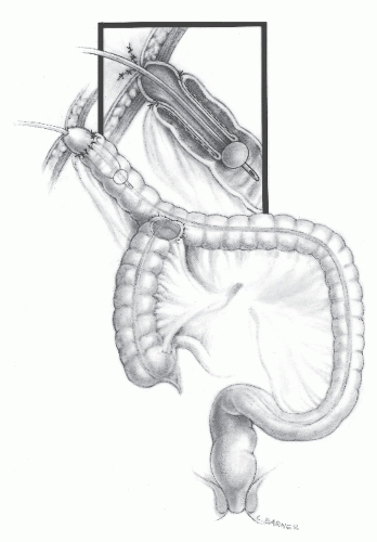

Anterior Resection

In low anterior resection, there are several factors that can impair continence. These include the loss of the rectal reservoir, damage to the anal sphincter mechanism, reduced sensation, and the effect of radiotherapy. This last factor may cause direct radiation damage to the sphincter if this is in the field of irradiation. If radiation is given postoperatively, it will reduce neorectal compliance (see later). Bowel resection designed to preserve the anal sphincter (e.g., low anterior resection, various pull-through procedures, coloanal anastomosis, abdominosacral resection) frequently results in discharge of mucus or incontinence for flatus and stool by the effect of anterior resection on the rectal capacitance, sphincter function, and neural regulation. Injury to the levator ani and external sphincter or to their innervation, and the decreased capacity of the neorectum, are contributing factors.192,295,327

Some degree of sphincter stretch occurs when a low coloanal or ileoanal anastomosis is carried out, either by transanal stapling or manually.197 Even after a stapled ileoanal anastomosis, resting tone falls in 50% of patients.68 This gradually improves with time (up to 2 years) but never fully recovers. In a prospective, randomized study of transanal-stapled anastomosis compared with the use of biofragmentable ring, 5 of 18 patients in the former group had ultrasound evidence of fragmentation of the internal sphincter that was associated with symptoms.193

Clinically, the patient will suffer from frequent smallvolume evacuations, often without warning and often associated with incontinence usually of the soiling type.345,497


Pelvic Radiotherapy

Radiotherapy will cause direct injury to the anal sphincter if this is in the field of irradiation as is the case of patients having treatment for anal carcinoma.425,473 Damage to the rectum can also occur following radiotherapy for prostatic carcinoma.272,365,445 When used for the treatment of rectal cancer, radiotherapy doubles the risk of poor function because
it is superimposed on the anterior resection.122 This is particularly true when given postoperatively.269 The long-term effects of adjuvant radiotherapy that was employed in the Swedish trials, especially those involving five 25 Gy fractions, have been reviewed by Birgisson and colleagues.33 Surgical options are limited, with every operative approach having a high complication and failure rate (see Chapter 24). Most patients are, therefore, usually managed conservatively by antidiarrheal agents and a bowel management program or by fecal diversion.179 Sacroneuromodulation has been used in four patients after anterior resection with chemoradiotherapy, of whom three appeared to benefit from permanent implantation.372


Activation of the Anorectal Inhibitory Reflex

Patients with fecal impaction often are incontinent owing to the reflex development of internal sphincter relaxation. This results in passive leakage of stool associated with a physiologically patulous sphincter.


▶ ASSESSMENT


Other Factors in the History

The second aim of the history is to determine the cause of the incontinence. It must, therefore, include details of previous obstetrical deliveries, anal operations, and symptoms that may possibly indicate neurologic, intestinal, or anal disease. The occurrence of rectal prolapse should be inquired about. Previous treatment such as rectal surgery or radiotherapy for cervical or prostatic cancer should be determined.272 The obstetrical history should include details of the type and number of deliveries, whether vaginal or cesarean, the duration of labor, any forceps or ventouse assistance, any tears whether episiotomy or inadvertent, and the birth weight of the infant.

The overall health of the patient, including an assessment of comorbidities such as pulmonary, renal, and cardiovascular disease, should be ascertained. A drug history is essential. A past or present history of psychiatric disease or dementia must be obtained and the social circumstances of the patient established.


Physical Examination

A general medical evaluation should include an assessment of the skin, oral cavity, lymph nodes, and a brief neurologic examination. This involves patient orientation and cognitive ability and motor and sensory defects in the perineum and lower limbs. A rectoscopy will identify proctitis if present. Anorectal examination by inspection and palpation should reveal the information that will dictate treatment.


Inspection

Fecal soiling around the anus may be noted. On spreading the patient’s buttocks, it will be possible to determine whether the anus is patulous, implying a weak internal sphincter from neuromuscular impairment. Inspection will determine whether the anal sphincter ring is intact or whether it is ruptured. This is one of the most important signs that will determine whether a sphincter repair is likely to help the patient. The presence of a scar alone does not indicate disruption if, for example, it represents the site of a previous repair or an episiotomy and when the sphincter ring is intact. A scar with a disrupted ring, however, may be a sign that a repair may be necessary or appropriate. The most obvious example of this abnormality is the presence of a cloacal deformity. This is usually caused by complete disruption of the anterior sphincter into the posterior vaginal wall (see Figure 16-6). A deliberate attempt to identify a mucosal ectropion (Figure 16-14) or a rectal prolapse (Figure 16-15) must be made because this will involve a different management plan from other causes of incontinence. If a prolapse is suspected from the history but is not initially evident during the examination, the patient should be asked to strain while sitting on the toilet (see Chapter 21). The presence of perineal descent during staining should be noted. Spontaneous opening of the anal canal or sphincteric relaxation is said to be indicative of anoreceptive sexual intercourse or it may suggest neurologic impairment.


Palpation

Palpation allows the resting tone of the anal canal to be assessed. Most of the effects are due to contraction of the internal sphincter. The patient is then asked to “tighten up,” and the degree of contraction or the “squeeze pressure” is evaluated. The examiner will be able to judge whether the contraction is normal or reduced. In extreme cases, it may be absent. The key question is whether any weakness felt is diffuse or localized. Diffuse weakness is a feature of pudendal neuropathy as occurs in idiopathic incontinence (see previous discussion). A localized weakness may be the site of a sphincter injury. In patients with disruption of the anal ring, an assessment of the quality or contractility of the divided external sphincter can only be obtained by palpation. With the finger in contact with the muscle, it is possible to gauge the power of its contraction. This is not possible by the physiologic tests currently available because pressure recording in a disrupted anal canal will not reflect the activity of either sphincter.


FIGURE 16-14. Mucosal ectropion.


FIGURE 16-15. Full-thickness rectal prolapsed.

In neurologic conditions, including lesions of the spinal cord and cauda equina, the tone may initially seem to be normal. However, when gentle traction is applied to the anorectal ring, the anal orifice is seen to gape (see Figure 16-3).177 Thus, Hill and colleagues performed a prospective study on 237 patients with idiopathic fecal incontinence and were able to demonstrate that an informed history and digital examination can predict the manometric findings and specialized anorectal physiologic studies with a high degree of accuracy.188 Others have reported the same conclusions.173,221 Sensory testing of the perineal skin is essential in patients with neurologic disease. Spinal and cauda equina lesions result in reduced sensation of sacral segments, S4 and S5, which will be apparent by soliciting numbness of the perineum and buttocks.

At the end of the examination, it should be possible to state whether the anal ring is intact, whether the anal muscle is diffusely weak, and whether the muscle is normal but interrupted by a defect. If a defect is present, the quality of the divided external muscle will have been assessed.


▶ INVESTIGATION

The clinical history and examination will give sufficient information to enable one to make the most suitable treatment choice for most patients but, unfortunately, they lack objectivity. Such information is obtained by tests of anorectal physiology and imaging. Physiologic testing is described in detail in Chapter 7. In current practice, almost all patients undergoing investigation for incontinence will have anorectal manometry and anal endosonography. In those with an evacuation difficulty, cineradiography, defecography, balloon evacuation,
and estimation of intestinal transit may also be performed— the first two particularly for patients with suspected rectal external or internal prolapse and the last for those with defecatory disorders (e.g., constipation, fecal impaction).46,106 If incontinence is believed to be due to a neurologic disorder, the opinion of a neurologist should be solicited.


Physiologic Studies

Physiologic evaluation of the gastrointestinal tract is now routinely performed in any specialized unit treating incontinence.134 This is useful not only for research but also for assisting in the management of the patient. The reader is referred to Chapter 7 for information on setting up such a laboratory. A working party comprising some prominent investigators who had contributed extensively to the literature on anorectal physiology was established in 1988 to determine indices of anorectal physiology.223 Their recommendations were useful at the time, but during the subsequent 20 plus years, there have since been changes in the value placed upon various aspects of these studies. This applies particularly to EMG and pudendal nerve latency, both of which have declined in importance in the evaluation of incontinence in the opinion of many investigators.

EMG was first studied by Beck in the 1930s for the investigation of the sphincter (see Chapter 7).24 The working party felt at the time that it was still a useful test. Since then, however, it has been less frequently employed owing to the pain caused by insertion of the needle electrode as well as the introduction of anal ultrasonography. This latter study is now the first-line investigation for assessing the state of the musculature.459 The recommendations also supported the value of pudendal nerve latency as an indication of the degree of denervation of the pelvic floor musculature.189,444 Since then, however, it has been felt by many in the field that the poor reproducibly and the unreliability of action potential recording have reduced its value. It is now more of research than of clinical interest in my opinion. Further details of this technique are given in Chapter 7.


Anal Manometry

Manometry involves measurement of the resting tone. This is mostly due to the contraction of the internal sphincter (resting pressure), the functional length of the anal canal, the increase in anal canal pressure due to the voluntary contraction of the external sphincter (squeeze pressure), and the RAIR.27 Computerized vector manometry was developed because conventional manometry is not able to determine whether a lowered anal canal pressure is due to a diffuse or to a focal sphincter lesion.40,357 As a research tool, this technique has been able to construct a three-dimensional anal pressure vectorgram from the data obtained by manometry and has allowed the anus to be viewed from all angles (see Figures 7-5,7-6,7-7 and 7-8). It was hoped that it would reveal occult anal sphincter injury to improve the selection of patients for sphincter repair, but despite its promise the technique has not become part of routine investigation.


Technique

Anal manometry can be undertaken using various techniques, such as open-tipped or closed-tipped catheters, perfused catheters, macroballoons, and microballoons (see Chapter 7). Most units today use an open-tipped perfused catheter (Figure 16-16). Variations in recording instrument diameter consistently affect measurements of resting anal canal pressure and the maximum squeeze pressure (MSP).296 Interpretation of physiologic studies is made difficult by incomplete knowledge of the physiology of defecation.80 Ambulant manometry is technically difficult to accomplish and remains a research tool.15


FIGURE 16-16. Open-tipped manometric catheter.


Normal Values

Pedersen and Christiansen studied 78 healthy volunteers to determine the range of normal physiologic variations with anal manometry.354 They found that the maximum intraindividual variations in the length of the anal high-pressure zone, the resting pressure, and the squeeze pressure were 10, 26, and 68 mm Hg, respectively. The median length of the pressure zone was 4 mm (14 mm Hg resting and 48 mm Hg squeeze). No gender difference was found in the length of the high-pressure zone, whereas resting pressures and squeeze pressures were higher in men than in women. One study has shown that male patients with so-called idiopathic fecal seepage may have a long anal sphincter with an abnormally high resting tone.347 Standard tests of anorectal sensorimotor function have been reproducible by different investigators, suggesting that comparison of data from different institutions is probably valid, provided the methods employed are clearly defined.382

The working party agreed that maximum resting anal pressure (MRP) should be used to denote the highest recorded pressure at any site in the anal canal at rest, and the MSP should be defined as the highest recorded pressure at any site in the anal canal during maximum voluntary (squeeze) contraction.223 Despite their recommendation to report pressure in kilopascals (100 cm of water = 9.8 kPa), most continue to use centimeters of water or millimeters of mercury.


Applications

Anorectal manometry is a useful objective measure to assess the power of the internal sphincter in patients with an intact sphincter ring and of the external sphincter during voluntary contraction. In patients with a disrupted ring, the pressure in the anal canal at rest or during maximal squeeze will not be a reflection of the intrinsic function of either sphincter.

Generally, patients suffering from problems with bowel control have lower anal canal pressures at rest and during maximum voluntary contraction than normal. There is also overlap of the value of squeeze pressures between asymptomatic
patients and those with impairment for bowel control. Many factors contribute to the mechanism for continence, and impairment of one may be compensated by the combined function of others.238


Pudendal Nerve Terminal Motor Latency

The PNTML used to be regarded as one of the most useful of physiologic parameters for the evaluation of incontinence, for determining prognosis, and for ascertaining response to treatment. The prevalence of increased latency (defined as a PNTML greater than 2.2 m/second) in 96 patients with fecal incontinence was 70% overall (75% female; 50% male) and was greater in patients with perineal descent or in those having had a difficult labor or excessive straining during defecation.383 Others have compared measurement of the PNTML with anal manometry in individuals with anal incontinence. In a study of 38 females and 14 males with fecal incontinence, the prevalence of an increased PNTML was 52%, being greater in females, but there was no relationship between increased PNTML and anal resting or voluntary squeeze pressures. PNTML might have been related to a shorter sphincter length, however.477 Manometry alone is, therefore, not helpful in identifying neuropathy, although Sangwan and coworkers found a good correlation between PNTML, single-fiber density estimation, and the RAIR. They felt that the RAIR was a good indication of the presence of neuropathy.398 Despite some slight discrepancy, the investigators believed that RAIR compared favorably with PNTML in diagnosing pudendal neuropathy.

PNTML requires training to reduce inaccuracy. Yip and colleagues compared the measurement of PNTML by students and teacher in 50 patients (41 females, 34 with fecal incontinence) and found a false-positive rate of about 20% among the students. The difference reduced, however, with experience and was abolished after 40 or more examinations.506 Despite this observation, no correlation between the PNTML and sphincter defect was found in a study of 124 females with late onset fecal incontinence, of whom 88 (71%) had a defect on ultrasound.340 Pudendal neuropathy was less common in those with (15% to 20%) than in those without (30% to 35%) a defect. The value of PNTML in the assessment of patients can be questioned further from the results of a study of 1,404 patients with fecal incontinence attending a pelvic floor outpatient clinic, of whom 83 were found to have an intact sphincter on anal ultrasound. Of these, only 28% had a raised PNTML (threshold >2.2 milliseconds), and although there was a correlation between PNTML and resting tone and fecal incontinence score, there was none with voluntary contraction pressure. This might have been expected if PNTML is an indication of pudendal neuropathy.373

In practice, PNTML is used less now than it had been 10 years ago for both diagnosis and assessment. It is susceptible to observer variation and may not correlate with the outcome after treatment. Anal ultrasound is a far more useful diagnostic modality because it gives an accurate picture of the anatomy of the anal sphincters as well as the possible occurrence of sphincter damage.


Sensation

Anal sensation is measured in the anal canal and the lower rectum by determining the current in milliamperes required to be felt by the patient on electrical stimulation—the higher the threshold, the greater the sensory deficit.380 The normal values of anal and rectal sensation are 2.0 to 9.4 mA and 7.0 to 36 mA, respectively. Anal sensation is reduced in neurologic diseases and in patients with injury to the spinal nerves supplying the perineum. Those with idiopathic incontinence associated with pudendal neuropathy have reduced sensation.


Rectal Sensation


Volumetry

Being a capacitance organ, the rectum is distensible. It can detect different degrees of distension from the first perception of the presence of a balloon introduced into the rectum to a sense of urgency with greater distension, and to a final point of maximal tolerable volume. The volumes at which these sensations occur will be a reflection of the capacitance of the rectum and also a reflection of its nerve supply.

The patient lies in the left lateral position. A balloon mounted on a catheter with a three-way tap is inserted into the rectum (Figure 16-17). The balloon is gently inflated with air or water from a 60-mL bladder syringe. The patient is asked to state when the balloon is first felt, and this volume is recorded. Inflation continues until the patient senses urgency and finally with further inflation, the patient is no longer able to tolerate the degree of distension. There is a large range of the threshold, urge, and maximal tolerable volumes as follows: 10 to 30 mL, 30 to 70 mL, and 120 to 250 mL. The volumes will be reduced where there is chronic inflammation of the rectal wall that leads to rigidity and loss of compliance, such as occurs in IBD and radiation proctitis. The volumes will also be reduced if the rectum has been replaced by colon or ileum in reconstructive surgery unless a reservoir has been constructed. They will also be increased with a megarectum and in neurologic disorders where denervation has occurred.


The Rectoanal Inhibitory Reflex

The patient lies in the left lateral position. A rectal balloon is inserted, and an anal balloon or open-tipped catheter is placed in the anal canal and fixed to the perianal skin in order to maintain its position. The anal pressure is continuously recorded. After a steady state has been achieved, 50 mL of air or water is introduced into the rectal balloon. This will induce a reflex fall of anal pressure. This maneuver is repeated, and the tracing is kept as a record of the investigation.


FIGURE 16-17. Balloon for estimation of rectal volume.


The reflex is absent in Hirschsprung’s disease; this is the most sensitive test in the diagnosis of this disorder. Sun and coworkers were able to demonstrate a close association between rectal distension and external sphincter contraction.443 Fecal incontinence occurred in some patients in their study as a result of delayed or absent external anal sphincter contraction when the internal sphincter relaxed. Bannister and colleagues analyzed the responses to rectal distension in 18 women with idiopathic fecal incontinence.19


Imaging


Anal Endosonography

This technique was developed and introduced into clinical practice by Bartram who modified the probe used for endorectal ultrasound, replacing the balloon with a sheath of adequate diameter to permit acoustic contact with the anal canal lining (Figure 16-18).249 The space between the sheath and the sensor on the probe, itself, was filled with degassed water. This technique has revolutionized the diagnosis of many anal and pelvic floor disorders, and in the case of incontinence, it allows identification of both internal and external sphincters. It can demonstrate the thickness of the former and the integrity of the sphincter ring. A normal anal sonograph is shown in Figure 16-1. Defects appear as amorphous areas of varying echogenicity that interrupt the normal striated pattern (see Figure 16-8).248

The technique was compared with electromyography in 15 women who had sustained an obstetrical injury, and the results demonstrated for the first time that an objective picture of the sphincter could be obtained, including the presence of a defect.247 The examination was well tolerated, much more so than electromyography, and a subsequent study revealed significant abnormalities in most of 44 patients with incontinence.248 Its ability to detect defects was confirmed in a second publication.52 Voyvodic and coworkers demonstrated a correlation between the size of the tear and anal pressures.483 Endoanal ultrasound has been used extensively for the identification and detection of defects in the anal sphincter as described previously.90,99,116,120,128,133,376,436 Sultan and colleagues have also used vaginal endosonography to image the anal sphincters.440 Anterior internal and external sphincteric defects can be clearly identified with both techniques, but obviously vaginal endosonography is limited to the anterior sphincter.


FIGURE 16-18. Two-dimensional endosound probe (7.5 MHz). (Courtesy Brüel & Kjær, Wilmington, MA.)


FIGURE 16-19. Brüel & Kjær Medical 2050 probe for three-dimensional endosonography. (Courtesy Brüel & Kjær, Wilmington, MA.)


Three-dimensional Endoanal Sonography

BK Medical Systems (Brüel & Kjær, Wilmington, MA) introduced the 2050 Transducer with built-in, three-dimensional imaging capability (Figure 16-19). The scanning head is moved along a 60-mm distance inside a fully encapsulated probe by using two control buttons on the handle of the transducer.169 Thus, the technique differs from conventional two-dimensional ultrasound in that multiple images are acquired during automatic withdrawal of the probe in short steps. Interpretation is not made in real time as it is with twodimensional ultrasound. The images of three-dimensional ultrasound can be obtained by a technician and interpreted by the radiologist or clinician at a convenient time later.

Gold and coworkers at St. Mark’s Hospital used this multiplanar imaging technique to reveal the length and radial extent of a sphincter tear (Figure 16-20).159 Twenty controls and 24 patients with fecal incontinence were studied. They were also able to clearly demonstrate the sex differences in sphincter configuration. Bollard and colleagues attempted to quantify the nature, characteristics, and frequency of variations in female anal sphincter anatomy.38 They observed that nulliparous women have a variable natural “defect” occurring along the anterior length of the sphincter, a factor that may contribute to overinterpretation of the existence of defects. Three-dimensional ultrasound imaging may ultimately prove to be the most useful diagnostic tool in the assessment of an individual with anal incontinence.


Magnetic Resonance Imaging

The application of high-resolution imaging of the anal sphincter mechanism has been achieved by means of an endoanal coil.100 The St. Mark’s Hospital group affirmed that an external sphincter injury can be readily assessed by means of endosonography.492 Williams who also compared this technique with that of three-dimensional endosonography analyzed the anal anatomy at similar levels by a graphic. Williams and colleagues used an overlay technique, whereby the images of three-dimensional ultrasound and magnetic resonance
imaging (MRI) were combined in the same patient.491 There was an excellent correlation with the images of the external and internal sphincters but not with the longitudinal muscle.


FIGURE 16-20. Three-dimensional ultrasound image of sphincter tear (arrows, internal sphincter; arrowheads, external sphincter).

Magnetic imaging is expensive and given the accuracy and easy availability of ultrasound, it has remained primarily a research tool. It may, however, be a method of quantifying atrophy of the external sphincter. Briel and colleagues were the first to use MRI to assess this.44 They studied 20 female patients aged 50 years (range, 28 to 75) and found atrophy in 8 (40%). Its presence was related to the clinical outcome. They subsequently studied 25 women with fecal incontinence who had a sphincter repair following obstetrical injury. Biopsies of the external sphincter were taken at operation. The MRI showed atrophy in 9 (36%), which was confirmed histologically in 8.45 Williams and colleagues studied 25 female patients, none of whom had a normal resting pressure and 16 had a low squeeze pressure. The respective cross-sectional area of the external sphincter was 240, and the mean fat content was 23%.492


▶ MANAGEMENT STRATEGY


Result of the Assessment

The clinical examination and investigations will allow the following to be determined:


  • Severity of incontinence from the history


  • Intact or ruptured anal ring by inspection and confirmed by anal ultrasound


  • Diffuse or localized sphincter weakness by palpation and anal ultrasound


  • Presence or absence of sensory loss by clinical sensation testing, electrical sensitivity, and balloon volumetry


TABLE 16-3 Fecal Incontinence: Treatment Options


Noninvasive


Conservative treatment


General measures


Drugs


Biofeedback


Irrigation


Anal plug


Invasive


Injectables


SECCA


Neuromodulation


Sacral nerve


Pudendal nerve


Posterior tibial nerve


Dorsal nerve of penis/clitoris


Surgical repair


Artificial sphincter


Graciloplasty


Artificial bowel sphincter (ABS)


Stoma


TABLE 16-4 Algorithm for the Management of Fecal Incontinence


Sphincter intact


Medical treatment


Failure → SNS


Failure → Artificial sphincter


Failure → Colostomy


Sphincter defect


Repair if large, SNS if small


Failure → Repeat repair or SNS


Failure repeat repair → SNS


Failure SNS → Artificial sphincter/colostomy


SNS, sacral nerve stimulation.


This will permit the surgeon to make a recommendation as to the management plan. The options available are shown in Table 16-3.


Algorithm for the Management of Anal Incontinence

An algorithm for the management of fecal incontinence based on current evidence is shown in Table 16-4.

The key to decision making depends on whether the sphincter ring is intact or not. This is determined on clinical examination by inspection and by anal ultrasound. Failure of all reasonable attempts to relieve incontinence will then require fecal diversion as the optimal surgical alternative.

A patient with a complete sphincter ring will not respond to repair because surgery cannot improve on the anatomical situation. Conservative treatment should be attempted. If this fails, operations include neuromodulation, anal canal injection, SECCA, or irrigation.

A patient with a disrupted sphincter ring will be helped by surgical repair if the displacement is large, such as may be present with a cloacal deformity. Where sphincter disruption is of a lesser degree, the patient may be suitable for a sphincter repair or for neuromodulation (sacroneuromodulation being the only approach that has been tried thus far). An individual who has had a failed sphincter repair, and in whom the muscle is still of good contractility, may benefit from a second attempt at repair. Irrigation is also an option in this group of patients when the aforementioned treatments fail.


▶ GENERAL APPROACH

The first question to answer is whether the patient should be treated conservatively in the first instance. The answer to this will depend on two factors, including the severity of the incontinence and the capability of other methods to improve the situation. In general, conservative treatment should be attempted unless it is obvious that some form of invasive treatment is inevitable. A cloacal deformity with severe incontinence is an example of a condition when repair is indicated. In most patients, however, conservative treatment should at least be tried.


When should conservative measures be abandoned in favor of invasive treatment? The answer to this question is the same for all functional conditions; it is when the patient feels, with full knowledge of the options, including their disadvantages, that another treatment should be tried. When this position has been arrived at, treatment options divide according to whether the sphincter ring is intact or not, such as is described previously.


▶ NONINVASIVE TREATMENT


Conservative Management

Unless the patient has a lesion that obviously requires surgery, such as occurs with acute perineal trauma, or a chronic traumatic lesion with wide displacement, conservative measures should be tried. This will comprise most patients. Conservative treatment includes a set of measures such as education, promotion of healthy living, dietary advice, and drug treatment. The patient may also feel improvement simply by receiving the attention of a therapist, having perhaps previously had little support, sympathy, and information.


General Measures

The measures to be taken will depend to a considerable extent on the type of incontinence and the age and social circumstances of the patient. General principles include education of the patient as to the anatomy and function of the pelvic floor as well as the mechanism of defecation. Weight reduction in the obese and daily exercise for everyone should be advised.23,402 Smoking may reduce intestinal transit time, a habit that may aggravate a tendency to urgency.406 If individuals are living in community-based accommodations, particularly when they require help from a caregiver, it is essential that a routine be established. This includes regular visits to the bathroom and the maintenance of a clean and well-ordered environment. Unfortunately, this is not always the case when it comes to many institutions. Therefore, education of the management personnel and caregivers is an important part of the program for improving the patient’s life. Such a regimen should emphasize the importance of avoiding constipation; clearly, if impaction develops, incontinence will ensue. It is also important to avoid urgency incontinence by anticipating the individual’s need for nearby bathroom access. In selected individuals, rectal irrigation may have a place (see later).69


Drugs that the patient is taking should be reviewed. This will include any nonstandard medications such as herbal treatments. Laxatives and products which thin the consistency of the stool should be identified. It should be recognized that patients with incontinence often manipulate their diets.176 This may involve avoiding meals or reducing the volume of food ingested. It is important to bear the possibility of nutritional deficiencies in mind and to check the blood count for anemia. In general, any dietary advice should be personalized with inquiry made as to foods that in the patient’s experience may tend to induce loose stools. There is some evidence that increasing dietary fiber may be beneficial,35 but in another study it was not found to be helpful.246

Drug treatment for incontinence includes antidiarrheal agents as well as those used to treat constipation. Loperamide is generally considered the drug of choice for diarrhea. It has no irreversible side effects, although it can be associated with bloating and abdominal cramps in some patients. In those with passive incontinence, phenylephrine theoretically might be beneficial through its direct effect on the smooth muscle of the internal sphincter. There is precious little information on this, however.


Perineal Exercises

In 1950, Kegel suggested an exercise regimen that appeared to be beneficial in both fecal and urinary incontinence.222 Since that time, numerous articles have been published attesting to the validity of this approach. Although it probably is not possible to increase internal anal sphincter tone by perineal strengthening exercises, muscle bulk and voluntary contractility of the external anal sphincter, puborectalis sling, and levatores may be improved by such a regimen.


Biofeedback

This is a difficult area because of the numerous subjective influences to which behavioral treatments in general are liable
and, in addition, to the particular effect the therapist is likely to have on the patient. Biofeedback involves the combination of monitoring of anal pressure, which the patient can relate to his or her own attempts at contraction of the pelvic floor and the attention of a therapist (Figure 16-21). An interesting observation on the value of exercise was made by Engel and colleagues in a report of six patients who had severe fecal incontinence from diverse causes.125 They inserted a 50-mL balloon into the rectum and recorded the response to sphincter contraction on a polygraph. By verbal reinforcement, each patient was able to sense the rectal distension and associated this stimulus with attempts to tighten the sphincter. This technique became known as biofeedback and has become one of the mainstays of conservative treatment. It is emphasized, however, that it is one part of a routine consisting of many features as outlined previously.


FIGURE 16-21. Patient in biofeedback treatment room with therapist and monitor.

There are useful reviews on biofeedback, one of the first being that of Heymen and colleagues.186 Norton and associates reviewed the published trials of biofeedback and found 70 noncontrolled and 11 controlled studies.334,336 All reported that biofeedback resulted in improvement, but the dropout rate was approximately 20%. The treatment groups often contained patients with various conditions, and few papers revealed long-term results. Most of the studies contained small numbers, and all were from single centers. In the 11 controlled studies, there were 592 patients; improvement occurred in 70%.

In a recent randomized, controlled trial aimed at determining whether the availability of biofeedback added value to pelvic floor exercises, there were 108 patients (83 females) with incontinence of at least 1 teaspoon of feces per week.187 They were randomized to pelvic exercises alone and pelvic exercises plus biofeedback. The patients entered a 4-week period of education and medical treatment. Those who experienced success were then excluded from the trial. The remainder went on to exercises (63) and exercises plus biofeedback (45). They each had six 1-hour training visits once in 2 weeks for 3 months. The patients were then evaluated (53 vs. 40) at 3 months. Those who had a successful result were then assessed at 1 year. The biofeedback included display of rectal and anal canal pressures to the patient, with the end points of perception of a 10-mL rectal balloon and a squeeze of 125 mm Hg maintained for 10 seconds. At 3-month assessment on an intention to treat basis, 20 (44%) of 48 biofeedback and 13 (23%) of 63 exercises only patients were free of incontinence. The frequency of days per week with fecal incontinence was 0.83 ± 1.5 and 1.6 ± 2.0 days per week (P = .083). At 12 months, 24 of the 45 biofeedback patients had “adequate relief” compared with 22 of the 63 exercises only patients.187


Electrical Stimulation

Electrical stimulation in incontinence has also been the subject of a Cochrane review.199 These authors found four eligible trials containing a total of 260 patients. The results were contradictory, although symptoms improved in the stimulation group in each report. The authors concluded, however, that it was not possible to determine that stimulation was the factor responsible.

Electrical stimulation with biofeedback was compared with biofeedback alone in a multicenter (six center) randomized trial. One hundred fifty-eight patients with incontinence for any reason (sphincter damage, rectocele, fistula surgery, previous hysterectomy, etc.) were randomized to receive amplitude-modulated, medium-frequency stimulation with EMG-biofeedback (“triple treatment”) or EMG-biofeedback alone twice daily for 9 months. At randomization, there were 79 patients in each group, but this had fallen to 52 and 62 at 6 months and to 19 and 43 at 9 months. Patients were lost to follow-up largely because they were satisfied with the results, because they had become discouraged, or did not have the time. The Cleveland Clinic scores at baseline were 10.9 ± 4.2 and 11 ± 4.8 in the EMG-biofeedback alone and the triple treatment groups, and these were 7.8 ± 5.1 versus 6.0 ± 5.3 at 6 months and 7.3 ± 5.2 versus 4.8 ± 5.7 at 9 months.403 In an invited editorial, Norton comments on the high attrition rate of the study.333 In addition, the patients had various causes for their incontinence, and the variance of the continence scores was quite considerable. Thus, the conclusions from the study that stimulation adds to biofeedback were not secure.


Anal Plug

In 1984, Prager described a device for control of feces from an end colostomy.368 Although this fell from use owing to the occurrence of local pressure necrosis in some cases, the balloon used was adapted for anal use. Mortensen and Humphreys investigated three different designs of an anal continence plug made of polyurethane sponge coated with a water-soluble surface in 10 patients (Figure 16-22).310 Patients were able to tolerate them for a median of 12 hours, indicating their potential usefulness in selected patients with anal incontinence.

Deutekom and Dobben reviewed the literature and found four trials of the anal plug.101 Two compared plug to no plug,472 one compared two plugs of the same brand,335 and one compared two plugs of different brands. There was no report of the outcome related to the severity of symptoms. Moreover, there were methodologic defects, including incompleteness of follow-up, failure to blind (three studies), and failure to assess on an intention to treat basis (three studies). Of 20 individuals tested, 14 were not able to tolerate the plug because of discomfort.335 There is a suggestion that plugs made of polyurethane function are better than those of polyvinyl alcohol.101


Colonic Irrigation


Antegrade

In 1990, Malone and colleagues described an antegrade colonic irrigation technique for the management of anal incontinence through the creation of a tube appendicostomy.280
This has been applied to the treatment of defecatory disorders in children with intractable constipation and for fecal incontinence. Levitt and colleagues developed a modification of this approach and reported their experience in 20 children with anal incontinence, for whom bowel management with conventional enemas was unsuccessful (Figures 16-23 and 16-24).262 Krogh and Laurberg reported 16 adults who underwent this approach, 10 of whom had fecal incontinence.237 Marked improvement was noted in 8, with all experiencing improvement in quality of life. Poirier and colleagues reported the results of the Malone operation in 18 patients with a defecation disorder followed for a mean of 18.5 months (range, 3 to 67).364 Five had incontinence, 4 of whom reported a satisfactory result.


FIGURE 16-22. Prototype conceal anal continence plug. The plug is wrapped in a water-soluble coat (left) and is inserted like a suppository with the gauze tape outside the anal canal. The expanded plug sits in the upper portion of the anal canal to facilitate bowel control (right). (Courtesy of Neil Mortensen, MD, John Radcliffe Hospital, Oxford, United Kingdom.)


FIGURE 16-23. Appendicostomy. Cecal plication around the native appendix. A: Appendix overlying cecum. B: Administration of an enema through the umbilicus. C: Completed plication. (From Levitt MA, Soffer SZ, Peña A. Continent appendicostomy in the bowel management of fecally incontinent children. J Pediatr Surg. 1997;32(11):1630-1633, with permission.)

Hughes and Williams described a colonic conduit that incorporated an intussuscepting valve to manage fecal
incontinence and disordered evacuation through an antegrade irrigation technique.201 The procedure involves division of the bowel at the level of the proximal transverse colon with the creation of the conduit (Figure 16-25). In their experience, after 1 month there was no leakage of solid or liquid feces from the anus between irrigations, nor was there stool or irrigating fluid refluxing to the abdominal wall. No appliance was required.


FIGURE 16-24. Neoappendicostomy. A: Flap of cecum with mesenteric vessels at its base. B: Tubularization of the cecal flap over a feeding tube. C: Completed neoappendix. D: Plication of cecum around the neoappendix (neoappendix suture line should not appose the cecal suture line). E: Completed plication. (From Levitt MA, Soffer SZ, Peña A. Continent appendicostomy in the bowel management of fecally incontinent children. J Pediatr Surg. 1997;32(11):1630—1633, with permission.)


Retrograde

Retrograde administration is easier to undertake because no surgical procedure is required and associated complications are avoided. In the last few years, the apparatus for delivery has improved from the simple catheter used for rectal washout. Closed systems are now available, which will allow irrigation that normally takes about 30 minutes per day to accomplish with minimal leakage and soiling.


Technique

The commercial systems available for transanal irrigation use either a rectal balloon catheter (Peristeen anal irrigation system; Coloplast A/S, Kokkedal, Denmark or Mallinckrodt, St. Louis, MO—Figure 16-26) or a cone-shaped colostomy tip (Coloplast A/S, Humlebæk, Denmark; Qufora irrigation system; Allerød, Denmark; or Biotrol irrimatic pump; Braun). The former is inserted into the anal canal, and the balloon is inflated to retain the catheter in place while a tepid tap water enema is administered. The irrigant is introduced by gravity or by using a pump, as in the Peristeen anal irrigation system.

Christensen and colleagues studied the completeness of retrograde irrigation by scintigraphic labeling of the stool and the irrigant in 19 patients, of whom 5 had a spinal cord lesion, 6 idiopathic fecal incontinence, and 8 idiopathic constipation.72 Irrigation was less effective in the constipated patients than in the other two groups. Bowel clearance was related to the proximal extent of the irrigating fluid. This was usually to a point just beyond the hepatic flexure. The irrigation was successful in clearing the colon in the incontinent patients.


Spinal Cord Injury

Patients with spinal cord injury pose a special problem in that their treatment options are limited. Christensen and colleagues conducted a multicenter (five centers), randomized trial comparing conservative bowel management with conservative bowel management plus retrograde irrigation over 10 weeks in 87 patients with this condition.69 At the end of this period, the St. Mark’s continence scores in the two groups were 7.3 (4.0) and 5.0 (4.6), being significantly in favor of the irrigation group. The American Society of Colon and Rectal Surgeons (ASCRS) fecal incontinence scores for depression and embarrassment also favored the irrigation group.


Anterior Resection Syndrome

Koch and colleagues studied the application of irrigation in 30 patients with incontinence following anterior resection.231 Data were available on 26, of whom 21 still used irrigation at the time of assessment. Of these, 5 had stopped; but of the remaining 21, 12 (57%) had complete continence, 3 (14%) had incontinence for flatus, and 6 (29%) had incontinence for liquid stool. The authors concluded that irrigation was a successful treatment option for these individuals.


Overall Assessment.

In a review, Tod and colleagues concluded that at the time, 2005, there was only limited evidence to support rectal irrigation for fecal incontinence and they indicated the need for further research.460 They summarized their position by demonstrating in the studies reviewed a continuation rate of irrigation of 40% to 69% over periods ranging from 6 weeks to 56 months. The most common reason for discontinuance was lack of effectiveness of the regimen, followed by perceived benefit but no need to continue, and third by technical difficulty.

In another review, 17 articles on irrigation in adults were analyzed.70 These included 1,229 patients, of whom 648 (53%) were said to have had “success.” Only one of these studies was randomized.69 In the incontinent patients, success was recorded in 47%. None of the studies addressed longterm outcome. The most recent review analyzed 25 eligible studies and reaffirmed that rectal irrigation improved symptoms in patients with spinal cord injury.121 It also indicated that it was particularly helpful in children with spina bifida.


Comment.

Retrograde irrigation is easy to perform and has a very low morbidity. The available evidence strongly indicates that it can be usefully applied to various forms of incontinence, including anterior resection syndrome, idiopathic incontinence, and neurogenic incontinence. One may anticipate a success rate of at least 50%. It may be sufficiently beneficial that one may be able to avoid a colostomy when all other treatment methods have been tried and failed. In current practice, this option is probably underused.


▶ INVASIVE TREATMENT


Bulking Agent Injection

Patients with passive seepage due to weakness of the internal sphincter have been treated by injection of bulking agents into the submucous and intersphincteric plane in the upper anal canal. Shafik was the first to report this approach using Teflon (polytetrafluoroethylene) paste (DuPont, Texas).409 He then tried autologous fat injection in 14 patients in whom complete continence was achieved at a follow-up of 18 months in all patients following up to three injections.410


Technique

The procedure can be carried out in the office or in a day center. The bowel is prepared with a Fleet enema (CB Fleet Co., Inc., Lynchburg, VA). The procedure is covered by a single dose of antibiotic such as a second-generation cephalosporin with metronidazole. With the patient in the left lateral position under mild sedation and after infiltration of a local anesthetic, a finger is inserted in the anus. An 18-gauge, 2.5-in. needle attached to a ratchet gun or other convenient syringe, depending on the viscosity of the material, is placed through the skin just to the side of the anus. It is advanced under digital control into the submucosa or intersphincteric space and also into any obvious defect (Figure 16-27).

Durasphere is delivered using a prepacked syringe with an 18-gauge, 4-cm long needle. Injections are made at four evenly spaced positions using about 2 mL at each site. The technique of injection through a proctoscope was modified by Tjandra and colleagues by the use of ultrasound guidance.457 This improves the accuracy of placement of injectable silicone PTQ implants (Bioplastique), with textured polydimethylsiloxane elastomer particles suspended in a bioexcretable hydrogel carrier of polyvinylpyrrolidone (PVP, povidone).


FIGURE 16-27. Injection of bulking agent.


Randomized Controlled Trials

Maeda and colleagues have carried out a Cochrane review273 and indentified four published randomized, controlled trials.278,416,455,457 In the trial by Tjandra and colleagues, 82 patients were randomized to intersphincteric silicone injections with (42) or without (40) endosound guidance.457 Of the 82, 71 had an intact internal sphincter, and 60% of the patients had an increase in the PNTML. At a mean of 6 months (range, 1 to 12), the respective proportions of patients who had more than 50% improvement in the Wexner score were 69% and 40%, a statistically significant difference. A high proportion of patients in each group (93% and 92%) had an increase in the quality-of-life score of more than 50%. The authors concluded that ultrasound guidance improved the results.457

Only gold members can continue reading. Log In or Register to continue

Stay updated, free articles. Join our Telegram channel

Jul 17, 2016 | Posted by in GASTROENTEROLOGY | Comments Off on Fecal Incontinence

Full access? Get Clinical Tree

Get Clinical Tree app for offline access