Chapter 42 Endoscopic Ultrasound–Guided Fine Needle Aspiration of Pancreaticobiliary Lesions
Endoscopic Ultrasound–Guided Fine Needle Aspiration in the Diagnosis of Pancreatic Tumors
Pancreatic Cancer
EUS is considered one of the most useful diagnostic procedures among the body imaging tools for detecting pancreatic cancer. EUS was shown to be superior (sensitivity of 98%) to other imaging modalities, including computed tomography (CT), in 146 patients with pancreatic cancer (Table 42.1).1 With the more recent introduction of spiral CT with dual-phase contrast, the detection rate for CT is improving. However, more recent comparisons between dual-phase spiral CT and EUS still favor EUS. The ability to obtain cytologic specimen by EUS-guided FNA has greatly aided in differentiating benign versus malignant lesions seen on EUS alone.
The application of EUS-guided FNA to the pancreas in particular has great clinical utility. CT-guided and ultrasound-guided percutaneous FNA have previously been the most commonly used methods for diagnosing pancreatic cancer. The sensitivity of percutaneous FNA ranges from 45% to 100%, with a specificity of up to 100%. However, obtaining a tissue diagnosis with CT or ultrasound guidance is limited by the ability to visualize the lesion. In our previous multicenter trial, 56% of patients with pancreatic carcinoma had CT scans that did not show a mass or revealed nonspecific enlargement of the pancreas.2 Endoscopic retrograde cholangiopancreatography (ERCP) with cytologic brushing also has historically had a relatively low yield, with sensitivities between 30% and 56%. The overall sensitivity, specificity, diagnostic accuracy, negative predictive value, and positive predictive value of EUS-guided FNA for pancreatic cancer in this study were 83%, 90%, 85%, 80%, and 100%. These values were superior to CT alone (without FNA): 56%, 37%, 50%, 28%, and 65% (P < .05). There were four complications in 164 patients (2%), including two major (perforation, bleeding) and two minor (fever) complications. Comparison among the four centers showed that institutions in which a cytologist was present during the procedure had a significantly higher cytologic yield, sensitivity, and diagnostic accuracy.
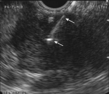
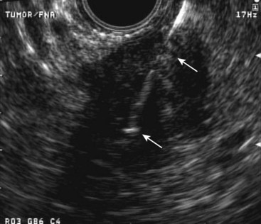
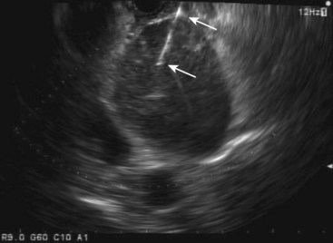
Advantages of EUS-guided FNA include procuring a tissue diagnosis while obtaining additional tumor and nodal staging information, the avoidance of additional diagnostic testing or surgery, and the prognostic information gained from the staging information (Figs. 42.1 and 42.4). Another report from a large single-institution study of 144 pancreatic lesions undergoing EUS-guided FNA showed sensitivity, specificity, and diagnostic accuracy of 82%, 100%, and 85%.3 More recently, helical or spiral CT has improved imaging of the pancreas. However, preliminary studies still show superiority of EUS compared with spiral CT.4
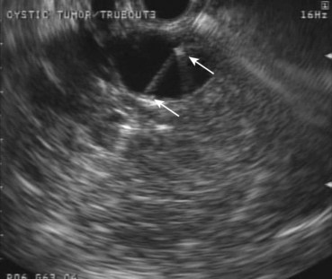
Fig. 42.3 Quick-Core (Cook Medical, Orange, CA) 19-gauge needle used to evaluate a pancreatic cyst.
(Courtesy of Cook Medical, Orange, CA.)
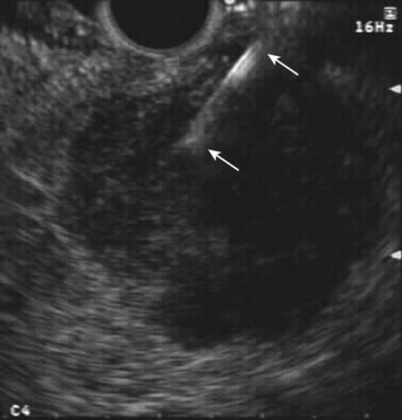
Fig. 42.4 Fine needle aspiration (FNA) (25-gauge) of metastatic liver lesion from pancreatic cancer.
Despite these data, EUS and EUS-guided FNA still possess limitations. The most difficult diagnostic problem for any imaging test, including EUS-guided FNA, is the differentiation between pancreatic carcinoma and chronic pancreatitis. Although a positive FNA is almost 100% accurate, a negative FNA is only about 80% accurate. A multicenter, retrospective study of 20 cancers missed on EUS by nine experienced endosonographers found that although 60% (12 cases) of the cancers were missed because of underlying chronic pancreatitis, other factors associated with missed lesions included a diffusely infiltrating carcinoma (3 cases), a prominent ventral or dorsal split (2 cases), and a recent episode (<4 weeks) of acute pancreatitis (1 case).5 A study of 116 pancreatic malignancies identified in patients with and without chronic pancreatitis found a sensitivity of EUS-guided FNA for malignancy of 89% in patients without chronic pancreatitis compared with 54% in patients with chronic pancreatitis.6 An additional study of 282 patients with pancreatic mass lesions undergoing 300 EUS-guided FNA procedures also found reduced sensitivity in patients with chronic pancreatitis compared with patients without chronic pancreatitis (73.9% vs. 91.3%).7 However, it was thought that this reduced sensitivity could be overcome with an increased number of FNA passes (median of five passes in patients with chronic pancreatitis patients vs. two passes in patients without chronic pancreatitis). An additional prospective study aiming to assess the number of passes to perform to maximize cytologic yield suggested that seven passes be made to optimize yield in pancreatic and miscellaneous (non–lymph node) lesions.8
These data have significantly affected the clinical algorithm of patients with known or suspected pancreatic malignancies. A clinical outcomes study was performed at a single center comparing the management and survival of 136 patients with pancreatic cancer between the pre-EUS and post-EUS eras.9 EUS detected carcinomas that were either not seen or only possibly seen by CT in 34% of patients, and there were 75% fewer required operations for diagnosis. The median survival without liver metastases was also longer during the EUS period (102 days vs. 205 days; P < .02, log-rank test), probably secondary to lead-time bias.
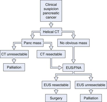
We believe that all patients thought to have operable disease based on initial CT imaging should undergo EUS with or without FNA before surgical intervention (Fig. 42.5). Considering the possibility of a false-negative result (up to 20%, especially in the setting of chronic pancreatitis), we also believe that surgical intervention should not be precluded in a patient with a high suspicion of resectable pancreatic carcinoma and a negative FNA cytology. EUS-guided FNA of pancreatic lesions is also worthwhile in patients with a prior negative tissue diagnosis by ERCP or CT of the abdomen. Gress and colleagues10 reported their experience with EUS-guided FNA of pancreatic mass lesions in 102 patients who had negative cytologic tissue diagnosis by ERCP sampling or CT-guided FNA. Among their patients, 57 of the 61 patients (93.4%) with a final diagnosis of pancreatic cancer had positive cytology results for adenocarcinoma by EUS-guided FNA. The false-positive results were zero.
The resulting changes in clinical algorithms can result in significant economic savings. In an earlier report, we reviewed a series of 44 consecutive patients who underwent EUS with or without FNA as part of their pancreatic cancer evaluation.11 Surgery and further diagnostic testing were avoided in 41% and 57% of patients. A substantial cost saving of $3300 per patient was calculated. In a series of 216 consecutive patients, Erickson and Garza12 studied the use of EUS with EUS-guided FNA as the initial approach to patients with obstructive jaundice. EUS and EUS-guided FNA not only proved useful as a diagnostic and staging modality but also served in directing the need for subsequent therapeutic ERCP, saving approximately $1007 to $1313 per patient. In addition, if EUS and EUS-guided FNA were not used at all, an extra $2200 would be spent per patient. In contrast to CT-guided FNA, EUS-guided FNA of the pancreas can be performed during the initial EUS procedure. The overall complication rate of EUS-guided FNA was reported to be 0.5% to 2.9%. An additional study comparing ERCP with brushing with EUS-guided FNA, laparoscopic biopsy, and CT-guided or ultrasound-guided FNA found that EUS-guided FNA is the best and most cost-effective initial method and the preferred secondary alternative method for the diagnosis of suspected pancreatic cancer.13
Several case reports have described malignant seeding of the needle tract after transcutaneous FNA. However, the true incidence has yet to be established. Theoretically, EUS-guided FNA of pancreatic cancers should have a lower chance of malignant seeding because of the short needle tract. A retrospective study comparing 46 patients diagnosed with pancreatic cancer via EUS-guided FNA with 43 patients diagnosed via CT-guided FNA seemed to confirm this hypothesis.14 All patients underwent neoadjuvant therapy, followed by restaging CT and attempt at surgical resection if disease progression was absent. Despite no differences in tumor characteristics between the groups, the frequency of peritoneal metastases was 2.2% in the EUS group compared with 16.3% in the CT group (P < .025).
Cystic Neoplasms
EUS can be helpful in distinguishing cystic neoplasms from pancreatic pseudocysts, although the specificity is not perfect.15 The more problematic discernment is between serous and mucinous cysts, with the latter considered premalignant. The interobserver agreement for the interpretation of cystic lesions in the pancreas is quite low. The interobserver agreement on 31 pancreatic cyst cases among eight expert endosonographers was shown to be “fair” between endosonographers for diagnosis of neoplastic versus nonneoplastic lesions (κ = 0.24).16 Agreement for individual types of lesions was moderately good for serous cystadenomas (κ = 0.46) but fair for the remainder. Accuracy rates of EUS for the diagnosis of neoplastic versus nonneoplastic lesions ranged from 40% to 93%.
EUS imaging alone is often inadequate for the clinical management of these patients. EUS-guided FNA of cystic contents can be analyzed for cytology, biochemistry, and tumor markers to aid in cyst classification. Because cytology is a relatively insensitive test, cyst fluid tumor markers such as carcinoembryonic antigen (CEA) have been employed to improve the sensitivity for the detection of malignancy. Cyst fluid CEA values are uniformly low in serous cystadenomas, higher in mucinous lesions, and markedly elevated in mucinous cystadenocarcinomas.17 A multicenter European study reported a series of 67 patients who underwent EUS-guided FNA of pancreatic cysts and subsequently underwent surgery.18 EUS alone (no FNA) correctly identified 49 cases (73%), whereas FNA correctly identified 65 cases (97%). Sensitivity, specificity, positive predictive value, and negative predictive value of EUS and EUS-guided FNA to indicate whether a lesion needed further surgery were 71% and 97%, 30% and 100%, 49% and 100%, and 40% and 95%. A value of the tumor marker for colorectal and pancreatic carcinomas (CA 19.9) greater than 50,000 U/mL had 15% sensitivity and 81% specificity to distinguish mucinous cysts from other cystic lesions and 86% sensitivity and 85% specificity to distinguish cystadenocarcinoma from other cystic lesions.
The most definitive data on cyst fluid analysis come from the Cooperative Pancreatic Cyst study, a multicenter U.S. trial that assessed 341 patients undergoing EUS-guided FNA of a pancreatic cystic lesion.19 Surgical resection was performed in 112 patients, with a final histologic diagnosis of the cysts as follows: 68 mucinous, 7 serous, 27 inflammatory, 5 endocrine, and 5 other. The results of EUS, cyst fluid cytology, and cyst fluid tumor markers (CEA, marker for gastrointestinal (GI) and ovarian carcinomas [CA 72.4], marker for ovarian and endometrial carcinomas [CA 125], CA 19.9, and marker for breast carcinoma [CA 15.3]) were prospectively collected and compared. Receiver operator curve analysis of the tumor markers showed that cyst fluid CEA (optimal cutoff of 192 ng/mL) showed the greatest area under the curve (0.79) for differentiating mucinous versus nonmucinous cystic lesions and was the most useful cyst fluid tumor marker. The accuracy of CEA (88 of 111 [79%]) was significantly greater than the accuracy of EUS morphology (57 of 112 [51%]) or cytology (64 of 109 [59%]) (P < .05). No combination of tests provided greater accuracy than CEA alone (P < .0001). The study concluded that of the tested markers, cyst fluid CEA is the most accurate test available for the diagnosis of mucinous cystic lesions of the pancreas.
Based on these data, we routinely send cyst fluid for cytology, amylase, and CEA (Table 42.2). Pseudocysts have very high amylase levels (often >50,000 IU/L) with normal CEA and benign cytology. Serous cystadenomas usually have benign cytology, normal CEA, and normal amylase. However, the specific detection of serous epithelial cells on cytology to make a definitive diagnosis in these lesions is rare (<20%) with EUS-guided FNA.20 Mucinous cystadenomas usually differ from serous cystadenoma in having a high CEA. Mucinous cystadenocarcinoma classically has malignant cytology, a low amylase, and a very elevated CEA. Although there is still some overlap of the CEA levels in these three entities, we have found the recommended cutoff value of CEA greater than 192 U/mL to be very helpful in stratifying patients to surgical versus conservative management.
Several more recent studies have aimed to characterize pancreatic cysts via use of molecular markers within the cyst fluid. A small study of 27 patients comparing CEA with the presence of k-ras-2 or loss of heterozygosity mutations using surgical histology or cytopathology as a “gold standard” found CEA to be the most predictive of histology, with concordance among the three tests in only 35% of patients.21 A larger study of 100 patients compared CEA with molecular criteria including DNA quantity, k-ras-2 point mutations, or two or more allelic imbalance mutations with pathology as the “gold standard.” The study concluded there was poor agreement (κ = 0.2) between CEA levels and molecular analysis for diagnosis of mucinous cysts. The diagnostic sensitivity for CEA was 82% compared with 77% for molecular analysis but improved to 100% when results of CEA levels and molecular analysis were combined.22
A more recent large, prospective multicenter study of 113 patients presenting for EUS-guided FNA of a pancreatic cyst assessed cytology and CEA levels compared with a detailed FNA analysis that incorporated DNA quantification, k-ras mutation and multiple allelic loss analysis, mutational amplitude, and sequence determination.23 Of 113 patients, 40 had malignant, 48 had premalignant, and 25 had benign cysts. Mucinous cysts were associated with a k-ras mutation (odds ratio 20.9) and with CEA levels greater than 148 ng/mL and allelic loss amplitudes of greater than 65%. Malignant cysts were associated with a DNA analysis that detected an allelic loss amplitude of greater than 82% and a high DNA amount (optical density ratio >10). The combination of a high-amplitude k-ras mutation followed by an allelic loss showed a specificity of 96% for malignancy, and all 10 of the malignant cysts with negative cytology were diagnosed as malignant by DNA analysis. The authors concluded that elevated amounts of pancreatic cyst fluid DNA, high-amplitude mutations, and specific mutation acquisition sequences are indicators of malignancy and that the presence of a k-ras mutation is also indicative of a mucinous cyst. They recommended DNA analysis should be considered when clinically suspicious cysts have a cytologic examination that is negative for malignancy.
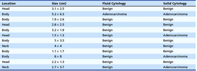
The cytology from the fluid of malignant cysts is usually nondiagnostic. For analysis of the cyst fluid to be most useful, several methods have been developed to improve the cytologic yield obtained from EUS-guided FNA. We have found that targeting any solid component, including the cyst wall, may enhance the yield on FNA cytology. We examined 42 pancreatic cystic lesions on which EUS-guided FNA was performed.24 The needle was advanced under EUS guidance into the cyst in the direction of the solid component and aspirated completely. Without withdrawal, the needle was advanced directly into the solid component. Fluid and solid cytologic samples were analyzed separately. All patients received prophylactic antibiotics. Of the 42 cysts, 12 were found to have a solid component (Table 42.3). Eight patients had both fluid and solid cytology showing benign cells. A single patient had malignant cells on both fluid and solid cytology. Three patients had benign fluid cytology but malignant (consistent with cystadenocarcinoma) solid cytology. The results of this study suggest an enhanced cytologic yield if the solid component is targeted.
In two small studies, the use of EUS-guided Tru-Cut needle biopsy (TCB) also seemed to enhance the diagnostic capability in cystic pancreatic tumors and lymphoepithelial cysts (see Fig. 42.3).25,26 In addition, the use of a cytology brush was found to be superior to conventional FNA in 7 of 10 patients undergoing EUS-guided FNA of cystic neoplastic lesions of the pancreas in a single-center study.27 Complications from this technique included one major and one minor intracystic bleed, with no infection or pancreatitis observed. Future techniques to enhance cytologic yield along with improved molecular methods of fluid analysis are needed to aid further our ability to characterize pancreatic cysts accurately.
Endocrine Tumors
EUS is very accurate in the detection of neuroendocrine tumors of the pancreas. Zimmer and associates28 reported their results in localizing and staging neuroendocrine tumors of the foregut in 40 patients examined by EUS, somatostatin receptor scintigraphy (SRS), CT, magnetic resonance imaging (MRI), and transabdominal ultrasound. EUS showed the highest sensitivity in localizing insulinomas compared with SRS, ultrasound, CT, and MRI. The authors suggested that ultrasound and EUS should be the first-line diagnostic procedures if insulinoma has been proven by a fasting test. Further diagnostic procedures were unnecessary in most cases. Further diagnostic procedures such as CT or MRI to search for distant metastases are necessary in large tumors or local invasive tumors. EUS shows the highest accuracy to detect or exclude pancreatic gastrinomas, but it fails to detect extrapancreatic gastrinomas in about 50%. The combination of EUS and SRS may give additional information. Zimmer and associates28 recommended that the first-line diagnostic procedures in patients with gastrinoma should be SRS and CT or MRI. If no metastases are detected, EUS should be the next preoperative imaging procedure. In nonfunctional neuroendocrine tumors, EUS provides the best information on local tumor invasion and regional lymph node involvement.
EUS has also been shown to be cost-effective in the preoperative localization of pancreatic endocrine tumors. Bansal and colleagues29 reported a case-control study of 36 patients who underwent preoperative EUS with a matched group of 36 patients who underwent surgical exploration immediately before the introduction of EUS. The EUS group had reduced charges for preoperative localization studies: $2620 versus $4846 per patient (P < .05). The lower cost was largely because of reductions in the number of diagnostic angiograms and venous sampling procedures performed. Surgical and total anesthesia times were decreased, as were the number of preoperative admissions for angiographic procedures. The cost-effectiveness ratio for the EUS group was $3144 per tumor localized compared with $5628 per tumor localized for the group treated before EUS became available (P < .05). The more specific utility of EUS-guided FNA in these patients was reported more recently (see Fig. 42.1).30 EUS-guided FNA was performed in 10 patients with clinically suspected functioning neuroendocrine tumors (hormonal disturbances) to determine the location and to confirm the diagnosis cytologically. EUS identified 14 tumors in these 10 patients. In all but one patient, CT did not show the tumor or missed at least one of multiple lesions. Mean tumor size was 12 mm (range 4 to 25 mm). Tumor locations were pancreas (n = 13) and duodenal wall (n = 1). Of the 14 detected lesions, 11 were aspirated under EUS-guided FNA with accurate diagnosis in all cases. Surgical confirmation of EUS-guided FNA findings was available in seven patients. There were no complications related to EUS-guided FNA.
More recently, an additional study of 30 patients with 33 lesions identified intraoperatively found sensitivity, specificity, positive predictive value, negative predictive value, and accuracy rate of EUS in conjunction with FNA of 82.6%, 85.7%, 95%, 60%, and 83.3%.31 EUS-guided FNA also seems to be highly effective and accurate in detecting the less common presentation of these lesions as cystic neuroendocrine tumors.32 In addition to diagnosing neuroendocrine tumors via EUS-guided FNA, EUS may also be useful in marking these subtle lesions using EUS-guided fine needle “tattooing” before surgery to assist in intraoperative localization.33 EUS-guided FNI may also be used in a therapeutic capacity for these lesions; a case of EUS-guided alcohol ablation of an insulinoma has been reported.34
Endoscopic Ultrasound–Guided Fine Needle Aspiration in the Staging of Pancreatic Cancer
In a prospective analysis, Mortensen and coworkers35 found a 30% overall impact of EUS-guided FNA on clinical management in 99 consecutive patients with pancreatic cancer of whom 20 patients underwent EUS-guided FNA for staging purposes: 5 liver lesions, 1 malignant ascites, 13 lymph nodes, and 1 aspiration from retroperitoneal tumor infiltration. The remaining 25 patients had diagnostic FNA: 22 pancreatic and 3 duodenal. EUS-guided FNA was performed only if positive results would have a clinically relevant impact on the subsequent management of the patient. The clinical impact of EUS-guided FNA was 12% (12 of 99) for staging purposes and 86% (18 of 21) for diagnostic purposes.
The economic impact of EUS-guided FNA in the preoperative staging of patients with pancreatic head adenocarcinoma was shown in a decision analysis model.36 The use of EUS-guided FNA prevented 16 surgeries per 100 patients compared with 8 surgeries per 100 patients if CT-guided FNA was performed for nonperitumoral lymph nodes. If the frequency of nonperitumoral lymph nodes was greater than 4%, EUS-guided FNA was the least costly procedure: $15,938 versus $16,378 for CT-guided FNA and $18,723 for surgery.
Lymph Node Assessment
According to a multivariate analysis, lymph node metastasis, intrapancreatic perineural invasion, and portal vein invasion are significant prognostic factors in patients with pancreatic cancer after curative resection.37 A retrospective analysis of patients who underwent curative resection was conducted. Of 193 patients, 38 (20%) survived for more than 5 years; 5-year survival rates for stages I, II, III, and IV disease were 41%, 17%, 11%, and 6%. Subsequently, a subgroup analysis of nodal metastasis and intrapancreatic perineural invasion was performed in 126 patients with records of these histologic findings. In the group of patients without nodal metastasis, the 5-year survival rate for patients without perineural invasion was 75%, whereas the 5-year survival rate for patients with perineural invasion was 29%; the difference in survival of these subgroups was significant (P < .02). In the group of patients with nodal metastasis, the 5-year survival rate for patients without perineural invasion was 17%, whereas the 5-year survival rate for patients with perineural invasion was 10%.
EUS imaging alone cannot fully distinguish malignant from inflammatory nodes, limiting its specificity in lymph node staging. Various EUS criteria have been described to distinguish malignant from benign nodes. These parameters, including size, shape, borders, and echotexture have lacked specificity, however. We previously conducted a study correlating EUS features of lymph nodes with the respective EUS-guided FNA diagnosis (Fig. 42.6).38 Computer analysis of EUS images of 48 lymph nodes in 47 patients using both linear array and radial scanning transducers was performed. Parameters included lymph node area, longest diameter, shape factor, and gray scale. There were 22 malignant and 26 benign nodes. When correlated with the FNA cytology results for each node, the only single criterion that was 100% specific for predicting malignancy was a longest diameter greater than 2.5 cm or an area greater than 2.5 cm2. However, using this size cutoff, the sensitivity declined to only 18%. No single criterion has an acceptable sensitivity and specificity to circumvent the need for a tissue diagnosis by EUS-guided FNA.
A multicenter study of 171 patients undergoing EUS-guided FNA of 192 lymph nodes (46 benign, 146 malignant) has been performed.39 The final diagnosis was ascertained by clinical follow-up (108 lymph nodes) or histopathology correlation (84 lymph nodes). The mean long axis dimension of benign lymph nodes was less than malignant lymph nodes (18 mm [5 to 37 mm] vs. 27 mm [5 to 80 mm]; P < .001). On average, two to three needle passes were made for each lymph node. The overall performance of EUS-guided FNA in lymph node assessment was sensitivity 92% (84% to 97% among four centers), specificity 93% (75% to 100%), and overall accuracy 92% (82% to 98%). If a long axis dimension of 15 mm was used for determining benign (≤15 mm) versus malignant (>15 mm) lymphadenopathy, EUS alone had sensitivity (67%), specificity (50%), and accuracy (63%) all inferior to EUS-guided FNA (P < .05). In 89 patients, a total of 101 lymph nodes underwent EUS-guided FNA for the staging of lung cancer (14 patients) or primary GI or pancreatic malignancies (75 patients). When comparing EUS-guided FNA with EUS size criteria (≤10 mm = benign), the sensitivity (90% vs. 91%; P = not significant) and accuracy (92% vs. 83%; P = not significant) for EUS-guided FNA were similar, whereas the specificity was superior to that of EUS size criteria alone (100% vs. 47%; P < .001).
Not only can EUS-guided FNA improve the specificity of lymph node metastasis with cytologic confirmation, but also more recently FNA has been used to detect genetic alterations in cytologic negative nodes. A prospective study was conducted to assess the clinical value of genetic staging of lymph node metastasis in patients with pancreatic adenocarcinoma who underwent curative surgery.40 In the primary tumors in 18 of 25 patients with pancreatic adenocarcinoma, k-ras gene mutations were detected. Among these 18 patients, a mutated k-ras gene was also found in at least one lymph node in 13 patients. Of these 13 patients, 7 had no evidence of histologic nodal involvement, and 6 had histologic lymph node metastasis. Although there was no significant difference in overall survival rates between the pathologic node-negative and node-positive patients, overall survival of the five patients with nodes negative for the mutated k-ras gene was significantly better than overall survival of the 13 patients with genetically metastasis-positive nodes (P < .001). Overall survival of the six patients with genetically metastasis-positive nodes limited to the peripancreatic area was significantly better than overall survival of the seven patients with genetic metastasis in lymph nodes beyond the peripancreatic areas (P = .018). These findings suggest that detection of k-ras gene mutations in lymph nodes may be clinically useful to assess the accurate tumor staging and to stratify patients who may be at higher risk for recurrence after curative resection.
Finally, in addition to abdominal lymphadenopathy, a more recent study of 160 patients with pancreatic and periampullary cancers undergoing EUS staging found that 5% had malignant mediastinal adenopathy.41 Only one of the eight patients with malignant adenopathy in the mediastinum had other sites of documented distal metastases by CT or positron emission tomography scan, although seven patients had a locally advanced cancer. These findings emphasize the importance of carefully examining the mediastinum for suspicious adenopathy routinely as a part of every EUS examination done to stage pancreatic cancer.
Liver Metastasis
EUS is not traditionally thought to be clinically applicable in liver imaging. However, more recent data have suggested otherwise. A prospective study was conducted in which 574 consecutive patients with a history or suspicion of GI or pulmonary malignant tumor undergoing upper EUS examinations underwent EUS evaluation of the liver.42 Focal liver lesions were found in 14 (2.4%) patients, and they underwent EUS-guided FNA (see Fig. 42.4). Before EUS, CT depicted liver lesions in only 3 of 14 (21%) patients. Seven of 14 patients had a known cancer diagnosis. For the other seven patients, the initial diagnosis of cancer was made by means of EUS-guided FNA of the liver. There were no immediate or late complications. This study showed that EUS can detect small focal liver lesions that are not detected at CT.
Findings of EUS-guided FNA can confirm a cytologic diagnosis of liver metastasis and establish a definitive M stage that may change clinical management. A retrospective questionnaire study regarding indications, complications, and findings of EUS-guided FNA of the liver was reported more recently, which included 21 EUS and FNA centers around the world.43 There were 167 cases of EUS-guided FNA of the liver. A complication was reported in 6 (4%) of 167 cases, including death in 1 patient with an occluding biliary stent and biliary sepsis, bleeding in 1 patient, fever in 2 patients, and pain in 2 patients. EUS-guided FNA diagnosed malignancy in 23 of 26 (89%) patients who had a prior nondiagnostic FNA under transabdominal ultrasound guidance. EUS also localized an unrecognized primary tumor in 17 of 33 (52%) cases in which CT had shown only liver metastases. EUS imaging characteristics were not predictive of malignant versus benign lesions. The authors concluded that EUS-guided FNA of the liver apparently is a safe procedure with a major complication rate of approximately 1%. Additional studies have confirmed that EUS can detect previously undetected or additional lesions not visualized by other modalities44–48 and correspondingly alter planned clinical management.44,45 In addition, EUS-guided FNA achieves results comparable to or better than CT-guided FNA of hepatic lesions.49
Liver lesions typically have a much higher cytologic yield (requiring fewer needle passes owing to less inflammatory and fibrotic reaction compared with a primary pancreatic neoplasm) and give the highest staging information (see section on FNA technique). A study supporting these statements found a 94% diagnostic yield with EUS-guided FNA (31 of 33 lesions confirmed positive) of hepatic lesions with a mean of only 1.4 FNA passes taken.50
Ascites
The utility of EUS-guided FNA was evaluated for detection and aspiration of scant ascites among patients undergoing EUS for diagnosis and staging of GI malignancies.51 EUS found ascites in 85 patients (15% of a series of 571 patients). CT performed before EUS identified ascites in only 18% of patients with ascites on EUS. Of the 85 patients, 31 underwent EUS-guided FNA paracentesis, and malignant ascites was diagnosed by EUS-guided FNA in 5 patients. The clinical impact was great in these patients because surgery was avoided.
Two additional studies have corroborated these findings. Kaushik and colleagues52 identified ascites in 25 patients undergoing EUS for suspected or proven malignancy and diagnosed 16 cancers (64% of patients). Six of the nine patients with negative ascites on EUS-guided FNA had a diagnosed malignancy, but only one false-negative ascites cytology result occurred in these patients, yielding 94% sensitivity and 89% negative predictive value. DeWitt and colleagues53 detected ascites on EUS in 60 patients being staged with known or suspected malignancy. Of these, MRI and CT detected ascites in only about 50% of patients who also had either of these examinations, and ascites was detected in only 27% of patients undergoing transabdominal ultrasound. Cancer was diagnosed in 16 (27%) patients. Of the eight patients who underwent subsequent surgery, three were found to have malignant ascites, illustrating the fact that a negative fluid cytology does not exclude the possibility of peritoneal carcinomatosis.
Two more recent articles have highlighted the fact that peritoneal implants may also be identified on EUS and that these can successfully undergo EUS-guided FNA with adequate specimen to confirm malignancy.54,55 Some investigators have suggested that in patients with a small amount of ascites, a transrectal approach may be more sensitive in identifying and diagnosing malignant carcinomatosis.56
Endoscopic Ultrasound–Guided Fine Needle Aspiration in Biliary Lesions
Diagnosis and Staging of Cholangiocarcinoma
Cholangiocarcinoma is associated with a high mortality, and it is often difficult to obtain an accurate tissue diagnosis, with ERCP and brushings of the bile duct being the preferred modality for this purpose. The currently reported diagnostic yield from ERCP ranges from only 30% to 60%, and the diagnosis of malignant biliary stricture remains a challenge. EUS-guided FNA is now being used to diagnose and stage cholangiocarcinoma (see Fig. 42.6).57,58 In the original case series describing the use of EUS in diagnosis of cholangiocarcinoma, 10 patients with bile duct strictures at the hepatic hilum, diagnosed by CT or ERCP or both, underwent EUS-guided FNA. Adequate material was obtained in nine patients. Cytology revealed cholangiocarcinoma in seven patients and hepatocellular carcinoma in one patient. One benign inflammatory lesion identified on cytology proved to be a false-negative finding by frozen section. Metastatic locoregional hilar lymph nodes were detected in two patients, and in one patient the celiac and paraaortic lymph nodes were aspirated to obtain tissue proof of distant metastasis.
These studies suggest that EUS with FNA is safe and effective in evaluating proximal biliary strictures. Several additional studies, totaling 142 patients, have reported on the effect of EUS-guided FNA in evaluating biliary (including hilar) strictures, most of which had prior negative cytology with other tissue sampling modalities.59–62 These studies have shown high sensitivities (80% to 90%) and overall accuracy rates (80% to 90%) with this technique. Even though the overall accuracy was high, this was due to the high percentage of malignancies in these series. One-quarter to one-third of patients with nonmalignant cytology on EUS-guided FNA were also ultimately diagnosed with malignancy. Given the low negative predictive values observed, a negative FNA result does not reliably exclude the possibility of malignancy. Nevertheless, this technique, when used in combination with ERCP, is extremely helpful in distinguishing benign from malignant strictures and in facilitating a definitive diagnosis by increasing tissue yield.
Another use of EUS-guided FNA is staging of cholangiocarcinoma via looking for perihepatic and distal lymphadenopathy. Assessing lymphadenopathy is not only important for staging patients before planned resection or chemotherapy, but it is also crucial in evaluating patients who are being considered for liver transplantation. A study comprising 44 patients with cholangiocarcinoma being evaluated with EUS before liver transplantation found 70 regional lymph nodes, with 9 of 70 nodes positive for malignancy63; this represented 8 of the 47 patients (18%). EUS detected 12 patients with lymph nodes not seen on standard imaging (CT or MRI). Of the 22 patients with negative lymph nodes by EUS and EUS-guided FNA, 20 (91%) had the EUS findings regarding their lymph node status confirmed. Two patients were found to have malignant perigastric lymph nodes at the time of surgery. An important additional finding of this study was that lymph node morphology and echo features did not predict malignant involvement and that EUS-guided FNA of all visualized lymph nodes in such patients is advised. Finally, in addition to cholangiocarcinoma, EUS-guided FNA has been found to be useful in diagnosing gallbladder masses, with sensitivity rates of 80% or greater for diagnosing malignancies.62,64
Diagnosis and Staging of Ampullary Cancer
Conventional abdominal imaging studies such as CT, MRI, and transabdominal ultrasound frequently fail to detect ampullary lesions. EUS is a sensitive modality for detecting and staging ampullary tumors. Accurate staging may be affected by biliary stent placement, which is frequently performed in these patients with obstructive jaundice. Combined data from two centers reported the accuracy of ampullary tumor staging with multiple imaging modalities in patients with and patients without endobiliary stents.65 Preoperative staging was performed in 50 consecutive patients with ampullary neoplasms by EUS plus CT (37 patients), MRI (13 patients), or angiography (10 patients) over a  -year period. Of the 50 patients, 25 had a transpapillary endobiliary stent present at the time of EUS examination. EUS was shown to be more accurate than CT and MRI in the overall assessment of the T stage of ampullary neoplasms (EUS 78%, CT 24%, MRI 46%). No significant difference in N stage accuracy was noted between the three imaging modalities (EUS 68%, CT 59%, MRI 77%). EUS T stage accuracy was reduced from 84% to 72% in the presence of a transpapillary endobiliary stent. This was most prominent in the understaging of T2 and T3 carcinomas.
A second retrospective study was published in which the role of EUS-guided FNA in the diagnosis and staging of ampullary lesions was reported.66 EUS-guided FNA was performed in 20 of 27 (74%) patients with suspected ampullary tumors. EUS-guided FNA made the initial ampullary tissue diagnosis in seven patients (adenocarcinoma in five patients, adenoma in one patient, neuroendocrine tumor in one patient). In addition, EUS-guided FNA resulted in a change of the diagnosis from adenoma to adenocarcinoma in one patient. In one patient, EUS-guided FNA detected a liver metastasis not seen on CT. Overall, EUS-guided FNA provided new histologic information in 9 of 27 patients (33%).
Another study of 35 patients who underwent EUS-guided FNA of ampullary lesions, with follow-up available in 27 patients, revealed 13 patients with adenocarcinoma, 6 with atypical cells (4 suspicious for cancer and 2 consistent with reactive atypia), 2 with adenomas, 1 with carcinoid, and 13 with no evidence of malignancy.67
Stay updated, free articles. Join our Telegram channel

Full access? Get Clinical Tree


