Disorders Associated with Granulomas and Macrophage Collections
Granulomas and collections of macrophages complicate various gastrointestinal conditions, as extensively discussed in Chapter 6. Macrophage collections take the form of both compact caseating or noncaseating granulomas or they appear as diffuse infiltrates. Structures that can simulate macrophage collections or granulomas include germinal centers of the lymphoid follicles, cross sections of smooth muscle, or tangential cutting of the pericryptal myofibroblast sheath.
Small mucosal macrophage collections are common and represent a nonspecific reaction to low-grade injury. The macrophages contain foamy vacuoles or basophilic granules and they are usually scattered singly or in small clusters in the upper or lower lamina propria (Fig. 13.147). The macrophages often stain with mucin stains (Fig. 13.148) and increase in number following mucosal injury. Small, loose collections of macrophages also lie in the lamina propria adjacent to sites of crypt rupture (mucin granulomas).
Occasional small granulomas can complicate almost any infection. Granulomas with central caseating necrosis constitute the histologic hallmark of tuberculosis or Yersinia infections (see Chapter 6). Well-formed granulomas occur in approximately 50% of cases of colonic CD (see Chapter 11) and their detection relates to the disease distribution. Xanthogranulomas and foreign body granulomas complicate the presence of foreign material, such as barium (Fig. 13.89), feces, talc, food, and sutures or reactions to the transmural inflammation associated with appendicitis or diverticulitis. Deep granulomatous lesions can involve the submucosa and contain foamy histiocytes and fibroblastic cells infiltrated by lymphocytes, plasma cells, and foreign material. These occur very commonly beneath invasive carcinomas (Fig. 13.149) or are associated with diverticulosis.
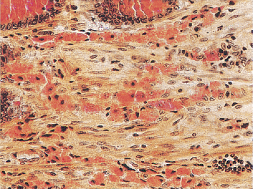 FIG. 13.148. Muciphages in the lamina propria stained with mucicarmine stain. These may be quite prominent, particularly in patients with previous mucosal injury. |
 FIG. 13.149. Granulomas associated with carcinoma. A: A colonic carcinoma invades into the muscularis propria. Its invasive front is surrounded by an inflammatory response, which consists of granulomas and chronic inflammatory cells. B: Giant cells at higher magnification. |
One also sees diffuse macrophage collections in Mycobacterium avium-intracellulare infections, Whipple disease, histoplasmosis, storage diseases, immunodeficiencies, and even algal infections. Another disorder characteristically associated with granulomatous inflammation is malakoplakia (see below).
Lamina Propria Muciphages
Muciphages, as first documented, are macrophages in the lamina propria that contain mucoproteins and are demonstrated by positive reaction with various mucin stains (462). They occur in up to 50% of rectal biopsies (462,463). These cells can be found throughout the colon in various situations and they reflect previous occult and clinically unimportant mucosal damage (463). Patients may present clinically with diarrhea, hematochezia, bowel habit change, constipation, hemorrhoids, and abdominal pain. Endoscopically, they present as nodules or polyps or they may be found incidentally (462,463). The mucosa may show regenerative or hyperplastic changes. In most cases the macrophages are located superficially (463). It may be prudent to stain for fungal or acid-fast organisms if the clinical situation warrants it.
Xanthomas
Related posts:


Stay updated, free articles. Join our Telegram channel

Full access? Get Clinical Tree


