Color Plates Diagnosis & Treatment: Gastroenterology Hepatology & Endoscopy


Color Plates


Plate 1.


Rectal mucosal biopsy specimen from a patient with dysentery caused by shigellosis. There is considerable mucosal inflammation caused by infiltration with polymorphonuclear leukocytes and mononuclear cells, as well as substantial damage to surface epithelial cells. However, mucosal architecture is generally preserved with straight, closely adjacent crypts. A crypt microabscess is seen on the right.
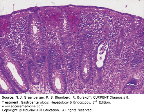


Plate 2.


Rectal mucosal biopsy specimen from a patient with dysenteric stools caused by a flare of chronic ulcerative colitis. The mucosa is heavily infiltrated with polymorphonuclear leukocytes and mononuclear cells. In contrast to Plate 1, mucosal architecture is markedly distorted, with substantial reduction in crypts and distortion of those that remain.
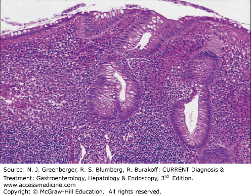
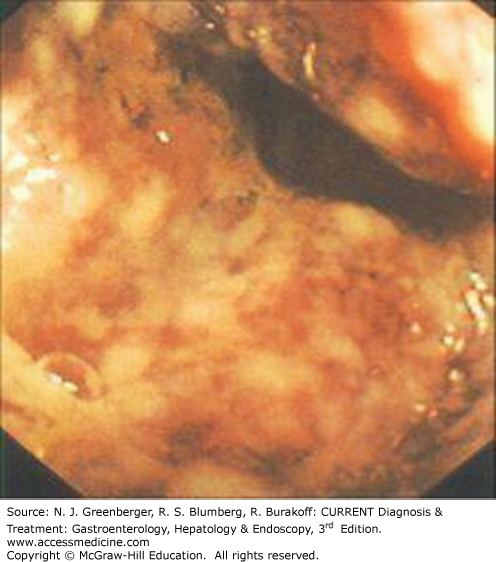
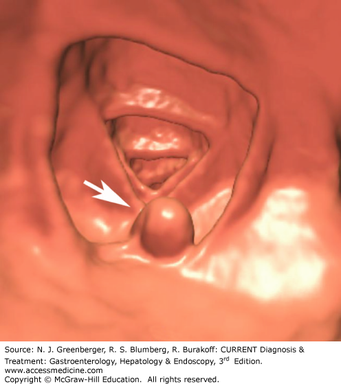
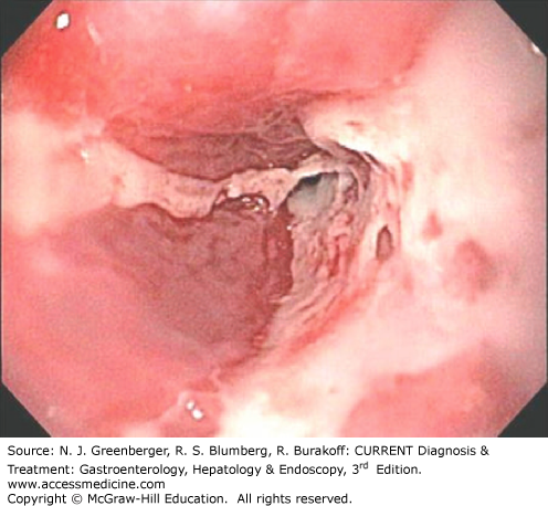
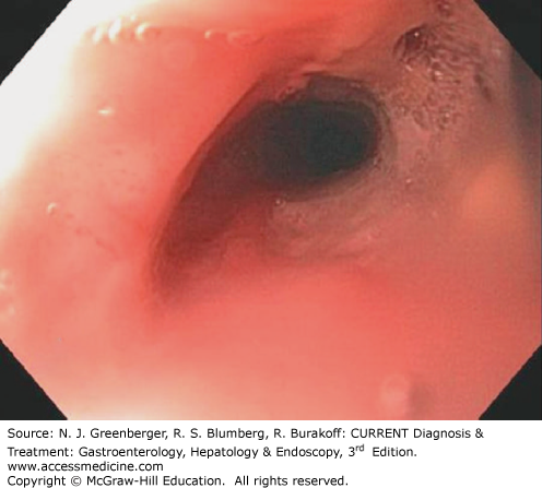
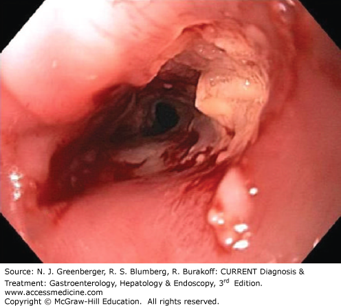
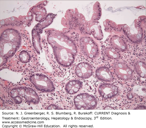
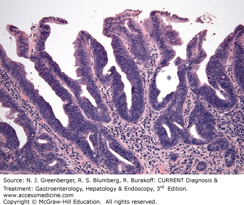
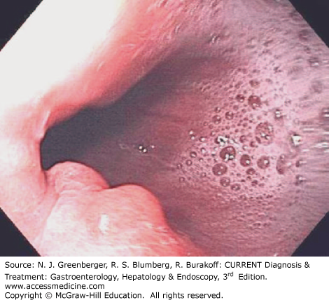
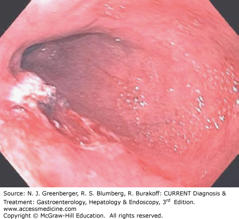


Plate 3.


Endoscopic appearance of colonic ischemia. (Used with permission from David Stockwell, MD.)


Plate 4.


Esophageal carcinoma. Fused PET-CT image shows a “hot spot” (black arrow) at the level of the distal esophagus corresponding to squamous cell carcinoma. Note the hepatic metastasis (white arrowhead).


Plate 5.


Colonic polyp. Three-dimensional endoluminal reconstructed image confirms the presence of a colonic polyp (arrow). (For corresponding axial MDCT image, see Figure 9–17 in the text.)


Plate 6.


Severe erosive esophagitis with peptic stricture.


Plate 7.


Esophageal stricture prior to dilation.


Plate 8.


Postdilation appearance of esophageal stricture shown in Plate 7.


Plate 9.


Endoscopic appearance of Barrett esophagus.


Plate 10.


Histopathologic findings in nondysplastic Barrett esophagus. Note the glandular epithelium containing goblet cells. (Used with permission from Jason Hornick, MD, PhD, Brigham and Women’s Hospital.)


Plate 11.


Histopathologic findings of low-grade dysplasia in Barrett esophagus. The surface epithelium displays nuclear stratification, limited to the lower half of the cytoplasm. (Used with permission from Jason Hornick, MD, PhD, Brigham and Women’s Hospital.)


Plate 12.


Histopathologic findings of high-grade dysplasia in Barrett esophagus. There is full-thickness nuclear stratification and the mucosa has a villous appearance. (Used with permission from Jason Hornick, MD, PhD, Brigham and Women’s Hospital.)


Plate 13.


Nodule of high-grade dysplasia in Barrett esophagus.


Plate 14.


Same area shown in Plate 13 after endoscopic mucosal resection.


Plate 15.


Endoscopic view of circumferential long segment Barrett esophagus with a few islands of normal pale pink squamous mucosa where prior surveillance biopsies have healed.


Plate 16.


Endoscopic view of the radiofrequency balloon catheter inflated to treat circumferential Barrett esophagus.


Plate 17.


Immediate postablation appearance.


Plate 18.


Endoscopic findings in eosinophilic esophagitis. Note trachea-like mucosal rings.


Plate 19.


Eosinophilic esophagitis. Note mucosal lacerations.


Plate 20.


Histologic findings in eosinophilic esophagitis. Note increased eosinophils in the squamous mucosa.


Plate 21.


The upper panel shows a characteristic severe lesion from a patient with untreated celiac sprue. Villi are absent, crypts are hyperplastic, and the lamina propria is infiltrated with many mononuclear cells. For comparison, the lower panel shows a biopsy sample from a normal volunteer showing normal mucosal architecture with tall villi and shallow crypts and just a few mononuclear cells in the lamina propria.
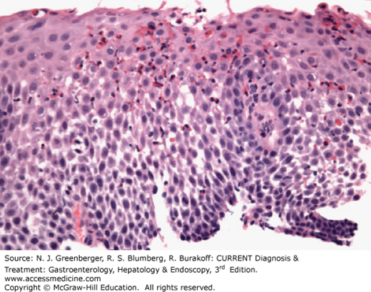


Plate 22.


The left panel shows at higher magnification the absorptive surface of the biopsy sample from the patient with celiac sprue shown in the upper panel of Plate 21. The surface absorptive cells are decreased in height and vacuolated, and the nuclei have lost their polarity. Numerous intraepithelial lymphocytes (IELs) can be seen between adjacent epithelial cells. The underlying lamina propria is heavily infiltrated with lymphocytes and plasma cells. For comparison, in the panel on the right is the tip of a villus from the biopsy sample shown in lower panel of Plate 23 from a normal individual. In contrast to the panel on the left, the absorptive cells are tall and have a well-developed brush border, with only occasional IELs evident between epithelial cells.
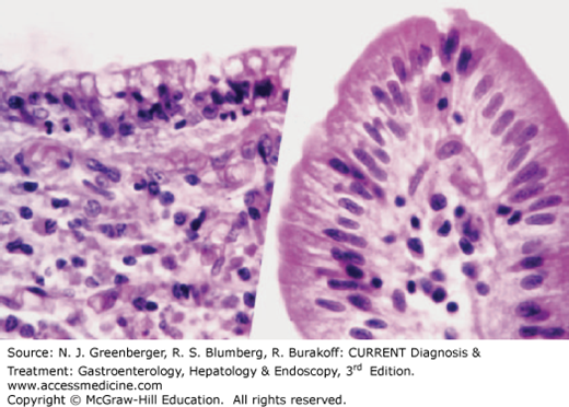


Plate 23.


Upper panel shows a biopsy sample obtained from a normal volunteer and stained with periodic acid–Schiff (PAS) stain. The glycoprotein-rich epithelial cell brush border and the goblet cell mucous are PAS-positive. The biopsy sample in the lower panel was obtained from a patient with untreated Whipple disease. The villus architecture is markedly distorted and the lamina propria is packed with large PAS-positive macrophages that virtually replace the lymphocytes and plasma cells that would normally be seen. Additionally, profiles of dilated lymphatics are evident in the lamina propria.
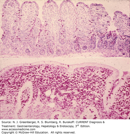
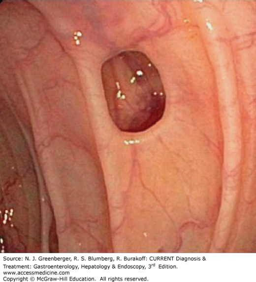
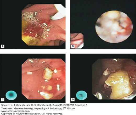
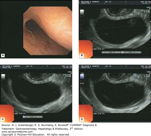
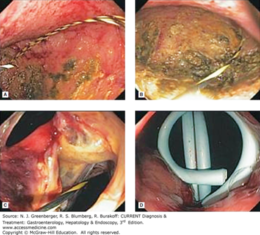


Plate 24.


Diverticulum.


Plate 25.


ERCP + ESWL: symptomatic pancreatic duct stones. A. area of papilla in the duodenum, B–D. stones. (Used with permission from David Leslie Carr-Locke, MD.)


Plate 26.


A. gastric bulge, B–D. EUS of pseudocyst. (Used with permission from Christopher Thompson, MD.)


Plate 27.


Stepwise images (Plates 26, A–D and 27, A–D) of endoscopic drainage of symptomatic pancreas. A. pseudocyst, B. wire in cyst cavity, C. cystogastrostomy site with wire, D. residual pigtail catheters. (Used with permission from Christopher Thompson, MD.)


Plate 28.


Endoscopic view of a diminutive adenomatous polyp before cold snare excision (narrow-band imaging mode).


Plate 29.


Diminutive adenomatous polyp after cold snare excision. Same patient as in Plate 28.


Plate 30.


Sessile adenomatous polyp.


Only gold members can continue reading. Log In or Register to continue

Stay updated, free articles. Join our Telegram channel

Jun 9, 2016 | Posted by in GASTROENTEROLOGY | Comments Off on Color Plates Diagnosis & Treatment: Gastroenterology Hepatology & Endoscopy

Full access? Get Clinical Tree

Get Clinical Tree app for offline access