Chronic Duodenitis Associated with Helicobacter Pylori Infection
Gastric Helicobacter pylori infections associate with various changes in the duodenal bulb. These include intraepithelial lymphocytosis (166), chronic duodenitis, chronic active duodenitis, gastric metaplasia-associated duodenitis (167), and duodenal ulcer (discussed in a later section). Endoscopically, the duodenum may appear normal or there may be mucosal edema, erythema, petechial hemorrhages, or erosions. These changes may be especially prominent in areas adjacent to peptic ulcers.
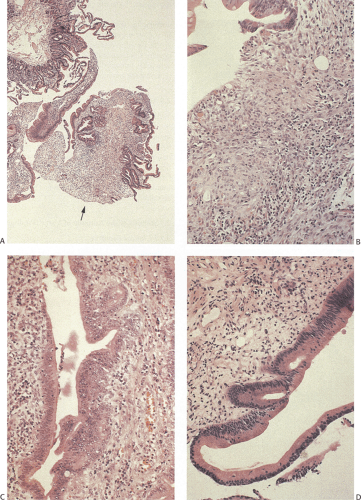
 FIG. 6.81. Sclerosing papillitis. A: Low magnification of tissue removed endoscopically as a polyp. It came from the area around the ampulla of Vater. In the upper portion of the photograph, one sees more or less normal duodenal mucosa. The three lower fragments are abnormal. The largest piece (arrow) consists of edematous tissue covered by small intestinal epithelium. In some places, glandular crowding is evident. B: Higher magnification of the edematous, inflamed tissue. C, D: Higher magnifications of the epithelium from these tissue pieces that, if examined in isolation, might be interpreted as representing an area of dysplasia. One might be worried about severe dysplasia in C due to the tangential cutting of the specimen. Features that suggest dysplasia are the glandular crowding, the nuclear palisading, and the high nuclear:cytoplasmic ratio. The intensity of the associated inflammatory changes and the gradual transition to more mature epithelium indicate that this is a reactive process, not a neoplastic one. D: Glands superficially resembling adenomatous epithelium lining the upper portion of the gland gradually merge with more normal-appearing epithelium.
Stay updated, free articles. Join our Telegram channel
Full access? Get Clinical Tree
 Get Clinical Tree app for offline access

|


