Changes Induced by Drugs and Toxins
The etiology of drug-induced colitis varies significantly, as does the underlying pathophysiologic disturbance that results from the drug ingestion. Since drug-induced colitis is likely underrecognized, its true incidence is unknown, especially since the changes induced are usually nonspecific and they mimic many other conditions. Drug-induced injury can cause acute diarrhea soon after the drug is started. Alternatively, chronic diarrhea may appear long after the drug administration. The histologic changes range from a normal-appearing colon to fulminant colitis and extensive necrosis. Drugs can induce ischemia, eosinophilic colitis, necrotizing colitis, microscopic colitis, IBD-like changes, and infectious colitis (Table 13.9).
Laxatives
Melanosis coli results from chronic ingestion of anthraquinones and laxatives derived from plants, including cascara sagrada, aloes, senna, frangula, and rhubarb. Evidence is beginning to emerge that melanosis coli does not result solely from laxative use. It can also occur in patients with IBD (both Crohn disease and ulcerative colitis) who have not used laxatives (132) and in individuals with diarrhea unrelated to IBD who have not used laxatives.
Anthraquinones concentrate in the colon, particularly in the right colon, where they are potent cellular poisons causing apoptosis, even when taken in small doses (133). The
apoptotic bodies are phagocytosed by macrophages and transformed into lipofuscin pigment by lysosomal enzymes (134). The mean apoptotic count is significantly increased in those with melanosis coli compared with a control population. It is said that it takes 4 to 12 months for melanosis coli to be visible and the same amount of time for it to disappear. It is possible that any condition associated with increased apoptosis may result in melanosis coli (135). When present in small quantities, anthraquinones probably stimulate neural tissues leading to their purgative actions. Anthraquinones and other laxatives also damage the myenteric plexus causing neuronal loss, Schwann cell proliferation (133), axonal fragmentation, axonal and dendritic swelling, and smooth muscle damage. This eventually leads to cathartic colon. Bisacodyl, anthraquinone purgatives, phenolphthalein, castor oil, and other agents also cause cathartic colon.
apoptotic bodies are phagocytosed by macrophages and transformed into lipofuscin pigment by lysosomal enzymes (134). The mean apoptotic count is significantly increased in those with melanosis coli compared with a control population. It is said that it takes 4 to 12 months for melanosis coli to be visible and the same amount of time for it to disappear. It is possible that any condition associated with increased apoptosis may result in melanosis coli (135). When present in small quantities, anthraquinones probably stimulate neural tissues leading to their purgative actions. Anthraquinones and other laxatives also damage the myenteric plexus causing neuronal loss, Schwann cell proliferation (133), axonal fragmentation, axonal and dendritic swelling, and smooth muscle damage. This eventually leads to cathartic colon. Bisacodyl, anthraquinone purgatives, phenolphthalein, castor oil, and other agents also cause cathartic colon.
TABLE 13.9 Examples of Colonic Toxicity Due to Medications | ||||||||
|---|---|---|---|---|---|---|---|---|
|
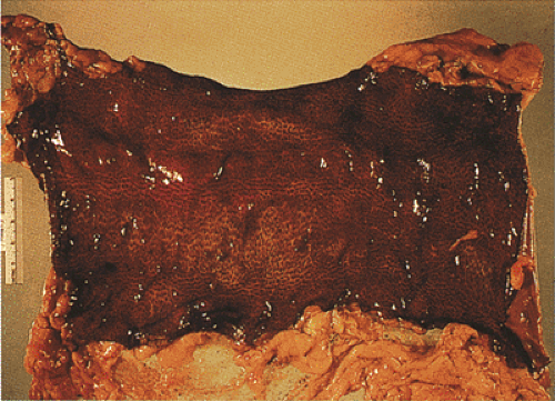
 FIG. 13.80. Severe melanosis coli. The bowel is dilated, has lost its haustral folds, and appears darkly pigmented. |
Many patients are symptomless only to be diagnosed following endoscopic examination for other reasons. Individuals with surreptitious laxative abuse typically present with unexplained chronic diarrhea. Some patients may have an obsession regarding their need to have a bowel “cleansing.”
The colon in patients with melanosis appears dark brownish (Figs. 13.80 and 13.81). Melanosis primarily affects the right colon, but in severe cases the discoloration extends to the left colon or diffusely involves the entire large intestine.
The appendix and terminal ileum may also become affected. Abnormal mucosal proliferations, such as adenomas or colon cancers, arising in a mucosa affected by melanosis coli retain their normal mucosal color rather than becoming pigmented. In severe cases, the entire lamina propria, the submucosa, and even the draining mesenteric lymph nodes contain pigmented macrophages. Migration of pigmented macrophages to regional lymph nodes results in sequential loss of the pigment from the superficial and deep lamina propria (134).
The appendix and terminal ileum may also become affected. Abnormal mucosal proliferations, such as adenomas or colon cancers, arising in a mucosa affected by melanosis coli retain their normal mucosal color rather than becoming pigmented. In severe cases, the entire lamina propria, the submucosa, and even the draining mesenteric lymph nodes contain pigmented macrophages. Migration of pigmented macrophages to regional lymph nodes results in sequential loss of the pigment from the superficial and deep lamina propria (134).
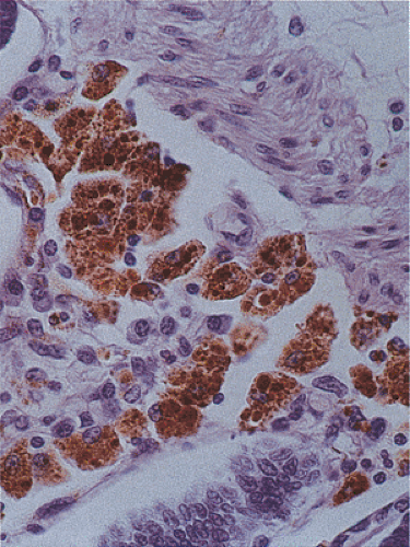
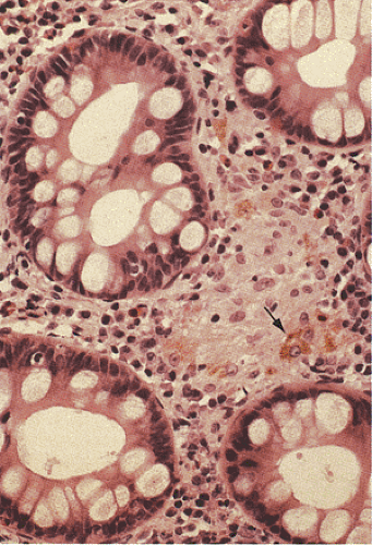
The histologic features of laxative use range from mild melanosis coli to severe cathartic colon. Because the autofluorescent pigment of melanosis coli contains melanin as well as glycoconjugates, it has been suggested that the pigment be termed melanized ceroid (136). The ceroid pigment develops from the abundant apoptotic epithelial cells, whereas the precursors of the melanic substance may derive from the anthranoids (136). Autofluorescent, refractile, golden brown, pigmented macrophages populate the lamina propria (Fig. 13.81). Occasionally, one also sees inflammation in the lamina propria, increased apoptotic bodies in the lining epithelium, superficial collections of apoptotic debris, and thickening of the muscularis mucosae. The associated inflammation probably represents a nonspecific response to an underlying injury or to stasis that may have been the cause for the laxative ingestion, rather than a direct effect of the laxative consumption. Subluminal microgranulomas often containing pigment may form (Fig. 13.82).
The pigment stains positively with periodic acid–Schiff (PAS), acid-fast, aniline blue sulfate, and Schmorl stains, but it is negative with the Perl reaction. The brown pigment may be confused with hemosiderin, which usually appears larger and more refractile than melanosis. Special stains for iron can be used in questionable cases.
Mucosal laceration or perforation with mucosal hemorrhage can complicate enema tube insertion. Devices other than the usual enema nozzle used to administer home enemas may cause erosions, mucosal tears, ulcers, or perforations. Patients utilizing enema solutions with various cleansing agents, including ethyl alcohol and hydrogen peroxide, may develop a severe proctitis (137,138). Hydrogen peroxide enemas, as used by some naturopaths, may induce a clinical picture resembling ischemic colitis, ulcerative colitis, or pseudomembranous colitis (137). The mucosal damage may result from ischemia secondary to the explosive entrance of the generated gases into the bowel wall. Histologically, one may see pneumatosis intestinalis, intense mucosal congestion, hemorrhage, or frank gangrenous necrosis.
Patients sometimes receive formalin enemas to cleanse the rectum either for health reasons or to sterilize the bowel wall during cancer surgery. Fortunately, this practice is very rarely used because 10% formalin causes severe, sharp pain and rectal bleeding within a few minutes of instillation. Within a week, the colonic mucosa becomes edematous, with multiple petechiae and superficial erosions. Histologically, one sees a nonspecific chronic colitis with superficial erosions. The lesions usually heal with a severe fibrosing process that progresses to stricture formation (139).
Kayexalate-Sorbitol Enemas
Kayexalate-sorbitol enemas, used to treat hyperkalemia after renal transplantation, cause intestinal necrosis. Since Kayexalate causes severe constipation, it is administered along with sorbitol, which acts as an osmotic laxative. The osmotic load from the sorbitol causes vascular shunting, resulting in mild colonic ischemia. Patients treated with Kayexalate-sorbitol enemas always have underlying renal disease and many are renal transplant patients (140,141). The colonic damage is potentiated by the presence of uremia. Patients present with an abrupt onset of severe abdominal pain within hours of enema administration. In some cases, elimination of the Kayexalate enemas causes resolution of the colonic manifestations.
At the time of resection, long segments of (or even the entire) colon and rectum may appear necrotic. Histologically, one sees transmural necrosis as seen in acute ischemic injury without reperfusion. The lesion mimics autolysis, although a mild neutrophilic infiltrate may be present. The presence of dark purple crystals that are PAS positive and stain with acid-fast stains establishes the diagnosis (140,141).
Stay updated, free articles. Join our Telegram channel

Full access? Get Clinical Tree


