Blood Supply
The duodenum is supplied by the celiac and superior mesenteric arteries (Fig. 6.11). The celiac trunk branches into the gastroduodenal artery. The superior mesenteric artery supplies the jejunum, cecum, and appendix, traveling through the mesentery in several major branches (Fig. 6.12). Ten to fifteen jejunoileal arteries arise from the left side of the
superior mesenteric artery, which originates 1 to 2 cm below the celiac artery. Each divides into two branches, which join adjacent branches to form a series of arcades. These in turn branch and form a second series of arcades before the vasa recta penetrate the intestinal wall (33). The ileocolic artery, which arises from the lower superior mesenteric artery, supplies the terminal ileum, cecum, appendix, and proximal ascending colon. It anastomoses with the right colic artery. This and subsequent branches form complex arcades.
superior mesenteric artery, which originates 1 to 2 cm below the celiac artery. Each divides into two branches, which join adjacent branches to form a series of arcades. These in turn branch and form a second series of arcades before the vasa recta penetrate the intestinal wall (33). The ileocolic artery, which arises from the lower superior mesenteric artery, supplies the terminal ileum, cecum, appendix, and proximal ascending colon. It anastomoses with the right colic artery. This and subsequent branches form complex arcades.
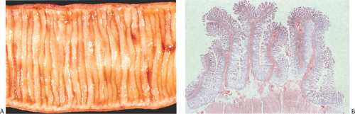 FIG. 6.10. Normal small intestine. A: The surface of the small intestine is covered by numerous regular folds (plicae circulares) with a submucosal core. B: Convolutions of mucosa and submucosa are the histologic equivalent of the folds of Kerckring. |
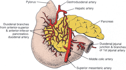 FIG. 6.11. Diagram of duodenal arterial supply. |
Intramural arteries enter the intestinal serosa and pierce the muscularis propria to form an extensive submucosal vascular plexus (Fig. 6.13). The submucosal arterial plexus gives rise to arterioles, which supply the mucosa, submucosa, and muscular layers. However, the mucosal capillary bed is isolated from that of the muscularis propria. The capillary bed in the muscularis mucosae has two layers (34). These two groups of arteries make their way into the mucosa. Some ramify on the luminal side of the muscularis mucosae and give rise to a capillary network that surrounds the crypts. Others continue into the villus, entering it at its base before arborizing into a dense capillary network. A rich network of blood capillaries ramifies through the lamina propria and is closely apposed to the epithelial basement membranes (Figs. 6.14 and 6.15). Villi are more highly vascularized than are the crypts (35). The mucosa receives approximately 75% of the blood flow. The bowel can autoregulate its blood flow, which means that it maintains a constant blood flow in the face of fluctuating arterial pressures. Following eating, small intestinal blood flow increases by over 100%, with the majority of the blood flow being diverted to the mucosa.
Related posts:


Stay updated, free articles. Join our Telegram channel

Full access? Get Clinical Tree


