Bacterial Infections in the Renal Transplant Recipient


Bacterial Infections in the Renal Transplant Recipient


Anna R. Thorner*,†

Robert H. Rubin†,‡


*Division of Infectious Disease, Massachusetts General Hospital, Harvard University, Boston, Massachusetts 02114; Division of Infectious Disease, Brigham and Women’s Hospital, Boston, Massachusetts 02115; and Center for Experimental Pharmacology and Therapeutics, Harvard University—Massachusetts Institute of Technology, Division of Health Sciences and Technology, Boston, Massachusetts, 02142


INTRODUCTION

Over the past 4 decades renal transplantation has evolved into the most practical means of rehabilitating patients with end-stage renal disease (ESRD) of diverse etiology, with 1 year allograft survival rates of >90% being achieved at leading transplant centers around the world. However, the two major barriers to success remain the same: rejection and infection, closely linked by the need for chronic immunosuppression and by the cytokines that are elaborated by the host in response to both processes. It is estimated that 50% to 75% of patients undergoing renal transplantation have evidence of invasive infection in the first year alone, with such infection having both direct and indirect effects. Of these infections, bacterial infections are the most prevalent and most rapidly progressive, occurring both de novo and as a complication of viral infection. As we approach the problem of bacterial infection in renal transplant recipients, certain general principles merit attention:


  • Because of the impaired inflammatory response caused by the immunosuppressive therapy, signs and symptoms of infection may be greatly attenuated. The consequences of this are several: a greater need for invasive biopsies, even in the face of unimpressive (although unexplained) lesions, and increased reliance on computed tomographic (CT) and/or magnetic resonance (MR) imaging in place of conventional radiography in patients with subtle signs and symptoms.


  • The potential effects of the impaired inflammatory response on the course of an infection are many (Fig. 26.1). When the clinical course is compared to that of a normal host, the transplant patient will usually have less pronounced symptoms for a sustained period, until there is a rapid deterioration which may be impossible to treat; the
    microbial burden is significantly higher in the transplant patient, requiring more intensive and/or sustained courses of antibiotics, with attendant risks of antimicrobial resistance and toxicity; and, for those diseases like tuberculosis (TB) in which person-to-person transmission is important, the efficiency of spread is greatly increased.


  • The prognosis for a patient is significantly influenced by how early in the process diagnosis is made and therapy instituted. The tension between the need for early diagnosis and the attenuation of clinical clues caused by immunosuppression is the essence of transplant infectious disease.


  • The range of organisms capable of causing clinical disease in transplant recipients is quite large. They can be divided into three general categories: (a) true pathogens, (b) sometime pathogens, and (c) nonpathogens. True pathogens are the classic plagues of mankind (e.g., influenza, bubonic plague, typhoid); they cause disease by crossing fascial planes, invading normal tissue; and/or by producing toxins. Innate immunity is not able to control these infections, and survival is dependent on the development of specific immunity and/or the institution of effective antimicrobial therapy. Sometime pathogens are usually present on the mucocutaneous surfaces of the body (e.g., Staphylococcus aureus on the skin and Bacteroides fragilis and Escherichia coli on the gut mucosa). As long as these surfaces remain intact, the colonizing bacteria have little impact. Damage to the surfaces provides the means for tissue invasion, with these sometime pathogens, when delivered to the wrong site, being quite capable of causing significant disease. Nonpathogens of importance are typically commensal organisms in the environment (e.g., Aspergillus sp, Mucor sp, Rhizopus sp, Nocardia sp, etc.), unable to cause disease except in immunocompromised individuals. The term opportunistic infection is used to describe invasive infection caused by nonpathogens or life threatening infection caused by an organism that causes only trivial disease in the normal host.


  • Because the consequences of infection in these patients are potentially so great, the emphasis should be placed on prevention rather than treatment of established infection. Thus, there is great interest in the deployment of preemptive and prophylactic antimicrobial regimens (1).


FIG. 26.1. A. The natural history of infectious disease in the transplant recipient compared to the immunocompetent host. The transplant recipient has an impaired inflammatory response, which causes a delay in the onset of signs and symptoms, and subtler clinical findings. While the normal host has a steady increase in the severity of illness over time, the transplant recipient has no clinical manifestations for an extended period, but then has a severe and rapid deterioration. B. Microbial burden in the transplant recipient compared to the immunocompetent host. The transplant recipient has a more rapid increase in microbial burden than the normal host, which increases the severity of disease and the efficiency of transmission to other patients.


RISK OF BACTERIAL INFECTION IN THE RENAL TRANSPLANT RECIPIENT

The risk of bacterial infection in the renal transplant recipient is determined largely by the interaction of three factors: the presence of anatomic/technical abnormalities; the environmental
exposures that the patient encounters; and the net state of immunosuppression.


Anatomic/Technical Factors

The anatomic/technical factors of importance include not only the transplant operation itself, but also those initiated during the perioperative period. Thus, the creation of devitalized tissue, the formation of fluid collections (be they due to urine leaks, blood, or lymphocele) is highly associated with the development of bacterial superinfection, and should be drained as quickly as possible. Similarly, bloodstream infection due to problems with vascular access, urinary tract infection (UTI) related to indwelling catheters, and pneumonia due to problems with the endotracheal tube are important problems in the first month posttransplant. Although perioperative antimicrobial therapy (e.g., cefazolin) will decrease the incidence of such infections, the most important determinant of the incidence of such infections is the technical skill with which the transplant procedure is accomplished (including the harvesting of the kidney from the donor) and the perioperative care provided (1, 2, 3, 4, 5).

In addition to an increased risk of infection associated with surgical and postoperative injury, damaged tissues from any cause, but particularly when such damage involves mucocutaneous surfaces, is associated with an increased risk of infection. Perhaps the best example of this is the occurrence of diverticulitis in renal transplant patients: the incidence is far higher than in the general population; complications such as perforation, peritonitis, and bacteremia are far more common in transplant recipients. In addition, the presentation can be quite occult (mild diarrhea and the absence of signs of peritonitis until late in the course). Surgical resection, together with antibiotic therapy, is almost invariably required to control this process, and minimal clinical symptoms (e.g., nonspecific abdominal pain or change in bowel habits) should trigger an evaluation (CT with contrast). Indeed, because of the gravity of this problem, our group advocates consideration of pretransplant sigmoid colectomy for patients who have had symptomatic diverticulitis in the recent past.


Environmental Exposures

Environmental exposures of importance to the transplant patient can occur within the community or within the hospital. Although more commonly thought of in terms of fungal infection, unexpected environmental exposures can also play a key role in the occurrence of certain bacterial infections in this patient population (Table 26.1). In the community, recent and remote exposures to such organisms as Mycobacterium tuberculosis can have a major impact on the immunosuppressed transplant patient. These infections can be categorized on the basis of the route of acquisition. Thus, bacterial infections of importance for the renal transplant patient that are due to excessive exposure include the following: those due to the ingestion of contaminated food (and, occasionally, potable water)—Listeria monocytogenes (from contaminated hot dogs, processed meats, and dairy products), and Salmonella and Campylobacter infection (from contaminated chickens and eggs); and those due to person-to-person spread from other patients or the hands of medical personnel—Clostridium difficile,methicillin-resistant Staphyloccus aureus, vancomycin-resistant enterococci, and resistant gram-negative bacilli. Finally, clinical disease due to excessive exposure to Legionella sp has had a significant impact on transplant recipients since the first outbreaks of legionellosis were first recognized (1, 2, 3, 4, 5, 6).


TABLE 26.1. Environmental bacterial exposures


Community exposures


Mode of acquisition


Pathogen


Person-to-person spread


Mycobacterium tuberculosis


Contaminated food


Salmonella sp


Listeria monocytogenes


Campylobacter jejuni


Soil


Nocardia sp


Hospital exposures


Mode of acquisition


Pathogen


Contaminated air or water system


Legionella sp
Pseudomonas aeruginosa and other gram-negative bacilli


Person-to-person spread


Methcillin-resistant Staphylococcus aureus


Vancomycin-resistant enterococci


Clostridium difficile


(Modified from Rubin RH. Infection in renal transplantation. In: Sweny P, Rubin R, Tolkoff-Rubin N, eds. The infectious complications of renal disease. New York: Oxford University Press, 2003:69-81, with permission.)


The Net State of Immunosuppression

The net state of immunosuppression is a complex function that is determined by the interaction of a number of factors (Table 26.2). The primary determinants of the net state of immunosuppression are the dose, duration, and temporal sequence in which the immunosuppressive regimen is deployed (7). The contribution of other factors is illustrated by the following observations: 90% of opportunistic infections occur in patients with preexisting immunomodulating viral infection (cytomegalovirus, Epstein-Barr virus, hepatitis viruses B and C, and the human immunodeficiency virus). Indeed, the occurrence of opportunistic infection in the absence of viral infection should suggest the possibility of an unrecognized environmental hazard. Metabolic factors, including uremia, iron overload and mobilization, and, perhaps, diabetes appear to also contribute to the net state of immunosuppression. Even more important is protein-calorie malnutrition. If one stratifies transplant patients on the basis of a serum albumin greater than or less than 2.5 g/dL, there
is a tenfold increase in the incidence of significant infection in those patients who are hypoalbuminemic (1,4,5).


TABLE 26.2. Factors contributing to the net state of immunosuppression


Host defense defects


Immunosuppressive therapy (dose, duration, nature, temporal sequence)


Technical factors (devitalized tissue, damage to the mucocutaneous barrier, foreign bodies)


Neutropenia


Metabolic factors (protein-calorie malnutrition, uremia, diabetes mellitus)


Infection with immunomodulating viruses (e.g., cytomegalovirus, Epstein-Barr virus, hepatitis B and C viruses, and the human immunodeficiency virus)


(Modified from Rubin RH. Infection in renal transplantation. In: Sweny P, Rubin R, Tolkoff-Rubin N, eds. The infectious complications of renal disease. New York: Oxford University Press, 2003:69-81, with permission.)


In recent years, it has become apparent that other genetically mediated mechanisms can impact on the risk of infection. For example, the metabolism of azathioprine may vary widely, depending on the polymorphism of the rate-limiting enzyme thiopurine methyltransferase, thus influencing the immunosuppressing effects of this compound (1). Even more compelling are reports that African-Americans receiving renal transplants have an increased rate of allograft rejection and a decreased rate of infection (8). Whatever the mechanisms involved here are, it is already clear that differing dosage schedules will be required.


TIMETABLE OF BACTERIAL INFECTION FOLLOWING RENAL TRANSPLANTATION

With the standardization of operative techniques, as well as immunosuppressive programs, a timetable for when different infections appear posttransplant can be defined; that is, although an infectious disease syndrome such as pneumonia can occur at any time in the posttransplant course, the etiology of the pneumonia will be different, depending on how much time has elapsed posttransplant. This concept of a timetable can be utilized in several different ways: in the development of a differential diagnosis for a patient presenting with a possible infectious disease syndrome; as a tool for infection control—exceptions to the timetable usually connote an excessive environmental hazard that mandates correction; as the basis for cost effective, targeted antimicrobial strategies to prevent infection. Indeed, the concept of the therapeutic prescription is derived directly from these considerations. The therapeutic prescription for the transplant patient has two components: an immunosuppressive program to prevent and treat rejection, and an antimicrobial program to make it safe. Changes in the immunosuppressive program mandate changes in the antimicrobial program as well (1).

The post-renal transplant timetable of infection is conveniently divided into three time periods: the first month; 1 to 6 months posttransplant; and the late period, more than 6 months posttransplant (Fig. 26.2).


BACTERIAL INFECTION IN THE FIRST MONTH POSTTRANSPLANT

There are three potential types of infection in the first month:


  • Despite the importance of the issues related to donor and recipient related infection, it must be emphasized that approximately 95% of infections in the first month posttransplant are the same bacterial infections of the wound, lungs, urine, and bloodstream seen in nonimmunosuppressed patients undergoing comparable surgery. The chief determinant of the incidence of these infections is the skill with which the transplant operation is performed and the postoperative care accomplished. Antimicrobial prophylaxis, aimed at skin flora, for 1 to 3 days will improve things further, but nothing takes the place of technically impeccable management.

    The first month posttransplant is characterized by two observations: the highest daily doses of immunosuppressive drugs, and a lack of opportunistic infection. Indeed, a single case of opportunistic infection during this one month “golden period” is prima facie evidence of a significant environmental exposure that must be identified and corrected. These observations also teach us that the prime determinant of the net state of immunosuppression is the sustained therapy (“the area under the curve”), not what was administered on a particular day.


  • Infection that was present in the recipient prior to transplant and that was not eradicated (a poor idea at best) prior to surgery and the initiation of immunosuppression—both of which will tend to amplify the extent and the impact of such infection. In the patient who has had multiple surgeries prior to the development of ESRD (typically, a child with significant congenital anatomic abnormalities), we have seen “cold abscesses” due to Staphylococcus aureus at a site of old wound infection reactivate posttransplant. More typically, the occurrence of aspiration pneumonia, peritonitis, and/or UTI merits attention prior to transplant. The biggest concern is bloodstream infection, as seeding of the vascular suture line can cause a mycotic aneurysm susceptible to catastrophic rupture.


  • Unrecognized bacterial infection in the donor can lead to devastating outcomes in the recipient, particularly if bloodstream infection is engendered. There are three potential sources of donor derived infection: the illness that led to organ donation could have been associated with bacteremic seeding; the organ could have been contaminated in the harvesting procedure; finally, most cadaveric donors have been subjected to critical care unit evaluation and treatment prior to organ donation, with all the attendant
    risks from vascular access, urinary catheters, endotracheal tubes, etc. Thus, we have reported Pseudomonas sepsis of donor origin leading to vascular anastomotic infection and catastrophic rupture in both recipients of kidneys from a single donor (9). What was notable in this case was that the donor had no signs or symptoms of infection prior to organ harvest, although blood cultures from the donor were positive for the same organism at the time of organ donation. A report of methicillin-resistant Staphylococcus aureus infection in the recipients of the kidneys, liver, and cornea of a single donor highlights the issue of transmission of active infection from the donor to the recipients (10). Similarly, an outbreak of Pseudomonas aeruginosa infection in the recipients of a liver, kidney, kidney-pancreas, and lungs from a single donor has been reported, which resulted in infection and disruption of the vascular anastomosis (11). In this case, direct contamination of donor tissue (including harvested donor vessels) by Pseudomonas-laden sputum was probably the mechanism of infection.


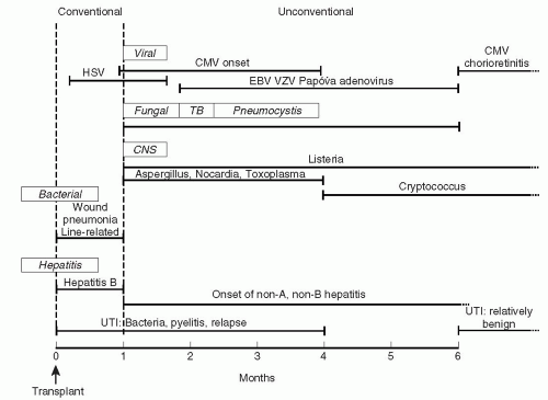
FIG. 26.2. Timetable for the occurrence of infection in the renal transplant recipient. (Modified from: Rubin RH, Wolfson JS, Cosimi AB, Tolkoff-Rubin NE: Infection in the renal transplant recipient. Am J Med 1981;70:405-411.)

Freeman et al have reported an extremely good outcome in a retrospective study of donor bacteremia. In a group of 95 donors with positive blood cultures, none of the 212 recipients became infected (12). However, the majority of the isolates were Staphylococcus epidermidis or diphtheroids, and the question of how many represented true bloodstream infection and how many skin contaminants needs to be raised. We believe that the problem of unsuspected donor bacteremia remains an important one, and will remains so until the development of rapid diagnostic techniques for the detection of occult sepsis becomes feasible.

When considering the issue of donor-derived infection, there is another group of considerations that merit discussion in this era of dire organ shortage. On occasion, patients may present with a mortal infection, but receive effective antimicrobial therapy before the hopelessness of the clinical situation becomes apparent. Under special circumstances, organs from such donors can be considered for transplantation. Thus, organs were successfully transplanted from a patient with enterococcal endocarditis, who had received several days of antibiotics prior to organ procurement. This effort to salvage extra organs should be undertaken with great care, with the limits to this approach being carefully compiled. The approach we advocate currently is the following:


  • The first consideration is the nature of the organism involved and the therapy that was prescribed up to this point. Some organisms with lesser virulence require a shorter duration and lower intensity of treatment prior to organ harvesting, provided there is evidence of clinical response (e.g., pneumococcal and/or meningococcal meningitis). In contrast, organisms that are virulent and tend to metastasize, particularly to endothelial surfaces, should be avoided. These include Staphylococcus aureus, Streptococcus pyogenes, Pseudomonas aeruginosa, and Salmonella sp.


  • Thus, organ transplantation may be considered if the infecting species is relatively bland (e.g., E. coli) or is rapidly cleared from the bloodstream with bactericidal antibiotics (penicillin-sensitive pneumococci and meningococci) for a minimum of 4 days, with rapid clearance of blood cultures following the initiation of therapy.


  • Organs from potential donors with bacteremia or invasive infection with the following organisms should not be used: Streptococcus pyogenes, vancomycin-resistant enterococci, methicillin-resistant Staphylococcus aureus, Streptococcus milleri, Salmonella sp, Nocardia sp, or mycobacteria.


  • All patients receiving organs from a donor with recently treated bacteremia should receive bactericidal antibiotics directed against the donor’s isolate, and this should be continued for a minimum of 10 to 14 days posttransplant.


  • These recommendations should be regarded as tentative, and an international registry should be established in order to monitor the success and failure of this approach.


Wound Infection

The reported incidence of wound infection after renal transplantation has ranged from 1.8% to 56% (13, 14, 15, 16, 17, 18). In the case of deep perinephric infection, 75% of cases require transplant nephrectomy, and there is a significant mortality from resulting sepsis or disruption of the vascular anastomosis (19, 20). Most patients who undergo renal transplantation are at significant risk of wound infection due to the combined factors of chronic uremia, protein malnutrition, immunosuppressive therapy and diabetes mellitus. These all contribute to poor wound healing and increased susceptibility to infection. Nonintact skin is also a significant risk factor for wound infection as well as bacteremia in the initial period following transplantation. Patients with skin conditions such as psoriasis and eczema should be treated aggressively in the time leading up to transplantation in order to minimize the risk of damaged skin serving as a portal of entry for bacteria.

The single most important determinant of wound infection is the technical success of the surgery. In a study of 439 patients from the University of Minnesota, there was a 6.1% incidence of wound infections following renal transplantation. When the patients who also had a hematoma or urinary fistula were excluded, the incidence of wound infection was only 1.6%, all of which were superficial. When diabetics and patients undergoing retransplantation were excluded, the incidence was 0.7%, with all being superficial (19). These rates are similar to those reported by other transplant groups (21, 22, 23, 24). The rate of wound infection in nonimmunosuppressed patients undergoing clean surgical procedures is 1.8% (25).

It is imperative that the transplant surgeon performs technically impeccable surgery in order to prevent urine leaks, wound hematomas and the development of lymphoceles. Additional procedures that convert the surgery from “clean” to “dirty” should be avoided. For example, a high rate of Bacteroides fragilis bacteremia has been observed when elective appendectomy was performed at the time of renal transplantation (26).

Prevention of urinary leaks is accomplished primarily by preservation of the blood supply of the donor ureter. When the blood supply is compromised, distal ureteral necrosis and fibrosis occur, which result in urinary extravasation or obstruction (27,28). In creating the urinary anastomosis, making a watertight nonobstructing connection is essential. At centers with active transplant programs, a urologic complication rate of less than 2% is seen (29, 30, 31).

Wound hematoma should also be aggressively prevented. Uremia and the use of heparin for posttransplant hemodialysis both increase the risk of hematoma formation. Wound hemostasis at the time of transplantation should be meticulous. When reexploration is required, either for bleeding or other complications, the incidence of wound infection increases tenfold (17,19,20,32).

Lymphoceles have an incidence of 2% to 18% in renal transplant recipients and may result in mechanical obstruction and/or secondary infection (33,34). Lymphoceles occur when lymphatic vessels are cut without ligation or when lymph nodes are removed. Lymph will then collect in the retroperitoneal space and must be drained surgically. Unilateral lower extremity edema on the side of the transplanted kidney is a sign of a lymphocele (20,33,34).

Several precautions are generally taken to prevent infection in the renal transplant recipient. The transplant wound is typically irrigated with antibacterial solution, although this has never been validated in a randomized trial. Open drains are avoided due to the chance that they may allow the introduction of bacteria from the skin (17,20,35). Rather, closed suction drains (e.g., Jackson-Pratt drains) are used for the first several days posttransplant to obliterate dead space and prevent fluid collections (1).

Perioperative antibiotics are an important cornerstone to the prevention of wound infections following renal transplantation. A regimen with activity against uropathogens and skin flora, such as staphylococcal species and diphtheroids should be chosen. Ampicillin-sulbactam, cefazolin, and other drugs with a similar spectrum of activity are reasonable choices. The prophylactic antibiotic should be given on call to the operating room and should be continued for no more than 24 hours postoperatively, unless there is another indication beyond basic prophylaxis (1,3, 4, 5,22, 23, 24,32,35,36).

In patients who have a fever following transplantation, imaging with either ultrasound or computed tomography should be undertaken to look for a fluid collection. Abnormal fluid collections should be promptly drained, and the patient should be given broad-spectrum antibiotics. The underlying defect that caused the fluid collection should also be corrected. Antimicrobial therapy should be continued for 10 to14 days or until the patient has been afebrile for 5 to 7 days. When perinephric infection is identified, transplant
nephrectomy should be considered, especially if there is any concern for a vascular anastomotic leak, which could have fatal consequences (1).

Only gold members can continue reading. Log In or Register to continue

Stay updated, free articles. Join our Telegram channel

Jul 26, 2016 | Posted by in NEPHROLOGY | Comments Off on Bacterial Infections in the Renal Transplant Recipient

Full access? Get Clinical Tree

Get Clinical Tree app for offline access