CHAPTER 19 Appendectomy
Step 1. Surgical anatomy
♦ The appendix is a finger-like evagination of the proximal wall of the cecum. There has been extensive debate as to its evolutionary significance, with many considering it a vestigial organ in humans. More recent thought suggests an immune regulatory role involving immune-mediated maintenance of the normal gut flora.
♦ The appendix is about 10 cm long and 8 mm wide and has a luminal diameter of 1 to 3 mm. The three taenia coli merge at its base. Its sole blood supply is from the appendiceal artery, which originates as a branch of the ileocecal artery. The artery runs in the mesoappendix, the mesentery attaching the organ to the wall of the cecum. The position of the appendix can vary considerably and may be retrocecal in up to 65% of adults. The terminal ileum is an adjacent anatomic landmark.
♦ Because of its position in the right lower quadrant, pain in this area is usually suspicious for appendicitis. Variations in anatomic location, because of such conditions as advanced pregnancy or intestinal malrotation, are infrequent but should be suspected in the appropriate clinical context.
Step 2. Preoperative consideration
Patient preparation
♦ Appendicitis is the usual diagnosis prompting appendectomy.
♦ Although the diagnosis has classically been based on symptoms, physical exam, and laboratory results, there is increasing use of contrast-enhanced computed tomography (CT) in making the diagnosis.
♦ After the diagnosis is made, each patient should receive intravenous fluid hydration, intravenous broad-spectrum antibiotic suitable to cover enteric organisms, and intravenous analgesics.
♦ Patients should be consented for appendectomy and both laparoscopic and open approaches should be discussed with the patient, as well as the usual complications including bleeding, infection, and damage to intra-abdominal structures.
♦ Expedient operation is advised, although the urgency of the operation is usually dictated by patient symptoms, laboratory results, availability of the operating room or anesthesia resources, and results of imaging studies.
♦ Having consumed nothing by mouth within the preceding 6 hours is usually ideal from an anesthesia standpoint but is not an absolute contraindication to proceeding with the operation.
Equipment and instrumentation
♦ Laparoscopic appendectomy is carried out using standard laparoscopic instruments, including atraumatic graspers and a hook electrocautery.
♦ A 5-mm or 10-mm, 30-degree or 45-degree laparoscope can be utilized. We usually use a 5-mm, 30-degree laparoscope for the procedure.
♦ Additional equipment includes the following:
 Endo GIA (Covidien Autosuture, Mansfield, Massachusetts) roticulating stapler with vascular (2.5 mm staple height) and gastrointestinal (3.5 mm staple height) cartridge loads; 45-mm cartridges are usually adequate and are easier to manipulate within the abdominal cavity than the 60-mm cartridges. Current versions of these staplers include a “lavender” (gastrointestinal) and “gold” (vascular) variant. These newer cartridges have changing staple heights along the length of the cartridge.
 The 10-mm laparoscopic clip applier should also be available in case there is a need to control a bleeding vessel or reinforce the staple line after deployment of the stapler.
 A pre-tied loop (Endoloop, Ethicon, Somerville, New Jersey) and a 10-mm laparoscopic clip applier are alternative options for controlling the appendiceal stump and artery if a stapler is not available.
Anesthesia
♦ General anesthesia is required.
♦ Placement of an orogastric (OG) tube will facilitate decompression of the stomach if there is evidence of significant gastric content based on recent oral intake or evidence from computed tomography (CT) scan.
♦ If antibiotics have not already been administered, this should be done prior to incision in the operating room.
♦ A Foley catheter is required in order to decompress the bladder and decrease the risk of bladder injury. In uncircumcised males, make sure to pull the foreskin back over the glans penis after inserting the catheter.
Room setup and positioning
♦ The patient is positioned supine on the operating table.
♦ After general anesthesia is induced and the preceding steps have been performed, the patient’s left arm should be safely tucked and padded. The right arm may be left “out” for anesthesia access.
♦ Ensure that the patient is secure on the table by testing in Trendelenburg and left-to-right roll positions.
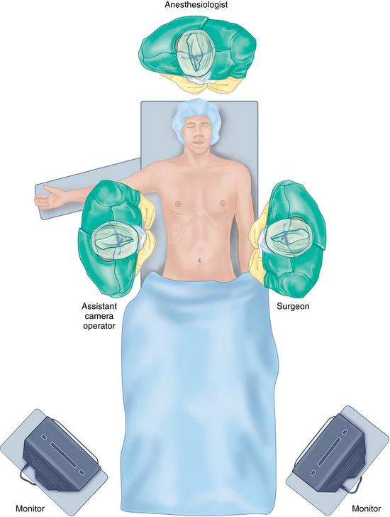
♦ The operating surgeon is positioned on the patient’s left side to the right of the scrub nurse or technician (Figure 19-1).
♦ The assistant is positioned on the patient’s right side.
♦ The main display monitor is positioned off the patient’s right leg directly in line with the appendix and operating surgeon’s line of sight. If an additional display monitor is available, it should be positioned off the patient’s left leg in line with the assistant surgeon. An additional monitor will reduce the assistant’s neck strain but may increase difficulty in using the laparoscope because of the viewing angle and fulcrum effect.
Step 3. Operative steps
Access and port placement
♦ The operating ports are a 12-mm and 5-mm port. The camera port can be 5 mm or 10 mm.
♦ The camera port is placed on the superior border of the umbilicus. Either a Veress needle or the Hassan technique may be used to gain access to the abdominal cavity.
♦ After establishing access, the abdomen is insufflated with carbon dioxide gas to 12 to 15 mmHg.
♦ A cursory laparoscopic examination is performed prior to subsequent port placement to assure the surgeon that there are no immediate contraindications to proceeding.
♦ There are a number of positions for the working ports of a laparoscopic appendectomy. We favor a 3-port (1 camera and 2 working) configuration with 2 variations:
 Our favored variation effectively hides the 12-mm incision low on the abdomen, just above the pubic symphysis. It is used when cosmesis is an important consideration and in cases of uncomplicated appendicitis. (Figure 19-2a).
Stay updated, free articles. Join our Telegram channel

Full access? Get Clinical Tree


