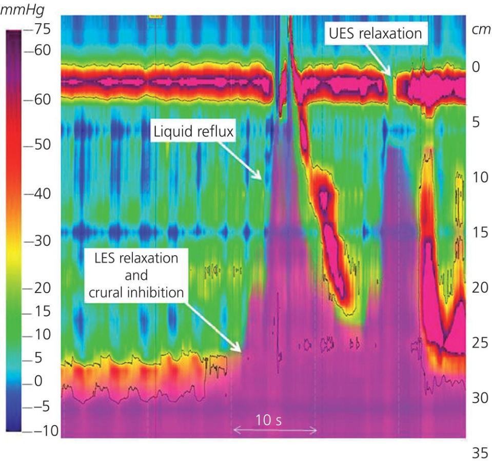
Joan W. Chen1, John E. Pandolfino2, and Peter J. Kahrilas2 1University of Michigan, Ann Arbor, MI, USA 2Northwestern University, Feinberg School of Medicine, Chicago, IL, USA The diagnosis and management of motility disorders of the esophagus have undergone a dramatic evolution over the last decade due to new technologies that have moved from the investigational realm into clinical practice. High‐resolution manometry and intraluminal impedance have enhanced our ability to visualize esophageal motor patterns and the dynamics of bolus transit through the esophagus. The functional lumen imaging probe is a novel technology that uses impedance planimetry to measure luminal diameter during volume‐controlled distension that has allowed for assessments of the mechanical properties of the esophageal wall and opening dynamics of the esophagogastric junction in various esophageal diseases. These techniques have improved our accuracy in assessing esophageal motor function, allowed us to better define and prognosticate clinical phenotypes, and expanded our ability to develop treatment paradigms for esophageal motility disorders. The role of contrast studies and endoscopy still remains crucial to the evaluation of esophageal motor disorders; however, they have become complementary diagnostic options in the evaluation of dysphagia, chest pain, and gastroesophageal reflux symptoms. With this background, the current chapter will focus on describing esophageal motility disorders using a complement of radiographic studies and advanced motility techniques that utilize esophageal pressure and diameter topography. This work was supported by R01 DK079902 (JEP) and R01 DK56033 (PJK) from the Public Health Service. Figure 9.1 Representation of a normal swallow illustrated with high‐resolution manometry (HRM) plotted in esophageal pressure topography (EPT). (a) Placement of a HRM catheter with closely spaced circumferential pressure sensors along the length of the esophagus. (b) HRM data can be displayed as pressure topography, also known as a “Clouse plot,” where pressure values between the closely spaced sensors are interpolated and the pressure magnitude indicated by color. Features of the topographic architecture of a peristaltic contraction are shown in (b). The four contraction segments (CS) and troughs between contractions, including the transition zone, are labeled on the EPT. UES, upper esophageal sphincter; EGJ, esophagogastric junction; CDP, contractile deceleration point. Source: Courtesy of the Esophageal Center at Northwestern. Figure 9.2 Normal peristalsis and bolus transit displayed on combined high‐resolution impedance manometry (HRIM) recording. The HRIM catheter and recording system provides bolus transit information in addition to esophageal contractile pattern. Impedance data, indicated by the magenta hue overlying the esophageal pressure topography, indicate the disposition of the swallowed bolus and demonstrate complete bolus clearance in this example. Source: Courtesy of the Esophageal Center at Northwestern. Figure 9.3 Reflux event seen on combined high‐resolution manometry impedance (HRIM) tracing. In this example, a transient lower esophageal sphincter relaxation (TLESR) is associated with liquid reflux from the stomach. This was followed by a swallow and repeated reflux with a microburp (UES relaxation). The bolus is then cleared by secondary peristalsis. UES, upper esophageal sphincter; LES, lower esophageal sphincter. Source: Courtesy of the Esophageal Center at Northwestern. Figure 9.4 Concomitant fluoroscopy and high‐resolution manometry esophageal pressure topography (EPT) plot of the oropharynx in a patient with a cricopharyngeal (CP) bar. The white arrow on the EPT plot indicates the high‐pressure zone at the noncompliant CP muscle. The fluoroscopic images are presented in sequence during a barium swallow. As in a typical swallow, glossopalatal junction opening occurs in synchrony with upper esophageal sphincter (UES) relaxation. This is followed by velopharyngeal junction closure, sealing off the nasopharynx to prevent regurgitation. Laryngeal vestibule closure and UES opening occur as the epiglottis is inverted, and the bolus is rapidly pushed through the UES. Bolus transit continues with pharyngeal stripping and clearance and concludes with laryngeal vestibule closure. The latter two fluoroscopic images in this patient demonstrate a prominent CP (white arrows) at the level of C5–6. Source: Courtesy of the Esophageal Center at Northwestern. Figure 9.5(a) Cricopharyngeal bar found on barium esophagram of a patient with oropharyngeal dysphagia. The posterior indentation of the barium column is caused by a noncompliant cricopharyngeus muscle (white arrow). (b) Zenker diverticulum originating above the cricopharyngeus muscle. (c) Large cervical spine fixation plates located at C5–6 level with associated esophageal luminal narrowing in a patient with dysphagia. Source: Courtesy of the Esophageal Center at Northwestern.
CHAPTER 9
Motility disorders of the esophagus
Acknowledgment


Stay updated, free articles. Join our Telegram channel

Full access? Get Clinical Tree


