CHAPTER 15 Total extraperitoneal (TEP) hernia repair
Step 1. Surgical anatomy
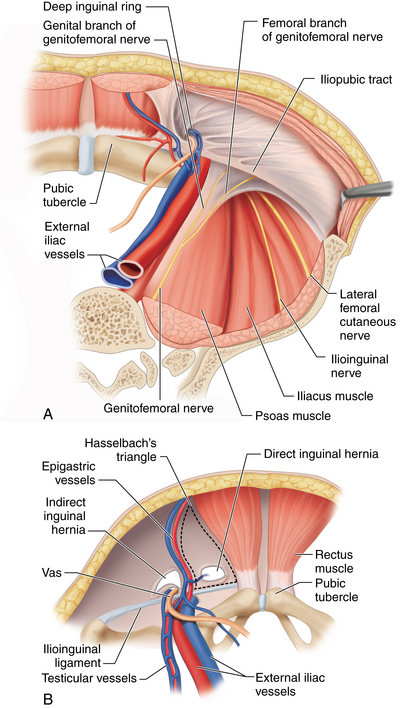
♦ A thorough knowledge of the extraperitoneal inguinal anatomy is required to avoid complications. The most important first step for successful completion of total extraperitoneal (TEP) hernia repair is correct entry into the preperitoneal space. Upon dissection of the preperitoneal space, proper orientation is confirmed by visualizing the rectus abdominis muscles superiorly, the preperitoneal fat and peritoneum inferiorly, the pubic symphysis and Cooper’s ligament in the midline, and the inferior epigastric vessels laterally (Figure 15-1).
Step 2. Preoperative considerations
Patient selection
♦ Relative to standard open inguinal herniorrhaphy, the laparoscopic approach provides superb laparoscopic visualization of the inguinal anatomy with magnified views, with the added benefit of being able to evaluate and repair the contralateral side without the need for additional incisions. Relative to open hernia repair, the laparoscopic approach is associated with improved cosmesis, reduced postoperative pain, faster recovery and return to work, and similar complication and recurrence rates. In the setting of a recurrent inguinal hernia following previous open repair, a laparoscopic repair is the preferred approach. Not only does it avoid dissection through old scar, but it is associated with reduced postoperative and recovery time and similar or improved recurrence rates compared with reoperative open herniorrhaphy.
♦ Laparoscopic repair can be performed either transabdominally (TAPP) or totally extraperitoneally (TEP); the choice largely depends on the surgeon’s experience and preference.
♦ Relative to TAPP, TEP, which is performed without violating the peritoneal space, may reduce the incidence of postoperative adhesions.
♦ Absolute contraindications to laparoscopic inguinal hernia repair include any medical condition that precludes general anesthesia, such as severe cardiac or pulmonary disease. Other contraindications include incarcerated and potentially strangulated hernia. Relative contraindications include previous prostatic surgery, which will distort anatomic planes. In addition, TEP repair may complicate future prostatic surgery, which should be a consideration for older male patients.
Preoperative preparation and anesthetic considerations
♦ Patients are instructed to void just prior to the procedure to minimize the use of urinary catheters. The combination of excessive intraoperative fluids and bladder catherization can be associated with an increased incidence of urinary retention. DVT prophylaxis is not required unless the patient is at risk for thromboembolism.
♦ Following general endotracheal anesthesia and muscle paralysis, the abdomen is shaved, prepped with antiseptic solution, and draped using a Steri-Drape. Prophylactic antibiotics are administered intravenously. First-generation cephalosporin or vancomycin in penicillin-allergic patients is typically given preoperatively. Patients are placed supine on the table with arms either positioned to the sides or tucked.
Surgical equipment
♦ Laparoscopic instrumentation includes a 30-degree, 10-mm laparoscopic camera; two 5-mm trocars; two blunt graspers; a 5-mm laparoscopic tacker; and one or two 6-by-6-inch pieces of polypropylene mesh.
♦ We prefer using a 10-mm dilating trocar for dissection, as opposed to blunt dissection, and a 10-mm structural trocar with pump for balloon inflation.
Step 3. Operative steps
Entry and dissection of the preperitoneal space
♦ A 2- to 3-cm transverse incision is made with a skin knife just lateral and below the umbilicus on the side opposite the symptomatic hernia. This incision avoids the midline where the anterior and posterior rectus sheaths merge. This incision is carried down through the subcutaneous tissues down to the anterior rectus sheath, which is incised with a 15-mm blade, just enough to expose the underlying rectus muscle. This incision is extended on either side with Metzenbaum scissors. The curved Mayo scissors are used to retract the rectus muscle laterally to expose the underlying posterior sheath (Figure 15-2).
♦ The surgeon carefully inserts the 10-mm dilating trocar in the space underneath the rectus muscle on top of the posterior rectus sheath and advances it inferiorly toward the pubic symphysis, making sure to stay away from the midline and maintaining downward traction on the trocar to avoid accidental posterior entry into the peritoneal cavity or bladder injury. The trocar is advanced inferiorly until it reaches the pubic bone in the midline (Figure 15-3).
♦ The laparoscope is introduced through the trocar, and the balloon is inflated using 40 squeezes of the pump, which instills approximately 1 liter of air. Balloon insufflation of the preperitoneal space is performed under laparoscopic visualization to ensure that the proper plane has been entered (Figure 15-4).
♦ An alternative for preperitoneal dissection is blunt rather than balloon dissection. The preperitoneal space can be dissected using the laparoscope itself by sweeping it side to side and then placing a standard 10-mm Hassan trocar through the subumbilical incision.
♦ If proper entry into the preperitoneal space has been achieved, several anatomic landmarks can usually be visualized, including the pubic symphysis in the midline, the rectus muscles anteriorly, and the inferior epigastric vessels laterally (Figure 15-5).
♦ Following balloon dissection of the preperitoneal space, the balloon is deflated, and the dilating trocar is removed and replaced with the shorter 10-mm structural trocar. The balloon at the tip of the structural trocar is inflated with four squeezes of the pump, and the trocar is locked into position and connected to carbon dioxide, which is insufflated to a pressure of 9 mmHg (Figure 15-6).
♦ The laparoscope is introduced through the 10-mm structural trocar, and two 5-mm trocars are inserted under laparoscopic visualization, one above the pubic symphysis and the other halfway between the subumbilical and suprapubic ports (Figure 15-7).


Stay updated, free articles. Join our Telegram channel

Full access? Get Clinical Tree


