Fig. 1
Penile blood flow scoring system devised by Malvar et al. [24]. Each artery was evaluated. 0 points were given for “no flow,” 1 point for a “low pitched dull sound with small elevation on the recorder,” 2 points for a “high pitched single sound with intermediate deflection,” and 3 points when the “flow sounds and signals approximated that of the radial artery”
Engel, Burnham, and Carter took this principle one step further and compared penile blood pressure to brachial blood pressure, using Doppler signal to confirm direction of arterial flow . They found a significant difference in penile-brachial index between impotent and normal patients, and concluded that ultrasound and blood pressure could be used as additional objective criteria to segregate organic from psychogenic impotence [25]. Velcek et al. further elucidated the concept of penile vascular insufficiency with their concept of penile-radial flow index. In this study, they compared arterial acceleration—defined as peak velocity over pulse rise time—between the penile arteries (averaged) and the radial artery. They found that impotent men had a flow index of 21.7 compared with normal men who had a flow index of 3.4. Penile vascular compromise was thus reflected by a decreased peak velocity, or prolonged pulse rise time with blunted velocity [26] .
In 1981, multiple investigators sought to better evaluate patients with Peyronie’s disease using ultrasound imaging. Altaffer and Jordan at the Naval Regional Medical Center, and Fleischer and Rhamy at Vanderbilt simultaneously published case reports totaling three patients where ultrasound was used to visualize echogenic lesions with posterior shadowing between Buck’s fascia and the tunica albuginea [27, 28]. Areas of normal penile shaft were easily discerned from areas affected by Peyronie’s disease, and noncalcified plaques could be discerned from calcified plaques based on the absence or presence of posterior shadowing. The authors of each study concluded that sonography can be used to better evaluate patients for potential medical therapy. The same year, Gelbard et al. reported a case series of 13 patients with Peyronie’s disease who were evaluated with ultrasound. In this study, they devised a complex penile water bath to remove the air interface, which resulted in a clearer resolution picture. Based on this study, they reported that precise measurement of plaques may not be necessary, but can be useful in tracking patients who undergo treatment for Peyronie’s disease [29] (Fig. 2) .
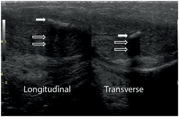
Fig. 2
Ultrasound of Peyronie’s plaque. An echodense plaque (solid arrows) is imaged both in the longitudinal and transverse projection. Note the posterior shadowing suggestive of a calcified plaque (open arrows)
Dierks and Hawkins used penile ultrasound in 1983 to assist with their management of penile fracture. In this case report, a 26-year-old male experienced a traumatic bending of his erect penis, resulting in a “pop” sound followed by rapid detumescence and swelling. After an initial course of failed observation, ultrasound was performed and a large hematoma was seen as an echolucent mass with internal echoes and average through transmission. No definite tears were seen, but the patient was taken to the operating room (OR) for exploration, where the hematoma was evacuated and a small tunica alubinea tear was found. The authors made four recommendations based on this case to be applied to any case of penile trauma : (1) The entire tunica albuginea should be imaged to evaluate for tears, including proximally, (2) The entire corpora should be imaged for breaks or cystic collections, (3) The urethra and spongiosum should be imaged, and (4) Dimensions of the hematoma should be described [30] .
History of Scrotal Ultrasound
One of the earliest uses of ultrasound for the scrotum was for the diagnosis of testicular torsion . In 1976, Perri et al. attempted to improve upon the contemporary overall testicular salvage rate of 25–37 % in cases of torsion with the use of their “Doppler stethoscope.” In this study, an ultrasound device that emitted Doppler signal was scanned over the scrotum, while the operator listened to the received signal with the attached stethoscope. This was done on 30 patients who presented with scrotal pain: 23 were diagnosed with epididymo-orchitis, as determined clinically and by increased blood flow on Doppler; the remaining 7 were explored, of which 3 had torsion and 4 had torsed appendix testes. The authors concluded that Doppler has an important role in the differentiation between infectious and vascular etiologies, and that in the future it will have a role in differentiating torsed spermatic cord from appendix testis. Moreover, in the three cases of true testicular torsion, two were salvaged and this was confirmed intraoperatively by Doppler. The single case where orchiectomy was performed was presumed to be an in utero torsion [31] .
Scrotal trauma was evaluated for the first time with ultrasound in the early 1980s. Albert reported three cases of direct scrotal trauma where ultrasound demonstrated increased echoes within the tunica albuginea, suggestive of hematoma and tunica disruption. All three cases were confirmed on exploration, with hematoma evacuation and primary repair of the tunica performed in two cases, and orchiectomy in one case where the testis was no longer viable presumably due to delay in presentation. The author reported that ultrasound was fast and easy to interpret in the diagnosis of scrotal trauma, given that normal testicles are homogeneous in echotexture while hematomas appear as areas of dense, heterogeneous echoes [32] .
The first case series reporting the use of ultrasound in the diagnosis of varicoceles in men presenting with subfertility came in 1977 out of the University of Pennsylvania. In this study, Greenburg et al. examined 46 men, divided into a group of patients with varicoceles found on physical examination, oligospermic men with no palpable varicocele, and patients who were status post-varicocele ligation. All men had “Doppler stethoscope” examinations performed. All patients with clinically obvious varicoceles had Doppler exams that confirmed these findings; large varicoceles were found to demonstrate continuous non-pulsatile “hums” as the patient breathed quietly, and regurgitant flow as the patient performed a Valsalva maneuver. Of the group with oligospermia but no clinically palpable varicocele, Doppler found patterns similar to those of the first group in 5 out of 13 patients. All five patients had sperm densities similar to the first group, whereas all of the remaining eight patients had sperm densities within normal limits. Finally, in the group of five patients who were status post varicocelectomy, three demonstrated normal semen parameters, and concordantly, had normal sonographic findings. The remaining two patients had persistent abnormalities in their semen analysis, along with flow patterns reflective of persistent varicoceles [33] .
The use of ultrasound for the diagnosis of scrotal masses was also explored in 1977, when three institutions reported their case series along with detailed sonographic findings. Kohiri et al. reported findings of 18 patients with scrotal masses, including 7 hydroceles, 3 tumors, 3 cases of epididymo-orchitis, 2 torsed testicles, and 1 traumatic injury [34]. Ultrasound correctly diagnosed 16 out of 18, failing to correctly diagnose both cases of torsion (Fig. 3).
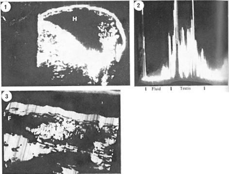
Fig. 3
Three ultrasound scans of testis and hydrocele [34]. Case 1: Longitudinal B-scan shows the clear echo-free large hydrocele (H) lying anterior to a testicle (T). Case 2: A-scan demonstrated that there are no internal echoes. On B-scan, the pattern was almost similar to case 1. Case 3: The testis (T) is surrounded by fluid (F). At surgery the cryptorchidism with hydrocele and varicocele was found
The same year, Shawker reported his series of 14 patients with scrotal masses palpated on exam. Ultrasound correctly diagnosed 11 hydroceles, one case of epididymitis with reactive hydrocele, one indirect inguinal hernia, and one hematocele [35]. Finally, Gottesman et al. described the combined series from University of California, Los Angeles (UCLA) and Rush, which included 27 patients, of which 20 were referred for palpable scrotal mass, 4 for pain, and 3 for a finding of enlarged retroperitoneal lymph nodes. Of the 54 testicles assessed in the study, 25 were normal, and Ultrasound was able to correctly diagnose 27 of the remaining 29 abnormalities. In all, 21 sonograms demonstrated extratesticular lesions, while the remaining 6 were testicular tumors. The two indeterminate cases were a hemorrhagic hydrocele and a large sarcoma [36] . Each series, along with a review article by Miskin et al. described similar sonographic findings in each type of scrotal pathology: (1) Hydroceles appear as a normal testis surrounded by sonolucent fluid, (2) Masses demonstrate normal echogenicity in a portion of the testicle, while the space occupied by the mass demonstrates a cluster of internal echoes, (3) Epididymitis appears as clustered echoes separate from the testicle itself, (4) Hernias demonstrate a lack of posterior wall echoes, indicating the presence of gas, (5) Abscesses show areas of lucency intermixed with areas of increased echoes within the testicle, (6) Varicoceles demonstrate clustered echoes with non-pulsatile waveform on Doppler, and (7) Spermatoceles appear as sonolucent cystic masses at the upper pole of the testicle [34–37] (Fig. 4).
Related posts:


Stay updated, free articles. Join our Telegram channel

Full access? Get Clinical Tree


