Since the 1960s, interventional radiology has played a role in the management of gastrointestinal bleeding. What began primarily as a diagnostic modality has evolved into much more of a therapeutic tool. And although the frequency of gastrointestinal bleeding has diminished thanks to management by pharmacologic and endoscopic methods, the need for additional invasive interventions still exists. Transcatheter angiography and intervention is a fundamental step in the algorithm for the treatment of gastrointestinal bleeding.
Key points
- •
Medical and endoscopic therapy has been very successful at diminishing rates of upper gastrointestinal bleeding as well as in its treatment.
- •
There will be a small but challenging subset of patients for whom endoscopy cannot achieve hemostasis or will not be able to identify the source of bleeding.
- •
For persistent slow or intermittent bleeding that cannot be localized by esophagogastroduodenoscopy, further diagnostic evaluation with computed tomography angiography or scintigraphy should be considered.
- •
Individuals with active bleeding that is refractory to endoscopic treatment should undergo transcatheter angiography and intervention (TAI).
- •
TAI’s technical and clinical success rates in most series are more than 90% and 70% for the treatment of upper gastrointestinal bleeding.
Introduction
Since the 1960s, interventional radiology (IR) has played a role in the management of gastrointestinal (GI) bleeding. What began primarily as a diagnostic modality has evolved into much more of a therapeutic tool. And although the frequency of GI bleeding has diminished thanks to management by pharmacologic and endoscopic methods, the need for additional invasive interventions still exists. Transcatheter angiography and intervention (TAI) is now a fundamental step in the algorithm for the treatment of GI bleeding.
Evolution
- •
1963: Selective arteriography for localizing GI bleeding
- •
1968: Selective arterial infusion of vasopressin to reduce blood flow in superior mesenteric artery (SMA) and to reduce portal venous pressure
- •
1970: Selective embolization of gastroepiploic artery using autologous clot (in conjunction with epinephrine) to stop active bleeding from a gastric ulcer
- •
1970s to 1990s: Arterial infusion of vasopressin used to treat GI bleeding
- •
1990s to the present: Superselective arterial embolization becomes primary endovascular method for treatment of GI bleeding
Materials and methods
In 1963, an angiogram was used as a means to help diagnose the presence of, as well as to help localize, a site of GI bleeding. The catheters available at that time allowed access to first-order and some second-order branches of the aorta but often not more distal. Embolic agents existed, but because they could not be deployed superselectively, the affected downstream territory was larger, with greater potential for ischemia. Sometimes, too proximal of an embolization did not treat direct collateral pathways to the bleeding site, resulting in ongoing or promptly recurring hemorrhage. Therefore, safe and effective treatment was somewhat limited.
Given the inability to reach sites of focal bleeding, more regional treatments were applied. Vasoconstrictive medications were administered intra-arterially to limit pressure and blood flow to sites of hemorrhage, facilitating hemostasis by patients’ autologous clotting mechanisms. These arterial procedures typically lasted from 12 to 48 hours ( Table 1 ).
| Tool | Use |
|---|---|
| Access sheaths | Continuous arterial access; conduit through which exchange of catheters can take place without compromising access site; can provide continuous arterial pressure monitoring |
| Catheters and wires, including microcatheters and microwires | Numerous shapes and sizes allow selective and superselective catheterization to maximize diagnostic evaluation and targeted delivery of therapeutic materials |
| Embolic agents | Temporary (Gelfoam and autologous clot) and permanent (particulate, coils, liquids ); to occlude or promote autologous occlusion of target blood vessel |
| Vasopressin | Vasoconstrictor diminishes pressure and flow to promote autologous hemostasis |
Although the use of vasoconstrictive medications can be effective, especially for the treatment of diffuse bleeding, it has some clear limitations. Because the medication’s effects cannot be confined to its site of administration, it poses significant systemic risks to anyone who has ischemic heart disease or prior stroke by further restricting blood flow to these areas. Additionally, because its effect ends on cessation of infusion, bleeding will resume if a stable clot has not formed. As a result, the rates of recurrence following therapeutic vasoconstriction were reported as high as 71%.
With continued evolution of endovascular tools, however, the treatment options and their safety and efficacy increased significantly.
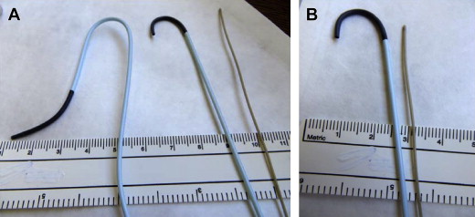
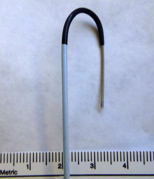
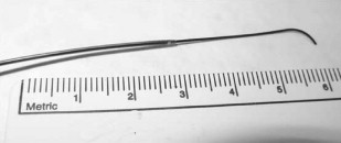
Probably the most important development that redefined the endovascular world was the development of the microcatheter. Microcatheters are small-diameter catheters (1.7–3.0 F catheters compared with 5–6 F diagnostic catheters) that can be navigated selectively into higher-order branch vessels, thereby allowing the targeting of very focal sites of vascular abnormality ( Figs. 1–3 ). The microcatheters serve as a conduit for the instillation of contrast material as well as a variety of embolic agents.
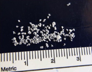
Embolic agents have also evolved significantly over the last few decades. Designed to fill spaces, embolic materials are used to help occlude blood vessels that are or may be supplying the sites of bleeding ( Figs. 4–8 ). Preventing blood from reaching the site of vascular defect eliminates the loss of blood through the site.



At the same time, however, embolic agents restrict blood from reaching downstream, which can have ischemic effects on those tissues as well as any nontarget sites of embolization, especially if the collateral vascular supply is limited. This effect is the main risk in performing embolization. Targeted, superselective embolization, however, has helped mitigate the risk.
With improved safety and control, the use of embolization as a therapeutic tool has expanded significantly. This use includes its application in the treatment of upper GI bleeding (UGIB).
Upper Gastrointestinal Bleeding
- •
Bleeding into the GI tract proximal to the Ligament of Treitz
Cause
- •
Peptic ulcer disease, duodenal and gastric
- •
Gastritis and esophagitis
- •
Mallory-Weiss tear
- •
Iatrogenic, biopsy, sphincterotomy
- •
Marginal (postoperative anastomotic) ulcer
- •
Trauma, including hepatic and pancreatic
- •
Postpancreatitis pseudoaneurysm
- •
Dieulafoy lesion
In the realm of GI bleeding, management algorithms are simplified by separating UGIB (sites proximal to the Ligament of Treitz) from lower GI bleeding (sites distal to the Ligament of Treitz). The distinction is important because of both anatomic blood supply considerations as well as accessibility to the various therapeutic modalities.
Typical vascular supply
- •
Celiac artery : distal esophagus, stomach, duodenum, ampulla (pancreaticobiliary) ( Fig. 9 )

Fig. 9
Celiac arteriogram.
- •
SMA : duodenum, ampulla (pancreatic and variant hepatic) ( Fig. 10 )

Fig. 10
Superior mesenteric arteriogram.
Upper GI bleeding arises in locations that are normally quite accessible to endoscopy. Additionally, these territories normally have richly overlapping blood supplies that can make hemorrhage difficult to control and recurrence rates higher when treated from intravascular locations, especially when the treatment locations are somewhat removed from the exact site of the vessel defect. For these reasons, endoscopy usually affords the best treatment option for most patients compared with TAI. Surgery is also a key component in the treatment algorithm, though it carries increased morbidity, especially in patients already in a tenuous condition.
For patients with UGIB in whom endoscopic treatment has not been successful, however, or for those who have had prior surgery preventing endoscopic access, TAI should be the next option to consider.
General upper gastrointestinal bleeding treatment considerations
- •
There is a rich overlap in blood supplies, so endoscopy is most effective in both treating and localizing the end sites of bleeding.
- •
For TAI, the collateral as well as primary blood supply must be evaluated and addressed, often necessitating treatment proximal and distal to the site of extravasation.
- •
Be aware of the patient’s surgical history and its potential impact on vascular supply.
- •
Surgery provides effective treatment ; but carries greater morbidity, especially in already acutely compromised individuals.
Upper gastrointestinal bleeding treatment options
- •
Watch and wait (most episodes are self-limited )
- •
Endoscopy will be (diagnostic, therapeutic, prognostic)
- •
Then TAI
- •
Surgery (endoscopy and TAI will be useful to temporize/optimize before surgery)
Because most episodes of GI bleeding are self-limited, it can sometimes be difficult to decide when to implement invasive treatment. To warrant the potential risk (and maximize the potential therapeutic benefit) of the invasive procedure, the patient’s hemodynamic stability can be a primary determinant. Multiple studies have suggested that hemodynamic instability correlates with positive findings during catheter angiography, both localization and successful embolization. The following parameters can serve as thresholds for the indication for TAI in GI bleeding:
- •
Systolic blood pressure less than 100 mm Hg
- •
Heart rate greater than 100 beats per minute
- •
≥4 units packed red blood cells (RBCs) transfused in less than 24 hours
It is extremely important to use these parameters as a guide rather than absolute trigger points. They need to be considered in conjunction with each individual’s overall clinical condition and comorbidities.
Diagnostic assessment
Endoscopy is usually successful in determining both whether there is active bleeding occurring as well as localizing the site of the bleeding. It should and will almost invariably be the primary diagnostic tool for UGIB. When additional diagnostic assessment is needed (often for difficult to localize or suspected slow or intermittent bleeding), multiple imaging modalities are available. Each one has different sensitivities for minimal bleeding rates and other advantages and disadvantages as well ( Tables 2 and 3 ).


