Vascular Disorders and Related Diseases


Vascular Disorders and Related Diseases


VASCULARIZATION OF THE DIGESTIVE TRACT—OVERVIEW

The splanchnic circulation is well developed and composed of major intramural and extramural pathways. Under resting conditions, the splanchnic circulation receives 30% of the total blood flow. During the process of digestion, the superior mesenteric artery blood flow increases by more than 100%.1 Conversely, exercise reduces both resting and postprandial blood flow. The blood flow through these pathways is subject to a complex interplay of extrinsic and intrinsic controlling mechanisms including hemodynamic factors (cardiac output, systemic arterial pressure), the autonomic nervous system, hormones (vasopressin, vasoactive intestinal polypeptide, somatostatin), and locally produced metabolites. Alterations of the various control mechanisms can lead to occlusive or non-occlusive disease and limited or extensive ischemic damage.


VASCULARIZATION OF THE SPECIFIC SEGMENT OF THE DIGESTIVE TRACT


Esophagus

The extramural arterial supply of the human esophagus is divided into three major segments, the cervical, thoracic, and abdominal esophagus, with numerous anastomoses. The pharyngoesophageal transition and cervical esophagus are supplied by the lower thyroid artery, a branch of the thyrocervical trunk. An additional, individually variable supply is provided by small branches of the subclavian, common carotid, vertebral, and superior thyroid arteries and the costocervical trunk. The thoracic esophagus receives blood from branches of the aorta, the bronchial arteries, and the right intercostal arteries. At the level of the bifurcation of the trachea, the main supply comes from branches of the bronchial arteries, which descend on the ventral side of the esophagus. Below the bifurcation of the trachea, the blood supply originates from two esophageal branches that arise directly from the ventral side of the aorta. Both branches run to the dorsal side of the esophagus where they anastomose with the descending branches of the lower thyroid and ascending branches of the left gastric and left lower phrenic arteries. The abdominal esophagus is supplied by esophageal branches of the left gastric and left lower phrenic arteries. An additional blood supply may be provided by branches of the aorta, the splenic artery, the celiac trunk, and an aberrant left hepatic artery.2, 3

The intramural arterial pattern is characterized by a well-developed subepithelial capillary network in the stromal papillae of the mucosa (intraepithelial channels), supplied by a prominent submucosal arterial plexus, composed of longitudinally oriented arteries with lateral anastomoses. Submucosal arteries are formed by penetrating branches arising from a minor extrinsic plexus in the adventitia. These branches pass through the muscle layer and give off branches to the muscle tissue and the myenteric plexus.4, 5

Esophageal veins are classified into three groups: (a) intrinsic veins including subepithelial and submucosal veins, which join the gastric veins below, and perforating veins, which pierce the muscular wall to join the extrinsic veins; (b) extrinsic veins formed by the union of groups of perforating veins, which join the left gastric vein below and the systemic veins above; and (c) venae concomitantes, which run longitudinally in the adventitia.6, 7

The subepithelial or superficial venous plexus drains the stromal papillae; lies in the lamina propria, close to the epithelium; and extends over the whole length of the esophagus.6 At the esophagogastric junction, the veins lie predominantly superficially in the lamina propria.7, 8, 9 Numerous small veins perforate the muscularis mucosae to join larger veins of the submucosal plexus (deep intrinsic veins) and constitute the palisades vessels seen endoscopically. The submucosal plexus consists of 10 to 15 longitudinal veins, evenly distributed around the circumference of the esophagus and connected by numerous anastomoses. In its proximal part, this plexus drains the longitudinally oriented veins from the ventral and dorsal pharyngoesophageal subepithelial plexus. At the distal end of the esophagus, the longitudinal submucosal veins increase in number but decrease in diameter. At the cardia, they become tortuous and aggregate in the longitudinal folds of the mucosa before joining the submucosal veins of the stomach. Perforating veins arise from the longitudinal submucosal plexus and pass through the muscle layer, which they drain also, at regular intervals.

The greater extrinsic periesophageal veins include two larger and several smaller veins. The larger veins run longitudinally on the outer surface in close proximity to the vagus nerves and connect the left gastric vein to the azygos or hemiazygos veins. Other veins are the cervical periesophageal veins draining into the inferior thyroid, vertebral, and deep cervical veins; small esophageal veins at the cardia joining the superior and inferior phrenic veins; and small abdominal esophageal veins draining into the left gastric vein as well as the vena phrenica inferior, gastroepiploica, and splenica.6


Stomach, Small Intestine, and Large Intestine

Intramural circulation. The gastric blood supply is derived from the common hepatic, left gastric, and splenic arteries arising from the celiac trunk (Fig. 2-1). The fundus and left margin of the greater curve are supplied by short gastric arteries derived from the splenic artery. The right gastric artery and the right gastroepiploic artery supply the lesser and distal greater curve, respectively. The proximal greater curve is supplied by the left gastroepiploic artery and arteries from the splenic artery. These vessels form two extrinsic arcading anastomotic loops. The loops give off a series of short branches to the anterior and posterior walls. They form a subserosal plexus.10, 11, 12 Perforating branches originating from this plexus pass through the external muscle layers to reach a richly anastomotic submucosal arterial plexus. Small side branches are given off to the external muscle layers and to Auerbach’s plexus en route, but the majority of arterioles to the external muscle come from the submucosal plexus. The mucosa is supplied by small branches from the submucosal plexus, which pass perpendicularly through the muscularis mucosae. In the lamina propria, the arterioles branch into capillaries, which run toward the lumenal surface between the gastric glands. There are frequent cross anastomoses between adjacent capillaries. Just underneath the surface epithelium, the capillaries form a polygonal network around the necks of the gastric pits.13, 14, 15, 16, 17 Mucosal venules drain into the submucosal venous plexus, which is continuous with a similar plexus in the esophagus and duodenum. In the gastric cardia the submucosal venous plexus is composed of a series of parallel veins oriented toward the esophagogastric junction. Drainage of the muscle layers occurs to the submucosal venous plexus and partly to perforating veins, which pass to subserosal veins. The latter drain toward the portal system.


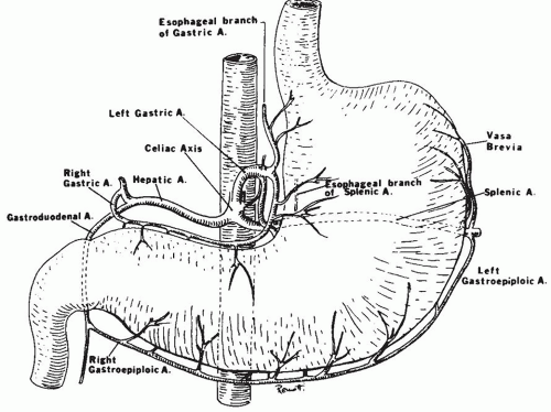
Figure 2-1. Arterial blood supply of the stomach.

The duodenum is supplied chiefly by the pancreaticoduodenal artery. The jejunum and the ileum are supplied by a dozen branches of the superior mesenteric artery. These branches divide and anastomose several times in the mesentery forming arcades. The last of these forms a marginal artery along the small intestine. The marginal artery is defined as the artery closest to and parallel with the wall of the intestine. From the marginal artery, blood reaches the intestine by way of short, straight branches or “vasa recta.” They penetrate the external muscle layers to reach a profusely anastomotic submucosal arterial plexus from which arterioles originate for the mucosa, sub-mucosa, and muscular layers. The submucosal plexus gives off two types of mucosal branches: long or villous arterioles and short or cryptal ones. Arterioles to the villi pass without branching in the lamina propria. At the tip of the villus, the arteriole splays into a network of capillaries that subsequently course down along the sides of the villus in a fountain-like pattern. The crypts receive their arterial supply adjacent to the muscularis mucosae. The lymphoid tissue is supplied by the submucosal plexus through interfollicular arteries between the lymphoid follicles and through follicular arterioles originating from the interfollicular arteries. Venous drainage of each of the small intestinal capillary beds passes to the submucosal venous plexus, which anastomoses both longitudinally and
circumferentially in the bowel wall. This plexus is drained by short veins, which penetrate the external muscle layers, chiefly along the mesenteric margin and then pass to branches of the superior mesenteric vein in the mesentery. The superior mesenteric vein receives venous drainage of the distal duodenum, the jejunum, the ileum, the appendix and cecum, the ascending and transverse colons and a right gastroepiploic vein draining the stomach, before joining the splenic vein to form the hepatic portal vein.13, 15, 18

The ascending and transverse colons are supplied by three branches of the superior mesenteric artery (ileocolic and right and middle colic arteries), whereas the splenic flexure, descending colon, and sigmoid are nourished by branches of the inferior mesenteric artery (left colic and sigmoid arteries). These vessels form arcades that are less numerous and complex than those in the small intestine. Within 2 cm of the colon wall, a single large anastomotic marginal artery is formed. This marginal artery runs retroperitoneally, extending from the ileocecal junction down to and into the sigmoid mesocolon close to its attachment to the colon wall, thus forming an anastomotic channel between ileocolic; right, middle, and left colic; and sigmoid arteries. The rectum has a richly anastomosing arterial system derived from the inferior mesenteric and internal iliac arteries. Through the superior rectal artery, it forms an anastomosis with the marginal artery of the colon. Short vasa recta pass from the marginal artery to the colon wall, with few or no anastomoses en route. Upon reaching the wall, they divide to form subserosal branches, which pass circumferentially around the bowel wall, and other branches, which form a subserosal anastomosing plexus. The subserosal plexus gives off branches that traverse the external muscle coat to reach the submucosal arterial plexus. There is extensive anastomosis in the submucosa both longitudinally and circumferentially. Arteriolar branches from the submucosal plexus penetrate the muscularis mucosae and then break up in a leash of capillaries. These capillaries ascend along the glands and reach the surface of the mucosa where they form a honeycomb pattern around the openings of the glands, just beneath the surface epithelium. The muscularis contains capillaries derived from both the subserosal and submucosal plexuses and is perforated by larger arteries coming from the serosa and subserosa. The venous drainage largely parallels the arterial supply.13, 19

Extramural (Splanchnic) circulation. The celiac trunk is a short (2 cm) but larger caliber (5-8 mm) artery, which arises from the front of the aorta. It divides almost immediately into three branches: the common hepatic, splenic, and left gastric arteries. There are however many variations of the typical origin. The most striking of these is a common origin of the celiac trunk and the superior mesenteric artery in a celiacomesenteric trunk (in ˜2% of cases). In this situation, a single artery is the sole source of vascularization of the supramesocolic organs. Collateral flow is possible only from the inferior mesenteric, phrenic, esophageal, and retroperitoneal arteries.10

The common hepatic artery arises on the right side of the celiac trunk, giving off branches to the stomach, duodenum, and pancreas. The right gastric artery arises from hepatic artery and less frequently from the gastroduodenal artery (8%). It descends to the pylorus along the lesser curvature of the stomach where it usually anastomoses with the left gastric artery. The right gastric artery frequently gives rise to the supraduodenal artery (of Wilkie).

The gastroduodenal artery usually arises from the common hepatic artery (75%) but may arise from the left or right hepatic artery or superior mesenteric artery. It divides into the right gastroepiploic and the anterior superior pancreaticoduodenal arteries. This artery anastomoses with the posterior inferior pancreaticoduodenal artery to form the pancreaticoduodenal arcade supplying the posterior surface of the entire duodenum and the head of the pancreas. The anterior pancreaticoduodenal arcade is formed by the anterior superior pancreaticoduodenal artery and the anterior inferior pancreaticoduodenal artery, which arises from the superior mesenteric artery. The right gastroepiploic artery is the final continuation of the gastroduodenal artery. After supplying one or more branches to the pylorus, it passes to the left along the greater curvature of the stomach. Ascending branches supply the greater curvature of the stomach and anastomose with descending branches of the right and left gastric arteries.

The left gastric artery courses toward the gastric cardia. It supplies part of the stomach and the inferior esophagus. The anastomosis with the right gastric artery may be absent.

The splenic artery from the celiac artery gives off branches to the pancreas and stomach. Often, a branch of the dorsal pancreatic artery descends below the inferior border of the pancreas to communicate with the superior mesenteric artery. Occasionally, this branch gives rise to the middle colic artery (artery of Riolan). The left gastroepiploic artery arises from the splenic artery prior to its terminal divisions or from a terminal division itself. It gives off the left epiploic artery, which anastomoses with the right epiploic artery, a branch of the right gastroepiploic artery, to form the arcus epiploicus magnus of Barkow, in the great omentum below the colon. Short gastric arteries originate from the distal splenic artery and supply the fundus and cardia of the stomach.

The superior mesenteric artery originates from the aorta at the level of the first lumbar vertebra, behind
the body of the pancreas (Fig. 2-2). It emerges from under the lower border of the pancreas, passes forward anteriorly over the upper border of the third portion of the duodenum, and descends anteriorly into the mesentery. Usually, the middle colic artery arises from the superior mesenteric artery just before it enters the mesentery. The middle colic artery can arise as a separate branch or be derived from a common right colic-middle colic trunk (53% of the cases). Occasionally, it arises directly from the celiac artery. When the middle colic artery has a large branch running parallel and posterior to it in the transverse mesocolon, this branch is often described as the arc of Riolan. The right colic artery can arise directly (38%) from the superior mesenteric artery. The middle and right colic arteries supply the right half of the transverse colon and the ascending colon. Within the mesentery, the superior mesenteric artery courses to the right iliac fossa, curving to the left to end in the ileocolic artery by forming an anastomosis. Major side branches of the superior mesenteric artery originating usually on the right side are the inferior pancreaticoduodenal arteries supplying the lower part of the duodenum. These arteries connect with the superior pancreaticoduodenal arteries. The inferior pancreaticoduodenal artery can also arise from or can be in common with the first jejunal artery. To the left, four to six jejunal arteries and nine to thirteen intestinal branches that supply the ileum can be identified. These are often called “intestinal arteries.”

They divide into two branches, forming a first-order arcading anastomosis with the neighboring branches. Subsequent anastomes form second- to fourth-order arcades. Branches of these arcades finally form the marginal artery. The marginal artery may thus be composed of arteries that range from third- or fourth-order arcades to the parent colic artery itself. The middle colic artery is often the marginal artery for the major portion of its distribution. Fine branches originating from the marginal artery reach the bowel wall as “vasa recta.” The vasa recta of the small intestine are shorter, closer together, and less straight in appearance than the large bowel vasa recta. The terminal branch of the superior mesenteric artery is the ileocolic artery. It distributes branches to the terminal ileum, the cecum, and the lower third or half of the ascending colon. In its distal distribution the ileal and colic branches of the ileocolic artery often form an “ileocolic loop.” The anterior and posterior cecal arteries and the appendicular artery arise separately from this loop.


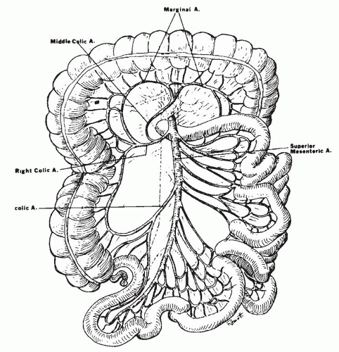
Figure 2-2. Distribution of the superior mesenteric artery.

The inferior mesenteric artery arises from the aorta anteriorly at the level of the third lumbar vertebra (Fig. 2-3). Major side branches are the left colic artery and the sigmoid arteries. The descending branch of the inferior mesenteric artery becomes the superior rectal artery. The left colic artery is usually an ascending branch from the inferior mesenteric artery. This branch bifurcates at the splenic flexure, its right branch joining the middle colic artery from the superior mesenteric artery and its left branch joining the marginal artery. Sigmoid arteries can originate from the ascending branch in common with the left colic or arise from a descending branch of the inferior mesenteric artery. A few sigmoid arteries may arise from a middle branch. The number of sigmoid arteries varies from one to five. The superior rectal artery divides
into two branches of unequal size at the level of the second or third sacral vertebra, commonly at the bottom of the pouch of Douglas. The larger right branch divides into several branches, which descend on the posterior and lateral surfaces of the rectal ampulla. The smaller left branch deviates to the left and supplies the lateral and anterior surfaces. In contrast to the small and large intestinal arteries, the branches of the rectal arteries do not form arcades but enter the gut wall directly and independently.


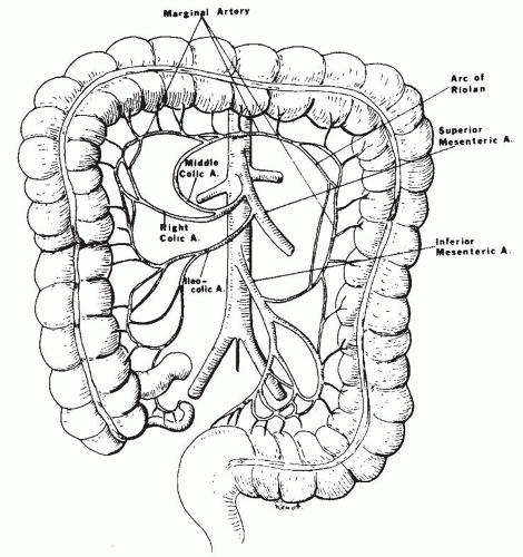
Figure 2-3. Arterial blood supply of the colon.

The branches of the superior rectal artery further anastomose with the middle and inferior rectal arteries originating respectively from the internal iliac and pudendal arteries.10, 11, 12

Venous drainage. The venous blood from the gastrointestinal (GI) organs and spleen is drained by the hepatic portal circulation through the liver before it returns to the heart. The hepatic portal vein is formed by the union of the superior mesenteric and splenic veins. The superior mesenteric vein drains blood from the pancreas, the stomach, the small intestine, and portions of the large intestine through the pancreaticoduodenal, right gastroepiploic, jejunal, ileal, ileocolic, right colic, and middle colic veins. The splenic vein drains blood from the stomach, the pancreas, and portions of the large intestine through the short gastric, left gastroepiploic, pancreatic, and inferior mesenteric veins. The inferior mesenteric vein, which passes into the splenic vein, drains portions of the large intestine through the superior rectal, sigmoid, and left colic veins. The right and left gastric veins, which open directly into the hepatic portal vein, drain large portions of the stomach. The cystic vein, which drains the gallbladder, also opens into the hepatic portal vein.

Collateral blood supply. The various pathways can be subdivided into six categories: channels between the celiac trunk and the superior mesenteric artery; channels between the celiac trunk and the aorta; channels between the different celiac branches; channels between superior mesenteric branches; connections between the superior and inferior mesenteric artery; and routes between the inferior mesenteric artery and parietal branches of the aorta. There are thus many anastomoses between the different arterial networks, but there are several weak points or “watershed” zones prone to vascular compromise in the setting of hypoperfusion. The collateral circulation between the hypogastric arteries and the inferior and superior mesenteric arteries is for instance most tenuous at the splenic colonic flexure (Griffith’s point) and in the ileocecal region (Sudeck’s point). These relatively underperfused sites are therefore preferentially involved in ischemic insults. Any vascular compromise from hypovolemia or splanchnic shunting during exercise can affect these zones.


ISCHEMIA OF THE GASTROINTESTINAL TRACT


Etiology and Clinical Manifestations

Esophageal ischemia. Esophageal infarction with or without perforation is extremely rare. It has been reported to occur secondary to traumatic aortic transection, as a complication of spontaneous rupture of the thoracic aorta and in the anticardiolipin antibody syndrome (Fig. 2-4).22, 23, 24

Gastric ischemia. This is also very rare, largely because of the protective collateral circulation. The rare cases of gastric infarction reported in the literature have usually followed severe occlusive three-vessel disease (celiac artery and superior and inferior mesenteric arteries), volvulus, and therapeutic selective transcatheter embolization of the right gastric artery for gastric bleeding (Fig. 2-5A-E). Patients usually present with severe gastric bleeding.25, 26, 27 Rarely chemoembolization beads that are injected into the hepatic artery for the treatment of hepatocellular carcinoma may accidentally enter gastric artery leading to gastric ischemia and ulceration.


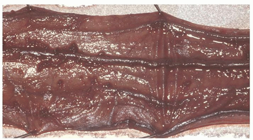
Figure 2-4. Acute esophageal infarction. Gross appearance of the opened esophagus showing a diffusely dark and necrotic mucosa.

Erosive gastritis due to chronic mesenteric ischemia has been documented in a number of case reports. The lesions occur primarily in the antrum. Biopsies will show epithelial desquamation, but otherwise the lesions are not very characteristic (see Fig. 2-5C,D).28, 29, 30, 31, 32

Ischemia can also occur in a less severe way resulting in lamina propria fibrosis, while the crypt epithelium is shed into the lumen of the pits. If the cells produce mucus, then this can resemble signet ring carcinoma, especially in antral mucosa, akin to the changes seen in pseudomembranous colitis. In the oxyntic mucosa the parietal cells collapse into the lumen, and the resulting disorganization can be mistaken for a parietal cell carcinoma (see Fig. 2-5A-E). The gastric changes are further discussed in Chapter 14. Rarely the stomach can be involved in severe NOMI. When very severe, it is found to affect the entire GI tract from the gastroesophageal junction to the rectum.

Acute mesenteric ischemia. Acute mesenteric ischemia is an abrupt decrease of mesenteric blood flow that may result in intestinal infarction. It can be caused by arterial (acute mesenteric embolism and thrombosis) or venous occlusion (Table 2-3). Venous ischemia results in vascular engorgement and hemorrhagic infarction. NOMI can be observed with systemic hypoperfusion, hemoconcentration including severe sepsis, and low flow states or as a result of vasoactive drugs. More exceptionally mesenteric ischemia is caused by vasculitis affecting the main mesenteric arteries. Venous mesenteric thrombosis can occur in patients with thrombophilia due to genetic or acquired coagulation disorders.33 All types of genetic coagulation disorders, including factor V Leiden, prothrombin mutation, proteins C and S deficiencies, and antithrombin-III deficiency, carry the risk of venous occlusion. In the western populations, factor-V Leiden is most prevalent.33, 34 Acquired coagulation disorders associated with mesenteric venous thrombosis include autoimmune disease with lupus anticoagulants, heparin-induced thrombocytopenia, polycythemia vera, and hemoconcentration. In general, in 50% of patients with genetic coagulation disorders, the venous thrombosis is triggered by an external cause (intercurrent disease, estrogen intake, pregnancy). Abdominal trauma is an established but rare cause of intestinal ischemia. Postoperative mesenteric ischemia is most typically associated with open aortoiliac aneurysm surgery and cardiac surgery.35, 36

Some 10% to 50% of all small intestinal ischemias are thought to be “nonocclusive,” indicating that upon diagnostic workup no critical stenosis can be found. The damage to the ischemic intestinal segments evolves rapidly and becomes irreversible within a few hours, which necessitates immediate diagnostic evaluation

and prompt therapeutic decisions. Intestinal ischemia accounts for an estimated 0.2% of all hospital admissions and 1% of all laparotomies. The true incidence of mesenteric ischemia is probably higher.37 The recent increase in acute mesenteric ischemia is most likely explained by improved abdominal imaging and by the aging population. Ischemia due to venous occlusion occurs in lower age groups. The symptoms and signs of acute mesenteric ischemia vary depending on the multitude of underlying vascular events. Arterial thromboembolism results in the most dramatic presentation, while the symptoms of NOMI and venous thrombosis tend to develop gradually over hours to days.38 The clinical picture of typical acute mesenteric ischemia can be subdivided into four stages.39 The hyperreactive state is characterized by severe abdominal pain in the absence of major abdominal findings at physical examination. In this stage there is hyperperistalsis and sudden vomiting or passage of loose stools. In the paralytic stage, the pain spreads over the abdomen and becomes continuous. The abdomen distends and loses its bowel sounds. The last two stages are nonspecific and are related to generalized peritonitis. In the third stage massive fluid and electrolyte losses occur due to capillary leak and a disturbed intestinal barrier. In the final stage shock rapidly evolves and the patient’s general state deteriorates rapidly. NOMI and venous thrombosis present more nonspecifically, and of all the early clinical signs only abdominal pain is seen in the majority of the patients. Recognition of vascular abdominal rest pain, defined as severe and persisting for more than 2 hours without recent caloric intake in the absence of peritoneal signs, is the key to early diagnosis.


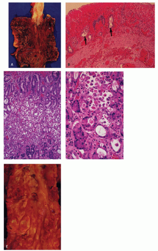
Figure 2-5. Acute gastric infarction. A: Gross appearance of the opened stomach and lower third of the esophagus. The mucosa is swollen, hemorrhagic, and partially covered by pseudomembranes. B: Section of mucosa and superficial submucosa showing intense congestion and hemorrhage. The outline of many atrophic gastric glands is still visible (arrows). C: Another area of gastric fundic mucosa showing desquamation of glandular epithelium. D: Higher-power magnification of part (C) showing desquamated parietal and chief cells lying within the space formerly occupied by the gland. Sometimes the desquamated epithelial cells may resemble malignant cells of early gastric cancers. E: Abdominal aorta from the same patient with gastric infarction showing virtual occlusion of the origin of the celiac axis and the superior mesenteric artery.


Table 2-3 Major Causes of Acute Mesenteric Ischemia


Nonocclusive mesenteric ischemia


50%


Superior mesenteric artery embolism


25%-30%


Superior mesenteric artery thrombosis


15%


Mesenteric vein thrombosis


5%-10%


Other


<5%


Laboratory analysis can be helpful in the diagnosis. Leukocytosis and metabolic acidosis with elevated lactate levels are common. Elevated serum enzymes include lactate dehydrogenase, creatine kinase, and amylase. However, no single enzyme is specific and they all carry low negative predictive value. Although abdominal imaging can help and is usually routine practice it should never delay medical and surgical treatment. Abdominal Doppler ultrasound is noninvasive and can be diagnostic for occlusive mesenteric ischemia. Emergency digital substraction angiography (DSA) is at present the gold standard for the detection of mesenteric occlusive disease. In selected cases, immediate endovascular revascularization can be performed. However, DSA cannot exclude transmural ischemia, and careful and repeated clinical examination of the abdomen is the key to performing a laparotomy or a laparoscopy. The other main value of DSA in assessing acute mesenteric ischemia is found in the handling of nonocclusive disease. In a patient with high suspicion of intestinal ischemia and with radiological signs of NOMI, the arterial catheter can be left in situ for vasodilator therapy. Computerized tomography (CT) scan carries a relatively short examination time and again in experienced hands has a sensitivity of 85% for proximal occlusion of the superior mesenteric artery and for mesenteric vein thrombosis. It is currently proposed as a standard test for the evaluation of acute abdominal pain. Magnetic resonance imaging (MRI) provides better anatomical resolution but is, due to longer examination times, in general not indicated in the acute management. Angiographic signs of nonocclusive disease include narrowing of the orifices of arterial branches, alternate narrowing and dilatation of intestinal branches (string of sausage), spasms of arteries in the arcade of Riolan, and impaired filling of intramural vessels.

When intestinal ischemia is being considered in an emergency setting, early fluid resuscitation is critical. When signs of generalized peritonitis are present, intravenous antibiotics should be started.40 Surgery with revascularization of the ischemic segment (embolectomy, thrombectomy, arterial reconstruction) should be performed as soon as possible and preferably within 12 hours. The surgeon will have to decide if the affected intestinal segment is viable or should be resected immediately.41

Intraoperative assessment of intestinal viability remains a problem. The usual criteria employed after vascular repair are the return of bowel coloration and the peristalsis and pulsation of the mesenteric vessels. In cases of questionable viability, second-look operations within 24 to 48 hours have been advocated in order to prevent complications.

When there is evidence of nonocclusive ischemia without signs of peritonitis, in a stabilized patient intra-arterial papaverine in the superior mesenteric artery should be administered during the angiography. The management of venous thrombosis is more controversial, although intravenous heparin has also been indicated. Surgery with venous thrombectomy has been attempted but experience is limited.

Acute mesenteric ischemia portends a poor prognosis. Although decrease in mortality from 80% to 50% has been reported with early diagnosis and emergency surgery, the mortality rates still remain very high. Mortality with venous thrombosis is estimated to vary between 20% and 40%. Part of the poor prognosis is associated with the advanced age of most patients and associated comorbidities. Interestingly,
most of this relates to superior mesenteric artery territory, and involvement of inferior mesenteric territory is less dramatic, possibly due to collateral circulation. For the clinician, a key to early diagnosis of mesenteric ischemia or mesenteric venous thrombosis is to keep a low threshold to consider it in the differential diagnosis since symptoms or signs may be nonspecific. If clinicians wait for dramatic signs of mesenteric ischemia like lactic acidosis, it may be too late.

Chronic mesenteric ischemia. Chronic mesenteric ischemia (also known as intestinal angina) is relatively uncommon. It is caused by atherosclerotic occlusion of the large mesenteric arteries. Due to the elaborate collateral circulation, usually two or three of the major mesenteric arteries have to be severely affected before patients become symptomatic. Even if abdominal imaging demonstrates severe stenoses in the large arteries, it can be hard to correlate these changes with symptoms. Patients with a suspected diagnosis of chronic mesenteric ischemia and generalized atherosclerosis are at increased risk for acute mesenteric ischemia with lethal prognosis. The true prevalence can probably be inferred only from angiographic studies of an atherosclerotic population.42, 43, 44 It is estimated that in such a population between 8% and 15% may meet the radiological criteria. Fibrodysplasia and large vessel vasculitis are rare causes. The existence of the celiac axis compression syndrome as a possible cause of chronic mesenteric ischemia remains controversial.

Clinically, it appears that 75% of the patients presenting with chronic mesenteric ischemia are women, generally heavy smokers and middle-aged. Postprandial abdominal pain and weight loss dominate the clinical presentation. The increased mucosal blood flow in the postprandial phase with a steal effect toward nonatherosclerotic vessels probably explains the postprandial ischemic pain. The pain can start as early as 15 minutes after the start of a meal and usually subsides within 1 to 2 hours. Weight loss may be a result of intestinal malabsorption due to villous atrophy, altered motility, or food avoidance due to pain triggered by meals. The classical triad, postprandial abdominal pain, epigastric bruit, and weight loss, is present in a minority of the patients. In more than 75% of patients, the celiac artery has an orificial stenosis, and ischemic gastritis with ulcerations can occur.

Although the final proof of chronic mesenteric ischemia lies in the response of the symptoms to successful surgical revascularization, abdominal imaging is helpful in evaluating patients with clinical suspicion of chronic splanchnic ischemia.45 Aortography and selective injection of all mesenteric arteries will allow to detect high-grade stenosis or occlusion.46

Superior Mesenteric Artery Syndrome This syndrome is thought to arise when rapid weight loss occurs. The fat surrounding the mesentery diminishes and the angle at which the superior mesenteric artery comes off the aorta becomes more acute. A loop of duodenum can become trapped in the angle and create an acute obstruction. Patients may report worsening of symptoms when lying supine and a relief when sitting upright and leaning forward (and hence bringing the superior mesenteric artery forward off the trapped bowel).


Pathology

Gross features. Ischemic lesions in the small intestine may be single, multifocal, or diffuse, depending on the cause of the vascular impairment and the state of the collateral circulation. Either occlusive or nonoc-clusive mesenteric ischemia can produce widespread infarction. In general, superior mesenteric vascular insufficiency involves the small and large bowels to the level of the transverse colon; inferior mesenteric insufficiency involves the splenic flexure and distal colon. Thrombi may extend into the mesenteric veins and extrude from the veins when transected. Ischemic lesions are, however, often patchy and irregular and do not necessarily conform to the area supplied by the major vessel. Extrinsic causes of intestinal ischemia may sometimes be apparent, for example, adhesion bands. In ischemia due to blunt trauma, there is often a hematoma in the mesentery and sometimes the bowel separates from the mesentery.

In early ischemia (including hemorrhagic enterocolitis), all that may be seen is purple discoloration of the bowel wall, the serosa retaining its normal glistening appearance. Later the viscus becomes dilated, thinned, and dark red or purple in color, and the serosa rapidly becomes dull and granular (Fig. 2-6). In more advanced ischemic injury, the bowel wall becomes thickened, rigid, and intensely hemorrhagic. Full-thickness infarction ensues leading to perforation and peritonitis. Most surgically resected specimens consist of bowel with only a thin rim of mesentery with vessels that are usually patent. Consequently, the patency of the larger, more proximal mesenteric vessels, which are more likely to be involved, cannot be examined, and the cause of the infarction (i.e., whether due to occlusive or nonocclusive vascular insufficiency) remains unclear on gross examination. However, sometimes atheromatous emboli can be identified in smaller arterioles, although by themselves they rarely give rise to anything other than minor transient lesions.

The appearances of the bowel following mesenteric vein thrombosis are similar to those of arterial insufficiency, except that there is more intense congestion and hemorrhage, and the thrombosed mesenteric veins are thickened and prominent. Lesions following mesenteric vein thrombosis tend to have indistinct borders compared to those due to arterial insufficiency.


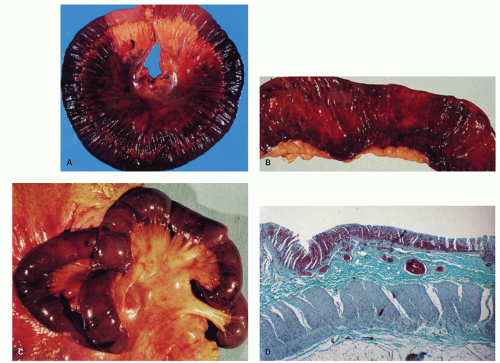
Figure 2-6. Focal segmental infarction of ileum. A: Intense congestion and hemorrhage of the loop of bowel and the attached mesentery. Note that the serosal surface has retained its shiny quality. B: Opened ileum showing the intense purple discoloration of the mucosa. Hemorrhagic enterocolitis. C: Autopsy specimen of small bowel showing diffuse purple discoloration. The serosa is uninvolved and maintains its sheen. D: Full-thickness section of intestine showing mucosal hemorrhage and submucosal congestion of vessels.

The appearance of the mucosa also depends on the severity and extent of the ischemic injury. At one end of the spectrum (acute hemorrhagic enterocolitis), the injury remains confined to the mucosa and submucosa, with intense congestion and hemorrhage and scattered superficial mucosal erosions. There is also extensive hemorrhage into the lumen.47 The more common pattern of ischemic injury consists of severe mucosal congestion, hemorrhage, and edema, often with deep linear ulcerations (Fig. 2-7). This results in boggy, coarse cobblestoning of the mucosa, which has a rather characteristic x-ray appearance, namely, blunt semiopaque projections into the intestinal lumen with a margin of half-shadowing, so-called thumbprinting (Fig. 2-8). The more severe forms of ischemia result in extensive mucosal necrosis, often with pseudomembrane formation. The necrotic mucosa is dark green or black in color due to bile staining or adherent altered blood. There is also thickening of the bowel wall due to marked interstitial hemorrhage. In full-thickness infarction, the bowel wall becomes gangrenous and friable and may perforate.

If the bowel is only partially infarcted, the ulcerated mucosa is replaced first by granulation tissue and later by an atrophic mucosa, which may contain superficial serpiginous ulcers. The submucosa and
muscle coats are replaced by fibrous tissue, resulting in strictures, which may be concentric or eccentric, depending on the extent of the ischemic necrosis.


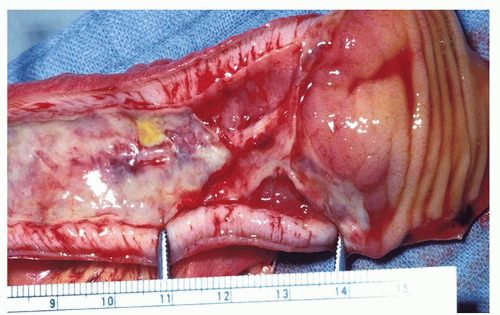
Figure 2-7. Segmental infarction of the terminal ileum showing a large mucosal ulceration. The demarcation between normal and involved segment is fairly sharp in arterial lesions as seen here.


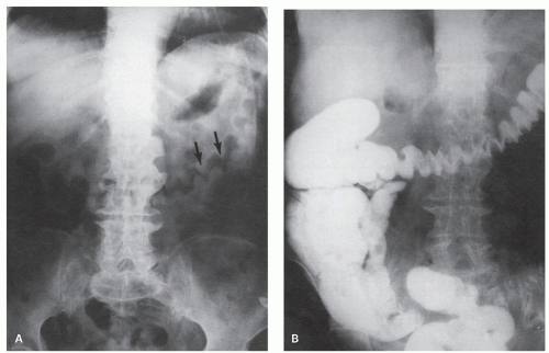
Figure 2-8. x-Ray appearances of ischemic colitis corresponding to the colon illustrated in Figure 2-9. A: Preliminary film showing the thickened mucosal folds, described as thumbprinting (arrows). B: Barium examination showing thickened mucosal folds. (Courtesy M. Weiner, M.D.)

Microscopy. There are two principal histologic patterns of intestinal ischemia: congestion, hemorrhage, and necrosis confined mainly to the (a) mucosa or (b) extending beyond mucosa and sometimes the full thickness of the bowel wall. Vascular injury affects the mucosa first. Marked mucosal and submucosal congestion, hemorrhage, and edema are the initial features. This may be all that is seen in some patients, and if the patient does not die from massive intestinal hemorrhage, the intestine is capable of complete resolution without scar formation when necrosis is limited to the mucosa.20

The microscopic mucosal lesions of acute ischemia have been studied in animal models mainly for the small intestine and were later confirmed in samples from patients.48 Fluid accumulation between the surface epithelial cells and the basement membrane may be found as early as 60 minutes after ischemic injury, but is best seen ultrastructurally.49 The earliest microscopic lesion is the formation of a subepithelial space underneath the epithelial cells lining the top of the villi, the Gruenhagen-Mingazzini space (Fig. 2-9). In clinical practice, this is difficult to differentiate from artifacts induced by handling of the specimen. The space progressively increases with lifting of the epithelial layer and separating it from the underlying lamina propria, followed by desquamation of the surface epithelium, with sparing of the deeper portion of the crypts. In the human small intestine, detachment and disruption of the lining epithelium at the villus tip are observed after 4 hours of absolute ischemia.50 Subsequently the basement membrane becomes damaged and ruptures with disintegration of the lamina propria, vascular congestion, and hemorrhage. The villi become shorter and progressively disappear (Fig. 2-10). When hypoxia/reperfusion is mild small intestinal villi may not disappear but just become shorter, in a diffuse way. The epithelial cells lining the
villi appear low cuboidal or flat with a basophilic cytoplasm. The crypts become mucin depleted, undergo coagulative necrosis, and eventually become detached from the basement membrane and extruded. Complete necrosis of the small intestinal mucosa in man is observed after about 44 hours of ischemia (Fig. 2-11).50


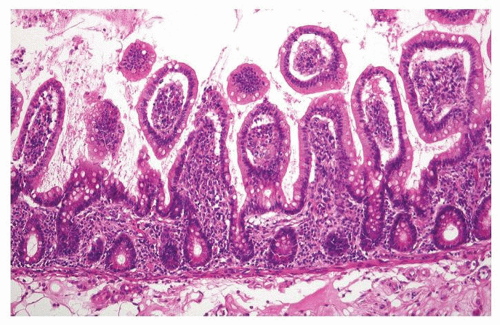
Figure 2-9. Early phase of small intestinal ischemia characterized by extensive lifting of the villous surface epithelial cells. This change cannot be reliably differentiated from artifacts that commonly occur in surgical or autopsy specimens.


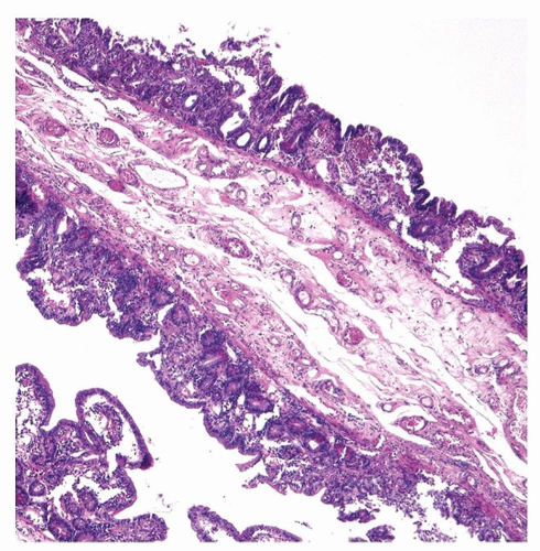
Figure 2-10. Diffuse flattening of the mucosa with loss of villi in the early recovery phase of small intestinal ischemic necrosis.

More extensive ischemic damage varies from superficial erosions to large ulcers to complete denudation of the mucosa. In severe cases, the sloughed mucosa is covered by a pseudomembrane composed of pus intermixed with mucus, blood, fibrin, and necrotic tissue, and frequently the devitalized tissue contains large numbers of mucosal bacterial colonies, both superficial and deep. Residual viable crypts often have extensive apoptosis, and epithelial cells slough into the lumen, sometimes producing a signet-ring appearance similar to that seen in the large bowel and stomach. Extensive hemorrhage is common, especially in the submucosa, and there may be platelet thrombi in capillaries. Occasionally, only vague outlines of the mucosa and the other layers of the bowel wall can be identified. In the early stages, there is only moderate leukocytic infiltration. Later, a secondary acute inflammatory infiltrate develops. The ischemic changes in the muscle coats are similar to those seen in myocardial infarcts. The earliest changes consist of poor staining of the muscle fibers, loss of nuclei, and thinning and separation of muscle fibers. The smooth muscle cells may show contraction bands, wavy fibers, thick waves, and coagulation necrosis.51, 52 “Contraction bands” are characterized by (hyper-) contraction of the smooth muscle cell with irregular eosinophilic bands and are a marker of cell death. With immunostaining, contraction bands are not reactive with antibodies against vimentin, desmin, actin, or myosin, indicating myofibrillar degeneration.53 Typical coagulative necrosis, often accompanied by a dense neutrophilic infiltration, is a late event.


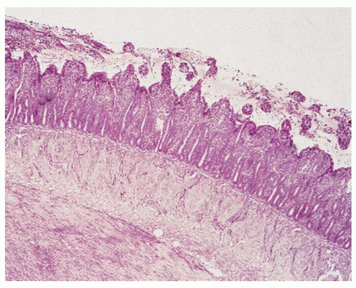
Figure 2-11. Extensive small intestinal ischemic necrosis in an experimental animal model.

The histologic changes in mesenteric venous thrombosis are similar, except that sometimes there is more intense transmural congestion and hemorrhage (Fig. 2-12A,B), and while the veins are thrombosed, the arteries are patent.54 However, even when thrombosed veins are found, it is not always certain that they represent the primary event. Presence of organization of the blood clot indicates a thrombus of some duration, which favors mesenteric venous thrombosis rather than an artifact or postmortem clot (Fig. 2-12C).

In the cases of resolution, fibrosis may result in stricture formation. Ischemic strictures are usually concentric. They can be short or long and single or multiple. They are relatively common in the left colon. They may be characterized by patchy mucosal ulceration. Histology shows irregularity of crypts without much inflammation. This may be confused with inactive or quiescent inflammatory bowel disease (IBDs). However, the degree of mucosal inflammation is still generally milder compared to IBD. Often one has to resort to clinicopathologic correlation with history suggesting predisposition to ischemia, endoscopic findings, presenting signs and symptoms, IBD serologies, and follow-up to rule out IBD. The submucosa is widened in the earlier phases but gradually becomes thinner and fibrotic. The fibrosis is hypocellular and extends downward between the smooth muscle cells of the circular muscle. Macrophages containing hemosiderin pigment are sometimes a prominent feature, but are not pathognomonic for ischemia and can also be seen in hemorrhagic IBD.55 Transmural lymphoid aggregated, fissuring, and fibromuscular obliteration of the submucosa are conspicuously absent, which help to differentiate it from Crohn’s disease.


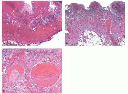
Figure 2-12. Low magnification in a case of mesenteric vein thrombosis (A) with marked vascular congestion and hemorrhage obscuring architectural details, which is more typical of venous obstruction compared with arterial obstruction. B: Another case of mesenteric venous thrombsis showing similar features. However, the hemorrhage is less intense and the thrombosis in veins is evident even at this magnification. C: Higher magnification showing varying degrees of organization of the thrombi in veins, differentiating this from a postmortem/postsurgical clot or an artifact.

Finally, it should be realized that sometimes transient ischemic episodes may occur in which no morphologic changes are found, although changes at the ultrastructural and biochemical levels could conceivably produce GI symptoms.


Ischemic colitis

Clinical Features The large bowel accounts for roughly half of all episodes of GI ischemia. Three major manifestations of colonic ischemia can be distinguished. Ischemic colitis (nongangrenous colitis) accounts for the large majority of the cases. Massive bowel infarction (gangrenous colitis) is noted in 15% to 20% of the cases and hemorrhagic enterocolitis is rare. Massive bowel infarction results from occlusive or nonocclusive vascular insufficiency, usually in association with small bowel infarction. Hemorrhagic enterocolitis is a variant of NOMI, with lesions confined to the mucosa and submucosa, occurring usually in the severely debilitated elderly individuals. Ischemic colitis is usually due to subacute colonic ischemia and can be reversible or irreversible. It can be mucosal (ulceroinflammatory pattern) or transmural (cobblestoning and stricture forming). It represents about 50% to 60% of all vascular disorders in the GI tract and 3% to 10% of lower GI tract bleeding.56 The incidence is underestimated because many patients have mild or transient disease.57 It occurs most frequently in the elderly and represents almost half of the colitides in patients aged over 50 years, but an increasing number of young patients are being identified. It affects both genders equally.58

In normal individuals, the rate of GI blood flow is the lowest in the colon. Colonic ischemia occurs when the blood flow is temporarily diminished in patients who already have a preexisting impaired blood flow because of arterial or venous thrombi, low flow states, diseases of the small vessels, or an elevated intraluminal pressure caused by colonic obstruction. Older patients with cardiovascular disorders; patients on various medications such as antihypertensives, oral contraceptives, or nonsteroidal antiinflammatory drugs (NSAIDs); people taking recreational drugs, for example, cocaine; patients with obstructing lesions of the colon such as carcinomas or diverticulitis; and patients with systemic conditions including vasculitis, infections (cytomegalovirus [CMV],
Escherichia coli [E. Coli] O157:H7), or coagulopathies are at risk. Intestinal vasculitis rarely occurs in the absence of systemic manifestations of vasculitis.

The etiology of ischemia in young people (<45 years) differs from that in the elderly (Table 2-4). In young patients, drugs, hypercoagulable states, infections, and hypovolemia/hypoperfusion constitute the major etiologies.59 Ischemic colitis has been reported following strenuous exercise such as running a marathon.60, 61, 62 About one-fourth of males and females show occult blood in stool after a marathon run and occasionally will present with overt ischemic colitis. The mechanism is likely diversion of blood away from the bowel. However, other factors are likely involved as young trauma victims with extensive splanchnic vasoconstriction rarely develop ischemic colitis. Infections like E. coli (O15:H7), C.difficle, and CMV that cause endothelial damage also cause ischemia in young. A variety of drugs that include nasal decongestants (pseudoephedrine), cocaine, ergot alkaloids, sumatriptan, oral contraceptives, kayexalate, and Alosetron have been associated with ischemic colitis.63, 64, 65, 66, 67, 68, 69, 70 Young patients should also be screened for protein C and S deficiencies, antithrombin-III deficiency, or activated protein C resistance, and possibilities of vasculitis should be considered in the differential.71, 72, 73 Colonic ischemia can also occur in young people following anorexic behavior.74 Other rare causes of ischemia include carbon monoxide poisoning, sickle cell disease, scuba diving, and even colonoscopy itself.75, 76, 77, 78

The most common presentation of ischemic colitis is where no clearly precipitating cause is identified other than older age. Colonic ischemia is frequently observed after aortic or cardiac bypass surgery.57, 79 The intestinal permeability is affected by the ischemia, which causes failure of the immunologic barrier and bacterial translocation.79

Any patient who has one or more of the above conditions and develops mild-to-moderate abdominal pain, diarrhea, or lower intestinal bleeding with minimal-to-moderate abdominal tenderness should be investigated for colonic ischemia. In cases of gangrenous ischemic colitis, the physical findings are more severe with acute abdominal pain, peritonitis, and hypovolemic shock. The clinical presentation, however, does not always correlate with the degree of ischemia. Colonic ischemia comprises a spectrum of disorders: (a) reversible colopathy (submucosal or intramural hemorrhage), (b) transient colitis, (c) chronic colitis, (d) stricture, (e) gangrene, and (f) fulminant colitis.57


Table 2-4 Etiology of Ischemic Colitis in Young Adults (n = 42)


Drugs


31%


Low-flow state (hypovolemia, dehydration)


9.5%


Vascular thrombosis


9.5%


Collagen vascular disease


5%


Unidentified


45%


Laboratory findings are nonspecific. Barium enema had been the primary diagnostic tool to diagnose colonic ischemia for a long time. Demonstration of “thumbprinting” (indentation of the barium due to submucosal swelling), transverse ridging (narrow contractions caused by spasm of the muscles in the colonic wall), loss of the colonic haustration pattern, ulcerations and spicula, intramural barium, strictures, sacculations, or pseudotumors reflect different stages of the disease. Serial studies are required to confirm the diagnosis.57, 79 Currently, this diagnosis is most often considered at colonoscopy first and confirmed by the clinical evaluation of disease and by typical biopsy findings. Sometimes CT scan is the initial investigation revealing evidence of colitis and followed by colonoscopy. The changes are not specific and may be similar to those seen in acute infectious colitis or acute presentations of IBD.58 Colonoscopy should always be carried out with caution as insufflation of air causes an elevation of intracolonic pressure with further impairment of blood flow.57, 79, 80 Occasionally ischemic lesions may masquerade as carcinoma. Biopsy-negative tumors require reendoscopy; ischemic lesions will change dramatically within a week or two.81

Treatment varies with the severity of the disease. Most cases of ischemic colitis resolve spontaneously. Such patients have what is called “transient or evanescent ischemic colitis.” In patients with more severe symptoms, general supportive measures, fluid replacement, bowel rest, and correction of possible precipitating conditions are recommended.82 There is no clinical evidence of beneficial effects of antibiotics, although they protect against bacterial translocation, which has been shown to occur with the loss of mucosal integrity. The progression of ischemic damage to gangrene is unpredictable and is another reason for the use of antibiotics at presentation.57


Pathology

Gross features. Ischemic colitis can affect all parts of the colon. In a study of 313 cases of biopsy-proven ischemia, the distribution of involvement was right colon in 25.2%, transverse colon in 10.2%, left colon in 32.6%, rectosigmoid in 24.6%, and pancolonic in 7.3%.83 Splenic flexure area appears to be the most common single site of involvement in the colon. The involved segments may be small or large, single or multifocal, or rarely pancolonic. The pathologic changes range from mild, reversible mucosal injury to severe, irreparable damage with fibrous scarring and stricture
or, rarely, gangrene and perforation (Figs. 2-13 and 2-14). In some cases, ischemia produces pseudomembranes (Fig. 2-15) characterized by yellowish mucosal plaques. However, most of these cases occur in patients on antibiotic therapy and are thought to be caused by C. difficile colitis, but can be seen in other severe infections especially verotoxin-producing organisms.


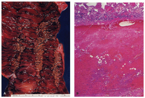
Figure 2-13. Acute ischemic colitis. A: Opened colon showing dilatation of the bowel wall and focal mucosal necrosis. Note the marked purple discoloration of the mucosa due to congestion and hemorrhage and the areas of greenish-brown discoloration. The latter result from bile staining of the necrotic mucosa. B: Full-thickness section of bowel showing transmural hemorrhage.

The initial changes consist of pinpoint petechiae and patchy areas of hyperemia alternating with pale areas. Later the mucosa becomes swollen, edematous, and purplish blue in color, and there may be contact bleeding. Multiple small superficial ulcers (<1 cm) follow; these may become large and serpiginous and resemble those of Crohn’s disease (see Fig. 2-13).

In resection specimens in the acute stage the colon is thickened and rigid. There is surface hemorrhage and a coarse cobblestoning of the mucosa due to linear ulceration and submucosal edema (Fig. 2-15A). In more advanced cases the necrotic mucosa shows a greenish discoloration, and the serosal surface varies in color from plum to black. In healed or chronic lesions, tubular or fusiform strictures with fibrous thickening of the submucosa, often with shallow ulcers, are seen.84, 85

Microscopy. The histologic changes range from mild mucosal and submucosal edema and hemorrhage to transmural destruction. The outcome of ischemia is also variable. About 50% of patients develop some degree of necrosis, followed by granulation tissue, scarring, and fibrous stricturing. The earliest mucosal changes appear to be mucosal congestion and hemorrhage, followed shortly afterward by lifting of the surface epithelial cells and coagulative necrosis of the surface and crypt epithelium. At this point, one may still see the ghost outline of the crypts or surviving crypt bases in the shape of tear drops, but shortly afterward the entire epithelium is sloughed, and one may then see crypt spaces devoid of epithelium (Fig. 2-16A,B). Similar changes may occur in an autolyzed bowel, though without the hemorrhage. This change may result in pseudomembranes composed of necrotic mucosa, fibrin, and blood. Histologically, there are subtle differences between genuine C. difficile-associated pseudomembranous colitis and ischemic necrosis. Hyalinization of the lamina propria such that the normal loose connective tissue punctuated with plasma cells, lymphocytes, and eosinophils is replaced with dense eosinophilic hyalinized matrix is characteristic of genuine ischemia (Figs. 2-17 and 2-18). Usually the residual glands become more closely spaced (lamina propria “collapse”). Atrophic-appearing microcrypts, lamina propria hemorrhage, full-thickness mucosal necrosis, and a diffuse microscopic distribution of pseudomembranes are more common in ischemia (Fig. 2-19). In genuine C. difficile-associated pseudomembranous colitis, neutrophils are more common, the upper parts of the
crypts are dilated, and necrosis is mainly present in the upper half of the mucosa.86


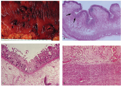
Figure 2-14. Acute ischemic colitis. A: Mucosal surface showing hemorrhage and swelling producing multiple polypoid nodules. B: Full-thickness longitudinal section of colon showing marked thickening of the bowel wall due to submucosal edema. The mucosa is focally ulcerated (arrows) and shows focal loss of glandular epithelium. C: Higher-power magnification of the mucosa in (B) showing focal loss of surface and glandular epithelium. D: Higher-power magnification of the muscularis propria in (B) showing the neutrophilic infiltration of the ischemic muscle coat.

It is also important to be aware that crypt epithelial cells can collapse into the lumen where they can become rounded up and resemble signet ring cells.87, 88 This trap is seen primarily in pseudomembranous colitis, but the resemblance to signet ring carcinoma can be striking; with only the realization that the signet ring cells are still confined to lumen of the crypts, an overall misdiagnosis can be prevented.

In the healing phase granulation tissue becomes prominent. The mucosa regenerates but is often atrophic, with shortened and branched crypts, and is subject to recurrent ulceration. The changes are usually patchy. At this stage, it may resemble chronic treated or quiescent IBD. However, despite the similarities in practice this is seldom a problem. Any mucosal destruction resolves with distorted crypts, but the chronic inflammation of IBD is not present. The sub-mucosa and muscularis propria are markedly widened initially by the granulation tissue and later replaced by fibrosis. This results in stricture formation, often with thinning of the viscus (Fig. 2-20). Sometimes the submucosal fibrosis may extend into the mucosa, frequently in a diffuse manner. Hemosiderin-laden macrophages may be found in the scarred tissue and have been described as a hallmark of ischemia (Fig. 2-21).89 However, in our experience, these are few and far between; they are also of little value in the distinction from other IBDs. Occasionally cholesterol emboli can be seen that may have traveled from proximally located ulcerated atherosclerotic plaques (Fig. 2-22). The overlying mucosa sometimes appears normal, and the patient can be asymptomatic, although GI symptoms occur in about one-third of cases. Limited ischemic bowel necrosis, occasionally with subsequent stricture formation, can however occur.90

Ischemic proctitis. It was thought that the rectum is spared from ischemic injury because of its ample collateral circulation. However, rectal ischemia does occur either in conjunction with colonic ischemia or
by itself and represents about 10% of all cases of large bowel ischemia. Rectal ischemia occurs in patients with occlusive internal iliac artery disease. Acute presentation may follow the ligation of the inferior mesenteric artery (operations of the lower aorta) or other iatrogenic interventions including sclerotherapy for hemorrhoids.91 The endoscopic features and pathology of ischemic proctitis are the same as those of ischemic colitis.92, 93 It must be differentiated from other causes of rectal inflammation such as the solitary rectal ulcer syndrome or rectal lesions due to abuse of suppositories.


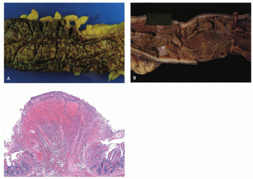
Figure 2-15. Ischemic colitis with pseudomembranes. A: Opened bowel showing green discoloration of the mucosa and adherent yellowish pseudomembranes. B: Opened colon from another patient showing loosely adherent greenish pseudomembranes. The underlying tissues are markedly inflamed. (Courtesy of Maureen Duffield, Johannesburg) C: Histologic section of mucosa and pseudomembrane. The mucosa is necrotic, and most of the glandular epithelium is gone. The pseudomembrane, which is in the center of the section, is composed of a fibrinous exudate mixed with necrotic tissue and inflammatory cells.


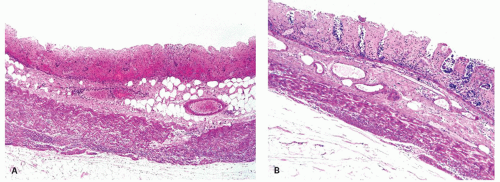
Figure 2-16. A: Extensive large bowel infarction with diffuse transmural necrosis and complete loss of glands. B: Epithelial cells can persist in some crypts.


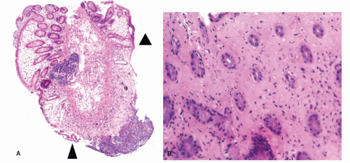
Figure 2-17. A: Endoscopic biopsy from ischemic colitis showing the well demarcated lesion with mucosal necrosis (arrowheads). B: Hyalinization of the lamina propria characterized by dense eosinophilic and paucicellular matrix and microcrypts are specific markers for a diagnosis of ischemic colitis.


INFLAMMATORY VASCULAR DISORDERS OF THE GASTROINTESTINAL TRACT (VASCULITIDES)


Introduction

Genuine “vasculitis” is characterized by fibrinoid degeneration (eosinophilic degeneration) of the vessel wall and infiltration of the vessel wall by leukocytes, with neutrophils, nuclear dust, and extravasated red cells in the vessel wall and the adjacent stromal tissue.94, 95

The term “vasculits” is applied to “noninfectious inflammatory” disorders of blood vessels. Vasculitis has many causes, which result in only a few limited histologic patterns (Table 2-5). Vasculitis can affect any caliber blood vessel in the GI tract (Table 2-6), and the frequency of GI tract involvement varies among different types of vasculitides (Table 2-7). It is characterized by general symptoms such as fatigue, fever, weight loss, and arthralgias and abdominal symptoms such as pain, nausea, vomiting, diarrhea, intestinal bleeding, and perforation. If GI manifestations occur in a patient with established vasculitis, endoscopy, angiography, and histology are generally sufficient to demonstrate the involvement of the GI tract. Endoscopy may reveal minimal abnormalities or ulceration and strictures. Angiography shows stenoses, aneurysms, and infarctions. Histology may show inflammation and necrosis of blood vessels and perivascular inflammation. In a patient, not previously diagnosed as suffering from vasculitis, diagnosis is more difficult. The physician should be alerted especially if general symptoms point toward systemic disease. In addition, other organ systems may show signs of inflammation such as the skin, eyes, lungs, nervous system, and kidneys.


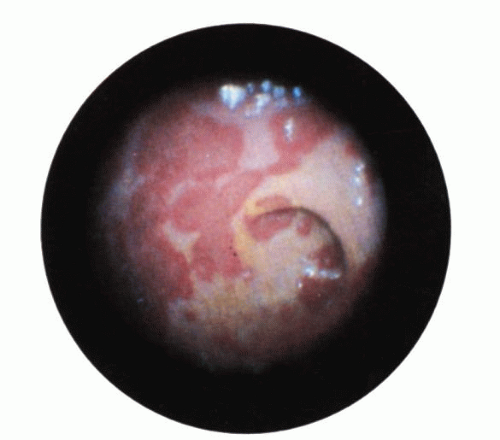
Figure 2-18. Ischemic colitis following aortofemoral bypass surgery. Endoscopic appearance showing a serpentine, white-based exudate in the rectum.

Complete biologic screening involves testing for evidence of inflammation and immunologic deficiencies; screening for immune complexes and complement, autoantibodies, infections, and factors that promote clotting; and testing for the evidence of vascular endothelial damage (Table 2-8). Involvement of large- and medium-sized vessels by vasculitis can be demonstrated by means of arteriography and other imaging techniques. Skin, muscle, nose, and kidney biopsies can reveal signs of vasculitis.96 Endoscopic
biopsies of the GI tract are more likely to be negative as very little submucosa is included, and they merely show features of mucosal ischemia. Rectal biopsies were indeed negative in many patients with established systemic vasculitis.97 The establishment of the involvement of larger vessels requires surgical samples. The presence of isolated GI vasculitis is extremely uncommon in the absence of other systemic features of vasculitis.98


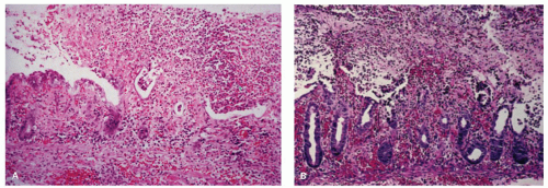
Figure 2-19. Comparison of ischemic colitis with pseudomembranes and pseudomembranous colitis due to Clostridium difficile toxin. A: Ischemic colitis with pseudomembranes shows full-thickness necrosis of the mucosa below the adherent pseudomembrane. B: Pseudomembranous colitis showing the characteristic explosive necrosis of the upper half of the mucosa with preservation of the lower half of the gland crypts.

The minimum criteria for a microscopic diagnosis of vasculitis remain controversial, but it is generally accepted that there must be two components: (a) an inflammatory cell infiltrate and (b) vascular injury. The absence of inflammation thus precludes the diagnosis of vasculitis, although in a late, healing state, inflammatory infiltration may be minimal. Like many other inflammatory conditions, vascular injury is a continuum. The spectrum ranges from endothelial cell swelling and leakiness to frank fibrinoid necrosis and fibrin deposition. Pathologists require a certain degree of histologic evidence of injury manifested by deposition of fibrinoid material, necrosis
of the vessel wall, or both. However, certain changes such as extravasation of erythrocytes and edema due to leakiness, thrombosis, and infiltration of the vessel wall can occur without fibrinoid necrosis of the vessel wall.


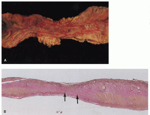
Figure 2-20. Ischemic stricture of the colon. A: Opened segment of the colon, showing stricturing and narrowing of the lumen. The central portion of mucosa is atrophic and surrounded by an irregular hyperemic margin. B: Full-thickness section of the colon showing marked thinning of the bowel wall and replacement of the muscularis propria by fibrous tissue (arrows).


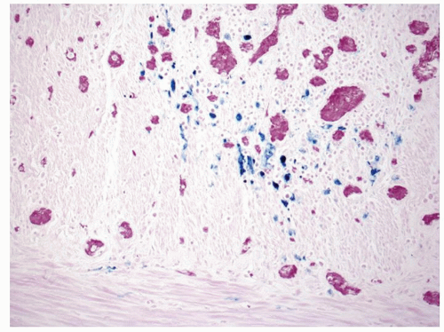
Figure 2-21. Recovery following ischemic damage can be characterized by abnormal crypt architecture and the presence of iron-laden, Perls-positive macrophages which are stained blue.

The term “vasculopathy” has been used to describe vascular alterations without clear inflammation. Another problem is the difference between primary and secondary vascular injury. Secondary vascular injury should be distinguished from secondary vasculitis, a term which is used by clinicians for vasculitis associated with rheumatic or other connective tissue disorders, malignant diseases, infections or exposure to toxic substances.99 Secondary vascular injury can occur in different inflammatory conditions, such as a peptic ulcer of the stomach and cholecystitis, when local blood vessels are affected by the inflammatory process. The occurrence of vascular lesions in otherwise severely inflamed tissue should therefore not be confused with a genuine vasculitis. Thus, it is always helpful to look in areas away from severe inflammation and necrosis to identify genuine vasculitis.


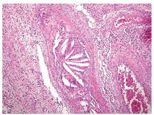
Figure 2-22. Cholesterol emboli that result from the release of material from atherosclerotic plaques in a submucosal artery.


Table 2-5 Classification of Vasculitis According to Histologic Pattern


I:


Vascular damage—vasculopathy: scant inflammatory cells


II:


Vasculitis:


Lymphocytes predominant


“Lymphocytic vasculitis”


Lupus erythematosus


Angiocentric lymphomas


Cytomegalovirus inclusion disease


Behçet’s syndrome


III:


Vasculitis:


Neutrophils predominant


Small vessel leukocytoclastic vasculitis


Polyarteritis nodosa


Behçet’s syndrome


III:


Vasculitis:


Mixed cell types: granulomas


Churg-Strauss syndrome


Wegener’s granulomatosis


Giant cell arteritis


Secondary vasculitis


There is also considerable overlap in the histologic appearances of different genuine forms of vasculitis. A reliable histologic classification is thus difficult because of lack of histologic specificity and the variability of histologic changes depending on the stage of the disease, its activity, and any prior treatment. In some cases a differential diagnostic scheme based on the histology can be helpful, but in general this type of classification is not very useful clinically (see Table 2-5).


Classification

Large vessel vasculitis was also the first type to be described.100 For years, vasculitis was known as “periarteritis nodosa,” a term initially used to characterize a nodular inflammatory lesion in medium- and small-sized arteries throughout the body. The name was later changed to “polyarteritis nodosa (PAN).” By the 1950s, it was realized that a variety of clinically distinct forms of vasculitis existed and that small vessels could also be involved. Small vessel vasculitis was referred to as either “hypersensitivity vasculitis” or “microscopic periarteritis.”101 The latter is now commonly known as microscopic polyangiitis (MPA). Since then the classification of vasculits has evolved and become more complex.

Despite many causes, vasculitis has limited histologic patterns.102 The histologic lesions vary with time,
and the clinical presentation depends upon the size of the vessels involved as well as distribution of the disease. The protean clinical manifestations, combined with the etiologic nonspecificity of the histologic lesions, complicate the diagnosis of specific forms of vasculitis. This is problematic because different vasculitides with indistinguishable clinical presentation (e.g., Henoch-Schönlein and MPA) may have different prognosis and treatment. A general classification covering all these aspects and easily applicable in clinical practice is at present not available. One approach is to categorize the noninfectious vasculitides on the basis of the predominant type of vessel affected. Such a classification has been proposed by an international conference, and attempts have been made to combine the classification according to the vessel size and etiology (see Table 2-6).103 This was followed by a proposal for uniform terminology and definitions.104 It is clinically important to identify the type and size of inflamed vessels and to determine
whether the inflammation is focal or extensive.102, 104 These variables have an influence upon the clinical presentation and the diagnostic techniques used.94, 95 Involvement of larger vessels is frequently associated with more severe clinical syndromes; for example, involvement of larger “mesenteric vessels” results in complications such as bowel necrosis, infarction, and perforation.

Only gold members can continue reading. Log In or Register to continue

Stay updated, free articles. Join our Telegram channel

Jul 20, 2016 | Posted by in GASTROENTEROLOGY | Comments Off on Vascular Disorders and Related Diseases

Full access? Get Clinical Tree

Get Clinical Tree app for offline access