Sensitivity (%)
Specificity (%)
Accuracy (%)
Physical exam
70
70
67
Color Doppler ultrasound
93
85
ND
Venography
100
60–70
ND
Thermography
84–98
81–100
ND
Physical Examination
Physical examination with the patient standing in a warm room is currently the preferred method for varicocele diagnosis. This method has a sensitivity and specificity of around 70 % compared with other diagnostic tools, such as venography and color Doppler studies [112, 184]. Based upon clinical examination, varicocele is generally classified according to Dubin et al. [185] into:
1.
Impalpable or subclinical type or grade 0 when it is not palpable or visible at rest or during Valsalva maneuver, but demonstrable by scrotal ultrasound and color Doppler examination.
2.
Palpable varicocele when it is clinically palpable at rest or with the aid of Valsalva maneuver. Such varicoceles are further divided into:
Grade 1: Palpable only during Valsalva maneuver
Grade 2: Palpable at rest, but not visible
Grade 3: Visible and palpable at rest
A grade III varicocele is easily identified, as shown in Fig. 4.1, while lower grade varicoceles may be difficult to recognize particularly in certain clinical situations such as prior scrotal surgeries, cryptorchidism, obesity and hydrocele [186].
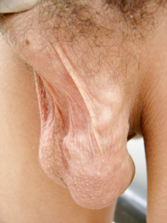
Fig. 4.1
Photograph of a grade 3 varicocele
Color Doppler Ultrasonography
Whenever physical examination is inconclusive or difficult to perform as in cases of low-grade varicocele, previous scrotal surgery, obesity, concomitant hydrocele, or scrotal tenderness/hypersensitivity, imaging studies are recommended. Among the non-invasive modalities, color Doppler ultrasound (CDU) has been shown to be the best non-invasive diagnostic tool.
The ultrasound study of the scrotum should be performed with high frequency linear probes and with devices able to evaluate blood flow. Blood vessels are first studied in a grey scale and then with the color Doppler and the pulse Doppler. For the correct detection of fluxes, CDU must be calibrated to detect a slow flow (7.5 kHz). The evaluation should be performed in the supine and then the upright positions, with and without a Valsalva maneuver, in order to obtain a complete evaluation of the fluxes in the seminal cord veins [187].
Using the commonly accepted CDU criterion of a 3 mm or greater vein diameter for varicocele, CDU was shown to have a sensitivity of about 50 % and specificity of 90 % compared to physical examination [188]. It means that CDU tests negative in approximately half of the patients with palpable varicocele (low sensitivity), while it is unlikely that a patient with a non-palpable varicocele will test positive by CDU (high specificity). To circumvent this matter, Chiou et al. [188] proposed a scoring system incorporating the maximal venous diameter (score 0–3), the presence of a venous plexus and the sum of the diameters of veins in the plexus (score 0–3), and the change of flow on Valsalva maneuver (score 0–3). Using a total score of 4 or more to define the presence of CDU-positive varicocele, the authors observed a sensitivity of 93 % and a specificity of 85 % when compared to physical examination. In their study evaluating 64 patients, all moderate to large varicoceles found on physical examination were positive by CDU diagnosis using the scoring system, but the same group had only a 68 % positive rate by traditional CDU diagnostic criteria. The scoring system for CDU diagnosis of varicocele, as proposed by Chiou et al., is shown in Table 4.2.
Table 4.2
Scoring system for color Doppler ultrasound (CDU) diagnosis of varicocele, as proposed by Chiou et al. [188]
CDU parameter | Score |
|---|---|
Maximum vein diameter (mm) | |
< 2.5-0 | 0 |
2.5–2.9 | 1 |
3.0-3.9 | 2 |
>/ = 4.0 | 3 |
Plexus/sum of diameter of veins | |
No plexus identified
Related posts:Stay updated, free articles. Join our Telegram channel
Full access? Get Clinical Tree
 Get Clinical Tree app for offline access

| |
