(a)
Temperature
≤ 36 °C or ≥38 °C
(b)
Heart Rate
≥90 beats/min
(c)
Respiratory
≥20 breaths/min
(d)
Respiratory Alkalosis
PaCO2 ≤32 mmHg
(e)
Leukocytosis
≤4 × 10⁹/L or ≥12 × 109/L or ≥10 % bands
3.
Criterium 3 Multiple Organ Dysfunction Syndrome (MODS) [1]
(a) | Cardiovascular | Systolic blood pressure ≤90 mmHg or |
Mean arterial pressure ≤70 mmHg | ||
(b) | Respiratory | PaO2 ≤75 mmHg breathing room air or |
PaO2/FiO2 ≤250 if mechanical ventilation | ||
(c) | Renal | Urine output ≤0.5 ml/Kg post fluid resuscitation |
(d) | Encephalopathy | Somnolent, agitated, confused, coma |
(e) | Metabolic Acidosis | pH ≤7.3 or Base excess ≥5 or lactate ≥1.5 × Normal |
(f) | Thrombocytopenia | ≤80 × 10 or ≥50 % ↓ in 3 days |
Based upon these criteria, the severity of sepsis may be determined [1]:
1.
Sepsis – Criteria 1 + ≥ 2 Criteria 2
2.
Severe Sepsis – Criteria 1 + ≥2 Criteria 2 + ≥1 Criteria 3
3.
Septic Shock – Criteria 1 + ≥2 Criteria 2 + refractory hypotension ≤90 mmHg
The associated mortality varies with number of criteria involved. Sepsis mortality varies between 7 and 17 % if 2 and 4 criteria 2 are satisfied respectively; severe sepsis mortality increases by 15–20 % for each organ system involved from criteria 3; mortality in septic shock is between 50 and 80 % [1].
Sources and Pathogenesis of Urosepsis
The genitourinary tract accounts for 20–30 % of sepsis [2]. Sources of urosepsis may include infections of any genitourinary organ: the kidney (i.e. Pyelonephritis, pyonephrosis, renal abscess), bladder (i.e. Severe cystitis), prostate (i.e. Acute bacterial prostatitis, post transrectal ultrasound guided prostate biopsy), testicular or scrotal (i.e. Epididymo-orchitis, Fournier’s gangrene). Sepsis from obstructive pyelonephritis by urolithiasis is the most common presentation of urosepsis, representing 43 % of cases, followed by prostatic etiology in 25 %, genitourinary malignancy in 18 % and other genitourinary diseases accounting for the remaining 14 % [3]. In urosepsis secondary to obstructive pyelonephritis, ureteral stones are the cause in 65 % of cases, malignant obstruction in 21 %, pregnancy associated obstruction in 5 %, anatomic abnormalities in 5 % and post urologic procedure in 4 % [1]. Bacterial etiology most commonly include: E. coli, Proteus spp., Klebsiella spp., Enterobacter, P. aeruginosa, and Staphylococcus aureus [1, 4]. In immunocompromised patients Candida spp. and Pseudomonas spp. should be considered [5].
Bacterial endotoxins, such as lipopolysaccharide (LPS) from the cell wall of gram-negative organisms appear to mediate the systemic manifestation of sepsis. These bacterial components activate the inflammatory, coagulation, and complement systems, stimulating the activity of monocytes, macrophages, neutrophils, and dendritic cells, amongst other inflammatory cell subtypes [6]. LPS-stimulated monocytes play a central role in mediating clinical sepsis, and produce tumour necrosis factor alpha (TNFa) and interleukin (IL)-1 at LPS concentrations of 25–50 pg/mL [7]. In addition to stimulation of inflammatory cells, endotoxin also directly binds receptors in the endothelial cell membrane, which also promotes pro-inflammatory mediators [8].
Early work in this space demonstrated that the rate of release of endotoxin in the blood stream can result in sepsis of different severities. Taudort et al. gave healthy volunteers a 3 ng/kg intravenous bolus of E. coli endotoxin versus an infusion over 4 h. The response of inflammatory mediators, specifically TNFa, IL-6 and neutrophil response, occurred earlier and was more severe in the bolus group compared to the infusion group [9]. This is directly relevant in the setting of urosepsis secondary to obstruction, as relief of obstruction often results in rapid levels of endotoxemia and thus rapid development of sepsis. The burden of inflammatory mediators in the blood can be prognostic as well. The serum level of TNFa has been shown to correlate with death from urosepsis [10, 11] (Table 4.1).
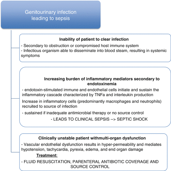
Table 4.1
The factors that cause a genitourinary infection to progress to sepsis are outlined below
Management
Managing urosepsis requires a prompt recognition, early goal directed resuscitation, broad-spectrum parenteral antibiotics, and source control [12]. Diagnosis is initiated with a focused history with the goal of identifying criteria 1 above. Enquiry on history includes assessment of systemic features: fevers, chills, rigors, mental status changes, malaise; urinary symptoms: difficulty voiding, dysuria, gross hematuria or pyuria, flank or abdominal pain, testicular, penile or perineal pain, perineal/scrotal skin changes, recent urologic instrumentation, and urologic history, Physical examination is mandatory, starting with review of vital signs and temperature as per criteria 2 above. Focused exam should assess flank and abdominal tenderness, and palpation of the scrotum and perineum for crepitus. The latter is of paramount importance for the early detection of Fournier’s gangrene.
Laboratory investigations should include a complete blood count, electrolytes and renal function tests, serum lactate, urinalysis, blood and urine cultures prior to antibiotic initiation. If history or physical examination identify a potential testicular, scrotal or prostatic source an ultrasound is warranted [1]. If clinical suspicion for a renal etiology, computed tomography (CT) scans are highly sensitive in detecting renal abscesses [13] in addition to hydronephrosis and urolithiasis [14].
Early goal directed therapy are required for reducing mortality and optimizing outcomes as described in Rivers Protocol [12]. This involves supporting the patients cardiovascular system with crystalloid fluid resuscitation and vasoactive or inotropic agents if refractory despite euvolemia. The respiratory system is supported with supplemental oxygen and possible mechanical ventilation to maintain tissue and organ oxygenation and perfusion; RBC transfusions are considered to maintain a hematocrit ≥30 % to ensure an adequate quantity of circulating RBC’s to perfuse tissue and organs. Sedation and paralysis may be considered if the patient is mechanically ventilated to reduce metabolic and oxygen demands in septic shock [12]. Early initiation of empiric parenteral antibiotics, ideally within 1 h of presentation, is essential to minimize mortality [15, 16]. The author recommends searching the patients past medical records for history of a resistant organism. Antibiotic selection should be initially broad to cover bacteria common to urosepsis (see above), consider local patterns of resistance and regional antibiograms, patient allergies, and pharmacokinetics & dynamics of urinary tract involvement and tissue penetration [17]. A general antibiotic strategy is to use a third generation cephalosporin combined with enterococcus coverage (i.e. ceftriaxone + ampicillin), or broad-spectrum agents such as piperacillin-tazobactam or a carbapenem, particularly if the local rates of extended spectrum beta lactamase (ESBL) producing organisms is high [18–24]. Once blood and urine cultures have revealed the offending organism and antibiotic sensitivities are available, the antibiotic may be tailored appropriately. If candiduria or candidemia is present, the addition of antifungal agents are necessary [20, 21].
< div class='tao-gold-member'>
Only gold members can continue reading. Log In or Register to continue
Related posts:
 Pathogenic Mechanisms of Uropathogens
BCG for the Treatment of Non-muscle Invasive Bladder Cancer
Bacteria in the Genitourinary Tract: The Microbiota and Probiotics
The Management of Infection Stones
The Use of Probiotic Bacteria to Treat Recurrent Calcium Oxalate Kidney Stone Disease
Pathogenic Mechanisms of Uropathogens
BCG for the Treatment of Non-muscle Invasive Bladder Cancer
Bacteria in the Genitourinary Tract: The Microbiota and Probiotics
The Management of Infection Stones
The Use of Probiotic Bacteria to Treat Recurrent Calcium Oxalate Kidney Stone Disease
 Struvite Stone Formation by Ureolytic Biofilm Infections
Stay updated, free articles. Join our Telegram channel

Full access? Get Clinical Tree


