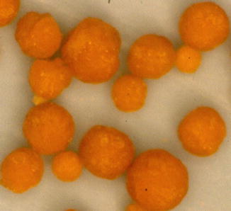
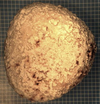
Fig. 6.1
(a) Uric Acid, (b) calcium oxalate monohydrate and a (c) mixed stone (50 % uric acid and 50 % calcium oxalate monohydrate)
Randall’s Plaques: Big Stones Have Small Beginnings!
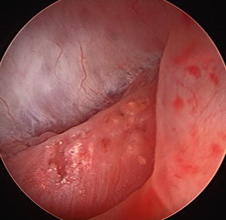
Dr. Alexander Randal demonstrated that stones begin from small calcifications that are present in the papilla of the kidney (Fig. 6.2). He referred to these small calcifications as Randall’s Plaques. During modern stone surgery these are often seen but are too small to be treated. Over time as they grow they detach from the kidney and become true kidney stones. It is from this that we know that large stones have small beginnings.


Fig. 6.2
Randall’s Plaques in a renal papilla
Calcium Stones
Simple Stone Facts
Calcium oxalate is the most common type of urinary stone.
Can be treated with surgical therapy.
Calcium containing stones are the most common types of stones formed within the urinary tract. They are most commonly composed of a mixture of calcium phosphate and calcium oxalate. Among the different calcium stones are calcium oxalate monohydrate, calcium oxalate dihydrate and calcium phosphate (Fig. 6.3). The etiology of calcium stone disease is very diverse.


Fig. 6.3
(a) Calcium oxalate monohydrate, (b) calcium oxalate dihydrate and (c) calcium phosphate stones (Courtesy of Louis C. Herring & Co., Orlando, FL)
The majority of patients with calcium stone disease will demonstrate hypercalciuria (40–75 %), which may be subdivided into one of three categories. These include absorptive hypercalciuria, renal hypercalciuria, or resorptive hypercalciuria. Other causes for calcium stones include hyperuricosuria (10–50 %), hyperoxaluria (8 %), and hypocitraturia (10–50 %). For a further detailed description of the metabolic factors associated with calcium stones, please refer to Chap. 8.
In 15 % of patients, calcium stones are secondary to a known underlying disorder. Primary hyperparathyroidism accounts for approximately 3 % of the calcium stone forming population. Less commonly, hereditary hyperoxaluria, renal tubular acidosis, sarcoidosis, Cushing syndrome, steroid treatment, Vitamin D intoxication, immobilization and medullary sponge kidney disease may be present. In the majority of patients, however, none of these disease processes are present and the stone disease is referred to as idiopathic or primary.
Treatment for calcium stones is surgical if the stones are too large to pass. A specific metabolic condition contributing toward calcium stone formation must be properly diagnosed so as to tailor the appropriate treatment for stone prevention.
Uric Acid Stones
Simple Stone Facts
Most common cause of these stones is low urine pH.
Associated with obesity, diabetes and metabolic syndrome.
Treatment is medical therapy by alkalinization of the urine.
Radiolucent (not visible on plain x-ray) stones.
The incidence of uric acid calculi is approximately 5–10 % of all renal stones (Fig. 6.4). Men are affected four times more often than women. Approximately 25 % of patients with gout will form uric acid stones, however, the majority of patients who form uric acid calculi have no detectable abnormalities in uric acid metabolism. Factors that may contribute or predispose to uric acid stone formation are divided into three categories: (1) high urinary uric acid levels or hyperuricosuria; (2) low urinary volume; and (3) low urinary pH. Low urine pH is the hallmark of uric acid stone formers. This will be described in more detail in Chap. 8.


Fig. 6.4
Uric acid stones (Courtesy of Louis C. Herring & Co., Orlando, FL)
Treatment is tailored toward increasing urine volume and raising urinary pH. If hyperuricosuria is present, it can be corrected with appropriate dietary management and/or administration of medical therapy (see Chap. 19).
Struvite Stones
Simple Stone Facts
Also called infectious stones or Magnesium Ammonium Phosphate stone.
Stones are caused by urease splitting organisms.
Primary treatment of stones is complete removal of stones and eradication of infection.
Medical management is recommended in patients who have incomplete removal of stones or poor surgical candidates.
Struvite stones, commonly called infectious stones, are composed of magnesium ammonium and phosphate (Fig. 6.5). Struvite stones represent 10–15 % of all urinary tract stones. Historically, these stones made up the majority of staghorn calculi. Presently, 56 % of staghorn calculi are metabolic and 44 % are infectious stones. These stones occur more commonly in females with a female to male ratio of 2:1. This female gender predominance is believed to be due to a higher susceptibility to infection.


