Fig. 10.1
Differences between female and male urethral sphincter mechanisms
Physiology and Function
Sensory innervation of the bladder is conveyed to the spinal cord in the pelvic and hypogastric nerves. This involves sensory nerves in the suburothelial plexus, myelinated (A) and unmyelinated (C) fibres. (A) fibres respond to passive distension and active contraction, whereas C fibres respond to noxious stimuli. The cell bodies are located in the dorsal root ganglia (DRG) at S2–3 and T11–L2 spinal segments. Bladder afferent activity thus ascends from the spinal cord to the pontine micturition centre (PMC) and cerebral cortex.
The motor innervation of the lower urinary tract (LUT) comes from both the parasympathetic and sympathetic branches of the autonomic nervous system. The parasympathetic preganglionic fibres are located in the S2–4 spinal segments, and these synapse with postganglionic cell bodies in the detrusor, bladder neck and urethra. Activation of the parasympathetic fibres causes detrusor contraction and relaxation of the smooth muscle of the bladder neck and urethra. Sympathetic cell bodies are located in T10–12 and L1–2. The preganglionic fibres synapse with postganglionic fibre in the hypogastric plexus. The predominant effect is inhibition of the parasympathetic pathways. The somatic supply to the pelvic floor muscles and external sphincter originates from S2-4 and conveyed by the pudendal nerve.
There are important gender differences with regard to bladder neck sphincter mechanism function. The male bladder neck has a well-developed layer of inner circular smooth muscle along with an outer layer of longitudinal smooth muscle. It has a relatively rich adrenergic innervation (contraction prevents retrograde ejaculation), and its functional competence is considered so reliable as to sufficiently maintain continence in the presence of damage to the EUS. The female bladder neck is less well developed with the predominant orientation of fibres being longitudinal and the principle innervation being cholinergic. Consequently its functional competence is less reliable.
Anatomical/Structural Versus Functional Disorders
A wide variety of pathology can affect the urethra and can be broadly classified in a variety of ways, e.g. congenital or acquired, male or female, anatomical/structural abnormalities or functional abnormalities. A structural and anatomical classification is detailed in Tables 10.1 and 10.2.
Table 10.1
Structural/anatomical classification of urethral disorders
Disorders of the male urethra | Disorders of the female urethra |
|---|---|
Congenital | Congenital |
Duplication of the urethra | Distal urethral stenosis in childhood (urethral spasm and dysfunctional voiding) |
Urethral stricture (rare) | |
Posterior urethral valves | |
Anterior urethral valves | |
Hypospadias | |
Urethrorectal and vesicorectal fistulas | |
Epispadias | |
Acquired | Acquired |
Urethral stricture | Urethritis (acute and chronic) |
Urethral stenosis | Urethral caruncle |
Urethritis and infections | Urethrovaginal fistula |
Urethral carcinoma | Urethral diverticulum |
Urethral stricture (rare) |
Table 10.2
Functional classification of urethral disorders
Functional disorders | Disorders of sensation | Hypersensitive sensation or pain due to inflammation |
Decreased sensation | ||
Disorders of motor function | ||
During storage | Incompetent or underactive/absent | |
During voiding | Obstruction due to non-relaxation or overactivity, e.g. detrusor-urethral dyssynergia (detrusor bladder neck dyssynergia or detrusor sphincter dyssynergia) | |
Whilst it is not possible to address all the different urethral pathologies, the common conditions will be discussed in detail in this chapter.
Male Urethral Disorders
Urethral Strictures
Introduction
The term “urethral stricture” refers to an abnormal narrowing of any segment of the urethra surrounded by the corpus spongiosum and implies varying degrees of ischaemic spongiofibrosis. The term “urethral stenosis” is reserved for narrowing of the membranous urethra, the prostatic urethra and the bladder neck as they are not invested by corpus spongiosum (in contrast to the term “urethral distraction defect” as seen with a pelvic fracture urethral injury) [1].
Acquired urethral strictures are more common in men. These can be subcategorised into iatrogenic, traumatic, inflammatory and idiopathic causes [1]. Fenton et al. found that the majority of strictures were idiopathic (34 %) and iatrogenic (32 %) in a review of 175 stricture patients with a total of 194 strictures [2]. Inflammatory and traumatic causes were found in only 20 % and 14 % of patients, respectively. The most commonly involved site was the bulbar urethra (52 %).
Clinical Presentation
Key points in the history include:
Presenting symptoms – obstructive symptoms (e.g. poor flow, urinary retention), storage symptoms, haematuria, dysuria
History of trauma, previous instrumentation (e.g. difficult urethral catheterisation) or transurethral procedures
History of sexually transmitted diseases or urinary infections
Current status of erectile and ejaculatory function
Previous treatment, e.g. urethral dilatation or urethrotomy, hypospadias repair, intermittent self-dilatation (ISD)
A directed physical examination should assess the following:
Difficulties with positioning of patient, i.e. lithotomy position (especially after trauma)
External genitalia – condition of tissues, previous fibrosis/scarring and lichen sclerosis (LS), previous circumcision, fistula, signs of ongoing infection
Abdominal examination – suprapubic scars, presence of a suprapubic catheter
Digital rectal examination
Evaluation and Follow-Up
Patients should undergo comprehensive symptomatic evaluation at presentation and follow-up [3, 4]. The recently published patient-reported outcome measure (PROM) for urethral stricture surgery provides a standardised and validated method of assessing outcomes and consists of a lower urinary tract symptom (LUTS) construct (6 questions), a separate LUTS-specific quality-of-life (QoL) question and Peeling’s voiding picture. The post-operative PROM is supplemented by 2 questions addressing overall patient satisfaction (Appendix A).
Retrograde urethrogram (RUG) is recommended as a reliable, accessible and versatile means to both diagnose and stage urethral stricture. It should be combined with voiding cystourethrography (VCUG) to achieve a synergistic approach to the evaluation of the entire urethra [5]. It remains the standard method to ascertain the location and length of a stricture and any associated urethral pathology, e.g. false passages, fistulae and calculi.
Cystoscopy is recommended by the SIU/ICUD committee as the most specific procedure for the diagnosis of urethral stricture. It is a good adjunct in the staging of anterior urethral stricture, particularly to confirm abnormal or equivocal findings on imaging studies. For pelvic fracture-related urethral distraction defect, cystoscopy is highly recommended for assessment of the bladder neck and posterior urethra [5].
Ultrasonography is another potential adjunctive investigational tool. In addition to ascertaining the length and location, ultrasound can also provide information regarding degree of spongiofibrosis and concomitant pathology, e.g. diverticula, fistulae, stones, false passages and periurethral abscesses [6–8]. Previous retrospective reviews have shown that intraoperative ultrasound changed the surgical approach in 19 % and was integral in deciding between two possible approaches in 26 % of patients [9]. This is suggested as a supplement to the standard approach for most reconstructive surgeons, i.e. an intraoperative decision based on the visual assessment of the anatomy, calibre of the urethra and its vascularity.
The value of uroflowmetry in assessing a stricture preoperatively or for the post-operative follow-up is of limited value. It is the norm to have a patient present with a very tight stricture at the first instance, as they usually have normal bladder function that compensates. In 1968, Smith reported that the effective diameter of the unobstructed male urethra was 11 Fr gauge. Hence, until the stricture narrowed beyond this point, there would be no significant interference with flow, and hence, patients would not be necessarily aware that there was a significant problem [10].
Treatment
In 1974, Sachse introduced direct vision internal urethrotomy (DVIU) to treat urethral strictures by incising with the use of a cold knife [11]. Both urethrotomy and dilatation work by disrupting the stricture, and rely upon adequate tissue vascularity for healing without re-narrowing.
A nationwide survey of practising members of the American Urological Association was performed in 2007 and found that the most commonly used procedures for treatment of urethral strictures were dilatation (used by 92.8 % of surveyed practitioners), optical internal urethrotomy (used by 85.6 %) and endourethral stents (used by 23.4 %). It was noted that for a long bulbar urethral stricture or short bulbar urethral stricture refractory to internal urethrotomy, only 20–29 % of respondents would refer to another urologist, whilst 31–33 % would continue to manage the stricture by minimally invasive means despite predictable failure [12].
Steenkamp et al. compared internal urethrotomy to dilation in a randomised controlled trial and found no difference in efficacy between the two methods. Recurrence rates were noted to be higher for strictures of increasing length. They thus recommended dilation or internal urethrotomy (DVIU) for strictures shorter than 2 cm, primary urethroplasty for those longer than 4 cm, and a trial of dilation or urethrotomy for those 2–4 cm in length [13]. Therefore, SIU/ICUD guidelines recommend that primary DVIU/dilation be used as first-line treatment for short (<1–2 cm), single bulbar urethral strictures (Grade A) [14].
Generally, a third DVIU/dilation is not recommended, except if necessitated by patient comorbidities, choice or economic resource (Grade A). This is evidenced by a previous study that showed that after a third dilation or urethrotomy for stricture recurrence at 3 and 6 months, the stricture-free rate at 24 months was 0 (p < 0.0001) [15].
Permanent urethral stenting is not recommended for patients with strictures who are considered to be candidates for urethral reconstruction. There is evidence that subsequent failure of a permanently implanted stent represents a major surgical challenge [16].
Urethroplasty for the anterior urethra consists of a range of operations such as excision and primary anastomosis (EPA) and the different types of substitution urethroplasty. The type of urethral reconstruction depends on several factors. The length, location and aetiology (e.g. lichen sclerosis) and previous interventions all have impact on the final outcome.
A recent analysis of 17 articles comprising a total of 1234 patients over 35 years (1975–2010) found that overall success of EPA was 93.8 %. Reported complications were <5 %, and there was no evidence of persistent loss of sexual function. Other possible complications include fistula, chordee, postmicturation dribbling and recurrence. Thus, EPA should be carried out for patients with short bulbar strictures, especially if expected success rates of other procedures are <90 % [17].
Care needs to be taken in the assessment and subsequently when performing an anastomotic reconstruction of the anterior urethra. In general, only strictures <3 cm were considered for anastomotic procedure. Mobilisation of the urethra and corporal separation allows gain of several more centimetres (Fig 10.2).
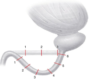
Fig. 10.2
Diagrammatic representation of the additional length (in cm) gained by separation of the corpora cavernosa [18]
Mobilisation of the bulbar urethra should not extend beyond the penoscrotal junction to prevent chordee. In this context, one should perform an augmentation (substitution) if the stricture is too long. Likewise, it is rare to be able to perform an anastomotic urethroplasty for the penile urethra due to the risk of producing a chordee. The exception would be in the case of an acute penile fracture.
Augmentation urethral reconstruction can be either one-stage or two-stage procedure. There are three potential options to a one-stage procedure – an onlay augmentation, an augmented anastomotic or a tube augmentation procedure. Whereas, a two-stage procedure includes excision of the stricture and reconstruction of the roof strip. The second stage tubularisation is carried out at least 3–6 months later, but further evaluation at 2 months is essential to identify what revision, if any, of the first stage is necessary [19].
A systematic review of graft augmentation urethroplasty techniques for treatment of anterior urethral strictures showed there was no significant difference between the average success rates for the dorsal and ventral onlay procedures, 88.4 % and 88.8 % at 42 and 34 months, respectively. The lateral onlay procedure has only been described in 6 patients and is reported to have a success rate of 83 % at 77 months. For penile stricture disease, the success rate for a two-stage procedure was significantly better than the one-stage technique (90.5 % versus 7.5 %, respectively) [20].
Oral mucosa is the most versatile augmentation (substitution) material. Oral mucosa has privileged immunology, with preclinical work showing less fibrosis compared to skin. This mucosa is wet and is unlikely to be affected by lichen sclerosis.
Bioengineered buccal mucosa grafts are likely to have a future role for patients with long and complex strictures. Particularly for redo cases where oral mucosa has already been used, then the supply may be limited. Autologous cell-seeded grafts are not dependent on ingrowth of epithelial cells and show some promise. Tissue-engineered oral mucosa was reported in five men. All patients had complex strictures secondary to lichen sclerosis. Initial graft take was 100 %. However, at 3 years, three patients had intact graft, albeit with the need for instrumentation [21].
Pelvic Fracture Urethral Injuries (PFUI)
Classic signs of blood at the meatus, inability to void and a distended bladder after a traumatic incident are the classic signs but may not always be present. Other signs include a butterfly sign or perineal/scrotal haematoma and a high riding prostate on per rectal examination. It is particularly important to exclude the coexisting rectal injury which is seen in up to 5 % of cases.
Posterior urethral injuries associated with pelvic fracture are best considered as pelvic fracture urethral distraction injury (PFUDD). This term was introduced by Turner-Warwick [22]. However, a large percentage of such injuries are partial and do not necessarily result in “distraction” or “separation”. Therefore, the terminology of PFUI has replaced PFUDD.
Some types of pelvic fracture are associated with a higher risk of posterior urethral injury. The highest risk of urethral injury was found in cases with straddle fracture when combined with diastasis of the sacroiliac (SI) joint (24 times more than the rest of the pelvic fractures); this was followed by straddle fracture alone (3.85 times) and Malgaigne’s fracture (3.4 times) [23].
Ideally a retrograde urethrogram should be performed in all patients suspected of having a PFUI. The urethrogram should be performed with 45° oblique views with the downside leg externally rotated and flexed at the knee. The downward obturator fossa should be completely closed on the scout film to confirm appropriate positioning. This is followed by the injection of 15–20 ml of contrast under fluoroscopic guidance. However, in polytrauma patients, this may be logistically difficult. In addition, extravagated contrast can confuse later CT findings.
There are two accepted treatment methods in the acute setting. Early closed realignment over a catheter can be done using blind or endoscopic techniques. The second and safest approach recommended for the majority of cases is the placement of a suprapubic catheter and an interval perineal urethroplasty 3–6 months later. It is well established that immediate open retropubic urethroplasty and open catheter realignment result in significantly higher incontinence and impotence rates and hence are not recommended (Grade A) [24].
The progressive perineal approach is the means to which an end-to-end bulbomembranous anastomosis (BMA) can be achieved in stepwise fashion. These are:


1.
< div class='tao-gold-member'>
Bulbar urethral mobilisation
Only gold members can continue reading. Log In or Register to continue
Related posts:


Stay updated, free articles. Join our Telegram channel
Full access? Get Clinical Tree
