Ureters
Nigel Cowan MD, FRCP, FRCR
Case 9-1
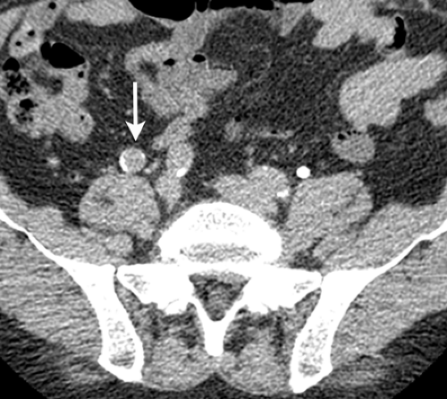
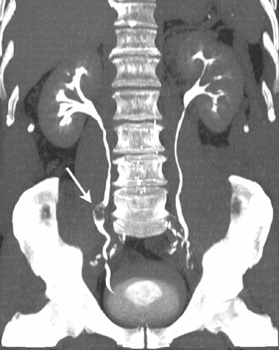
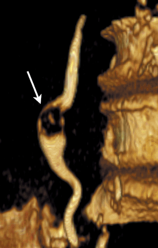
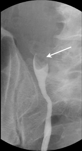
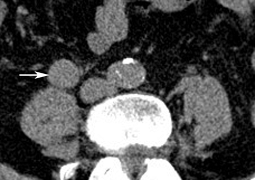
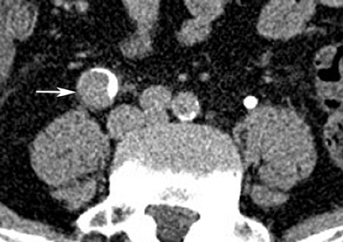
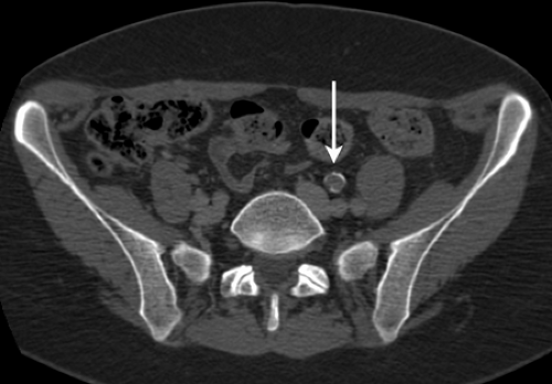
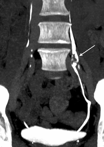
History: 60-year-old male with gross hematuria.
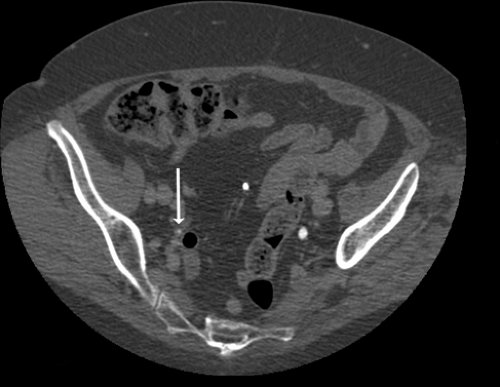
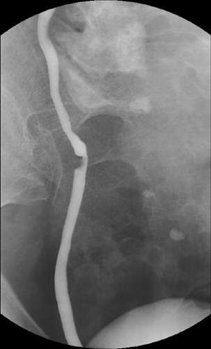
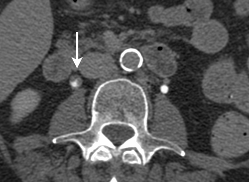
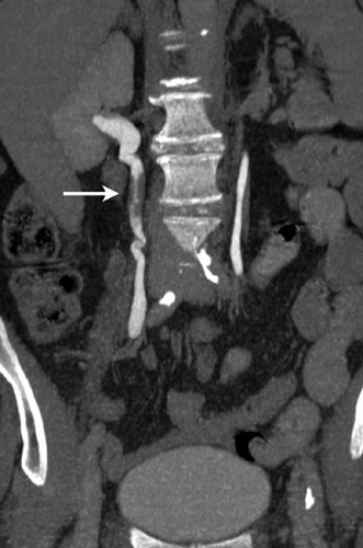
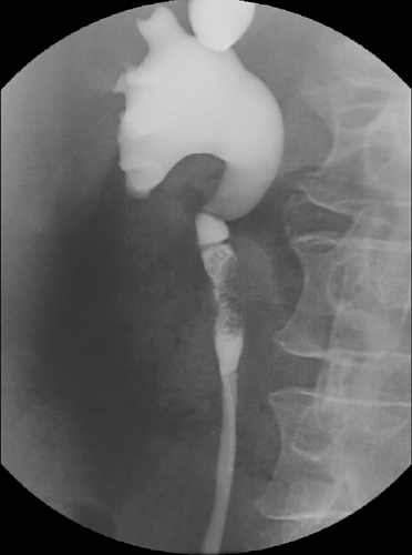
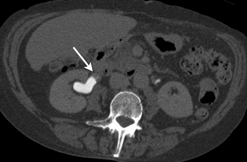
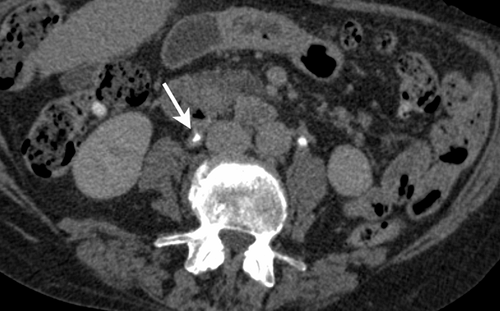
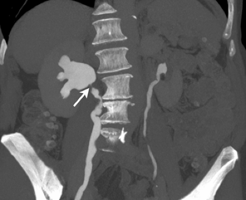
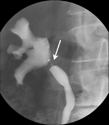
Findings: Axial excretory phase image (A) obtained during a CT urogram demonstrates a soft tissue mass (arrow) filling most of the lumen of the mid right ureter just lateral to the proximal right common iliac artery. Maximum intensity projection (B) and volume-rendered (C) images show that the ureteral mass (arrows) is both polypoid and stippled in appearance. There is minimal, if any, hydronephrosis and hydroureter. On an image from a retrograde pyelogram (D), the distal portion of the mass appears as a large filling defect (arrow) protruding into the dilated portion of the adjacent ureter.
Diagnosis: Transitional cell carcinoma of the ureter
Discussion: Transitional cell carcinoma (TCC) may present as a polypoid mass that arises from a portion of the ureteral mucosa and slowly grows over time. As the tumor grows, the tumor extends into and expands the lumen of the ureter. Consequently, there is little obstruction, if any. As a result, the ureter both above and below the tumor is widened. In cases of ureteral calculi, clots, and fungus balls, the distal ureter is typically narrowed. TCC can be further differentiated from other entities because TCC typically enhances after contrast material administration. On images obtained with retrograde pyelography, the widened ureter distal to the tumor and the interface of the contrast material with the tumor has been called the “goblet sign.” CT urography can be helpful in defining TCC size, location, and extent. In this case, the reformatted images clearly depict the upper and lower extent of the tumor. This case was treated by local surgical excision.
Case 9-2
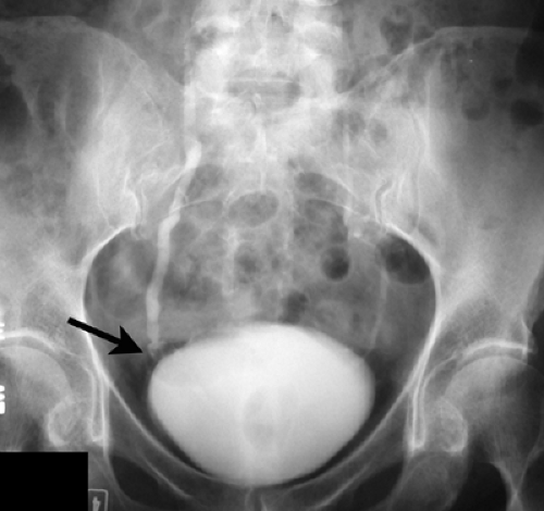
History: 56-year-old female with microscopic hematuria.
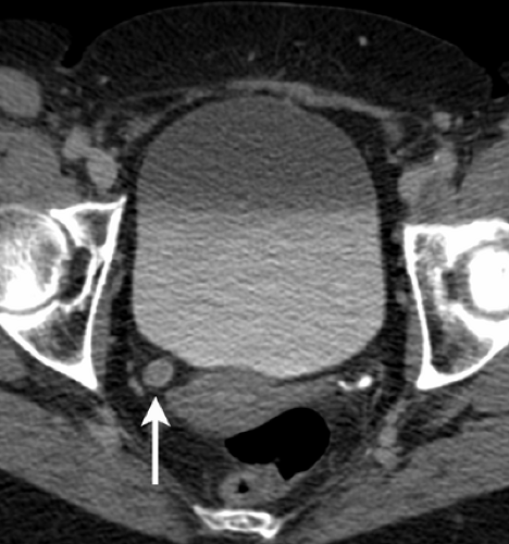
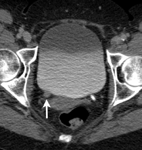
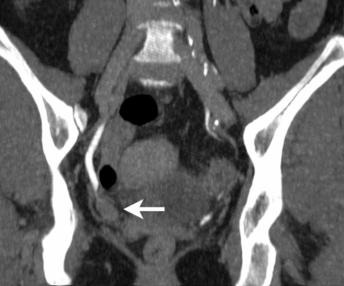
Findings: On an image from an intravenous urogram (A), obtained 15 minutes post injection of intravenous contrast material, the right ureter is wider than the left. Bowel gas overlies the distal right ureter, which may contain a small filling defect (arrow). Axial images (B, C) from the excretory phase of a CT urogram show a soft tissue mass (arrows) in the distal right ureter with a thin rim of contrast around its periphery (A). An oblique coronal image (D) demonstrates the upper and lower extent of the large soft tissue mass (arrow) extending close to the ureterovesical junction.
Diagnosis: Transitional cell carcinoma of the ureter
Discussion: As previously stated, most urothelial neoplasms originate in the bladder; however, they may occur anywhere where there is urothelium. When the ureter is involved, most tumors are located in the distal ureter. Overall, approximately 73% of ureteral tumors are located in the distal ureter, 24% in the middle ureter, and only 3% in the proximal ureter. This patient underwent a distal right ureterectomy. This case illustrates an advantage of CT urography relative to IVU in detecting urothelial tumors. On the IVU image, the transitional cell carcinoma (TCC) is not visualized in its entirety, in part because the distal right ureter is obscured by the overlying bladder. With CT urography, overlying structures, including the bladder, bowel gas, and bones, do not interfere with the evaluation of the ureters.
Case 9-3
History: 77-year-old male status post transurethral resection of transitional cell carcinoma of the bladder.
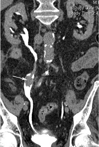 Figure 9.3 C (See color image.) |
Findings: Axial unenhanced (A) and excretory (B) phase images from a CT urogram demonstrate a 2 × 2-cm enhancing soft tissue mass (arrows) in the mid right ureter. The mass measured 29 HU on the unenhanced images and 59 HU on the excretory phase images. Contrast material is present along the periphery of the ureter. Curved planar reformatted image (C) illustrates that the mass (arrow) is pedunculated and involves a 6-cm segment of right ureter.
Diagnosis: Papillary urothelial carcinoma
Discussion: Upper urinary tract transitional cell carcinoma (TCC) occurs in 2% to 4% of patients with bladder cancer. Approximately one-third to three-fourths of patients with upper tract TCC have bladder tumors at some time. Several reasons have been proposed to explain the low incidence of subsequent upper tract tumors in patients with bladder cancer, including the larger surface area of the bladder, downstream seeding of tumor cells, and the fact that the bladder mucosa is exposed to carcinogens for a longer duration because it acts as a reservoir. Exposure to a variety of noxious stimuli, such as cigarette smoke, phenacetin, dyes, and cyclophosphamide, is associated with an increased risk of development of urothelial metaplasia and neoplasia. TCCs of the upper urinary tract are divided into papillary (≥85%) and nonpapillary (the remainder) types. TCC occurs typically in patients 60 years or older. Males are affected more than females, and most present with hematuria. Dysuria and frequency occur more frequently with ureteral tumors. Upper tract tumors are frequently found during the workup of bladder cancers. TCC should be differentiated from benign lesions such as fibroepithelial polyps (see Case 9-17) and blood clots (see Case 9-19). (Case courtesy of Lisa Zorn, MD, and Stuart G. Silverman, MD, Brigham and Women’s Hospital, Boston, MA.)
Case 9-4
History: 56-year-old female with gross hematuria status post radical hysterectomy and radiotherapy for cervical carcinoma 9 years ago.
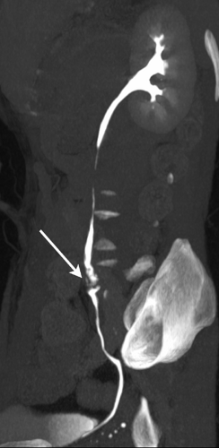
Findings: Axial excretory phase image (A) obtained during CT urography shows a polypoid soft tissue mass (arrow) in the left ureter. Coronal maximum intensity projection image (B) and sagittal thick-slab maximum intensity projection image (C) also show the lesion (arrows). Thin internal streaks of excreted contrast material extend into the mass. The remainder of the ureter is normal.
Diagnosis: Transitional cell carcinoma of the ureter
Discussion: This case illustrates another example of a transitional cell carcinoma (TCC) presenting as a polypoid soft tissue mass (see Cases 9-1 and 9-3). In this case, the papillary nature of the mass can be suspected since some of the excreted contrast material extends into the interstices of the mass. Ureteroscopy and biopsy were performed for histological confirmation of the diagnosis. CT urography was helpful in identifying the tumor, delineating its extent, and excluding other urothelial lesions.
Case 9-5
History: 77-year-old female with gross hematuria.
Findings: Axial image from the excretory phase of a CT urogram, obtained at the level of the kidneys (A), shows a hydronephrotic upper moiety of a duplex left renal collecting system. There is also a soft tissue mass (arrow) in the proximal ureter of the upper moiety. Axial image obtained at a more caudal location (B) demonstrates abnormal soft tissue (arrow) medial to the lower pole moiety renal collecting system. Low-attenuation areas along the lateral aspect of the kidney represent the inferior extent of the dilated upper pole renal collecting system. Coronal maximum intensity projection image (C) displays the dilated upper pole moiety renal collecting system and the soft tissue tumor mass (arrow) involving the upper pole moiety ureter.
Diagnosis: Transitional cell carcinoma in an upper pole ureter of a duplex collecting system
Discussion: In the past, patients with hematuria were frequently assessed with intravenous urography and/or ultrasound. When these studies did not show a cause for hematuria, conventional CT or MRI was performed. CT urography alone is often sufficient in patients with hematuria. In this patient, a large infiltrative transitional cell carcinoma (TCC) was both diagnosed and staged with CT urography. In addition to the urothelial mass, enlarged para-aortic lymph nodes and pulmonary metastases were identified.
Case 9-6
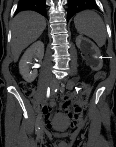
History: 80-year-old male with carcinoma of the colon and hydronephrosis. Retrograde pyelography could not be performed due to inability to pass the cystoscope into the bladder secondary to hypospadias.
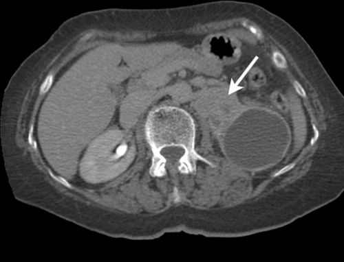
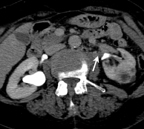
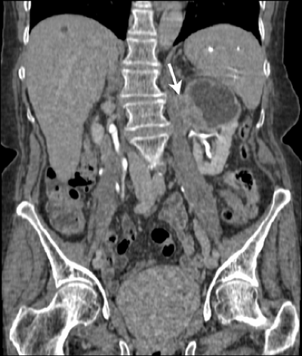
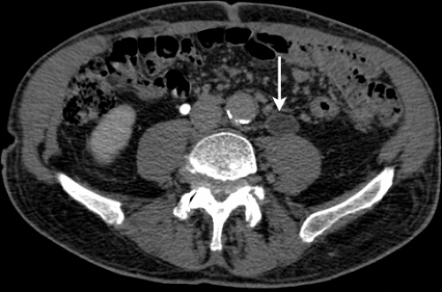
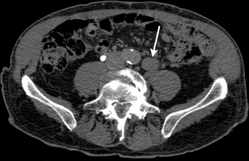
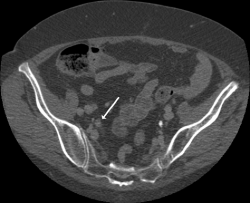
Findings: Coronal image (A) from the excretory phase of a CT urogram shows left-sided hydronephrosis with atrophy of the renal parenchyma. At 15 minutes after intravenous contrast material injection, there is only a tiny amount of excreted contrast in the left renal collecting system (arrow). A soft tissue density mass (arrowhead) in the dilated mid left ureter is detected in part because it is demarcated by the natural contrast between the urine and the mass. Axial excretory phase image just proximal to the mass (B) demonstrates marked dilatation of the unopacified mid left ureter (arrow). Axial phase excretory phase image obtained slightly more caudally (C) shows the large obstructing ureteral mass (arrow).
Diagnosis: Transitional cell carcinoma of the ureter
Discussion: With IVU, obstructing transitional cell carcinoma (TCC) may be difficult to diagnose. In the setting of high-grade obstruction, excretion is often delayed markedly. IVU is frequently nondiagnostic because the ureter is not opacified and therefore not visualized. With CT urography, when ureteral obstruction is present, urothelial tumors are usually detected even though the urinary tract may not be opacified at the time that the excretory phase images are obtained. The neoplasm is visible because its attenuation is higher than the adjacent unopacified urine. When the urinary tract is not opacified, the entire intrarenal collecting system and ureter should be searched carefully for portions that are of soft tissue attenuation. Urothelial masses may also be detected if early nephrographic phase images include the ureters. TCC typically enhances after contrast material administration (see Case 9-1). Therefore, the attenuation will be even higher and contrast between the enhancing tumor and the unopacified urine even more pronounced. This case shows that CT urography can be used to visualize the dilated unopacified ureter, the urothelium, and the tumor, and that CT urography can be used to diagnose urothelial neoplasms even when IVU is nondiagnostic. Furthermore, in this case, retrograde pyelography was technically not possible.
Case 9-7
History: 79-year-old female with gross hematuria for 2 months.
Findings: Axial image (A) from the excretory phase of a CT urogram shows that the lower right ureter is enlarged slightly but unopacified (arrow). Comparison can be made with the opacified normal caliber lower left ureter. Axial image obtained at a slightly more caudal level (B) demonstrates only minimal right ureteral opacification (arrow). A discrete filling defect can be seen in the right ureter at this level on a fluoroscopic spot film obtained during retrograde pyelography (C).
Diagnosis: Transitional cell carcinoma of the ureter
Discussion: Detection of intrarenal collecting system and ureteral neoplasms with CT urography is frequently dependent on differences in attenuation between the tumor and adjacent high-attenuation opacified urine. However, the ureters may not be fully opacified when excretory phase images are obtained, either as a result of obstruction (which produces delayed excretion) or due to ureteral peristalsis in
an unobstructed system. When obstruction is responsible for nonopacification, renal collecting system and ureteral tumors can often be detected because there is resulting hydronephrosis and hydroureter. In these instances, the soft tissue-attenuating tumor is outlined by the water-attenuating unopacified urine in the distended urinary tract. However, if the collecting system and ureter are not opacified, and not dilated, detection of small neoplasms can be difficult. In this case, only the slight dilatation of the right ureter suggests that an abnormality is present. This case demonstrates that if the ureter is not completely opacified during CT urography, it should be followed carefully to look for any focal dilatation. In some cases, additional images or retrograde pyelography may be needed. In this patient, a polypoid right ureteral tumor was removed subsequently at ureteroscopy.
an unobstructed system. When obstruction is responsible for nonopacification, renal collecting system and ureteral tumors can often be detected because there is resulting hydronephrosis and hydroureter. In these instances, the soft tissue-attenuating tumor is outlined by the water-attenuating unopacified urine in the distended urinary tract. However, if the collecting system and ureter are not opacified, and not dilated, detection of small neoplasms can be difficult. In this case, only the slight dilatation of the right ureter suggests that an abnormality is present. This case demonstrates that if the ureter is not completely opacified during CT urography, it should be followed carefully to look for any focal dilatation. In some cases, additional images or retrograde pyelography may be needed. In this patient, a polypoid right ureteral tumor was removed subsequently at ureteroscopy.
Case 9-8
History: 77-year-old female with gross hematuria and mild right-sided hydronephrosis detected with ultrasound.
Findings: Axial excretory phase obtained during a CT urogram (A) shows an irregular soft tissue mass (arrow) in the anterior aspect of the proximal right ureter. Coronal image (B) shows that the mass (arrow) involves a several-centimeter-long segment of ureter, and that the ureteral lumen engulfed by the mass is irregular. Retrograde pyelogram (C) also shows a mass with a stippled surface in the proximal right ureter and hydronephrosis.
Diagnosis: Transitional cell carcinoma of the ureter
Discussion: CT urography can be used to demonstrate the surface of sessile transitional cell carcinoma (TCC) almost as well as retrograde pyelography. The interstices of the surface of sessile, papillary TCC fill with contrast material. The spatial resolution of CT urography using multidetector CT scanners is adequate enough to display these subtle changes. Unlike retrograde pyelography, CT urography can also be used to show the extent to which the ureteral wall is thickened and the extent to which the periureteral fat is involved, if at all. CT urography also allows the remainder of the urothelium to be searched for synchronous tumors. This case was treated with nephroureterectomy. The extent of tumor noted at surgery correlated well with the findings at CT urography.
Case 9-9
History: 72-year-old female with pelvic pain and right-sided hydronephrosis detected with ultrasound.

