Fig. 6.1
Troubleshooting for a blurry image. With permission from Intuitive Surgical Inc., Sunnyvale, CA
With its high-definition, three-dimensional (3D) optics, which requires the use of a binocular endoscope and camera, the da Vinci Surgical System can experience unique difficulties with image acquisition that result in unclear or “foggy” images obtained from one side of the system (right or left). This problem can arise at one of several points along the vision system: the endoscope lens, the camera, or the cable connecting the camera to the Vision Cart. Rapidly diagnosing the source of such a problem requires a dedicated, experienced robotic surgical team within the operating room and allows a robotic surgical case to begin or proceed with little interruption.
In order to troubleshoot a problem resulting in a unilateral unclear image, one must first determine which side (right or left) is having image difficulty as the camera is currently assembled. To determine this, toggle between the right- and left-side images displayed on the Vision Cart touchscreen, as this screen displays only a two-dimensional (2D) image acquired from one side of the system. This is done by accessing the menu in the lower left corner of the Vision Cart touchscreen and toggling between the right and left channels. Once it is determined that the image difficulty or “fogginess” is limited to one particular side, disconnect the endoscope from the camera, and rotate it by 180° prior to reconnecting it to the camera head. If this maneuver results in a switch of the image defect from the right to left channel (or vice versa) on the Vision Cart touchscreen, then the problem is most likely related to the endoscope itself. The endoscope should be removed from the camera and its lenses cleaned properly prior to reconnecting to the camera head. If simple cleaning alone fails to improve the image, then the endoscope may require repair or replacement, and an alternate endoscope should be obtained and used for the remainder of the robotic case [1, 2].
If rotating the endoscope 180° does not result in a switch of the image defect from one side to the other, then other sources for the unilateral image problem must be sought. At this point, the issue is likely related to either the camera itself or the cable connecting the camera to the Vision Cart. While the camera itself should be cleaned and reconnected to the endoscope first, either the camera or cable may need to be replaced with an alternate camera or cable to resolve the issue. This can be performed rapidly by the robotic surgical team in the operating room, as problems with the camera or cable are frequently first noticed during the preoperative setup of the surgical case, prior to draping.
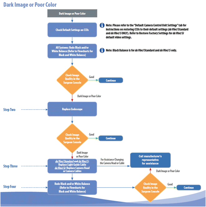
6.2.2 Problem: Unbalanced Color or Image Brightness (Fig. 6.2)

Fig. 6.2
Troubleshooting for a dark or poor color image. With permission from Intuitive Surgical Inc., Sunnyvale, CA
At the start of a robotic surgical case, the surgeon seated at the Surgeon Console may note image differences between the right and left eye despite having clarity of the image itself. This is frequently perceived as unbalanced levels of brightness between the two sides or may be perceived as discoloration such as a red- or blue-tinged hue to the image on one particular side. This usually arises due to a failure to perform white balancing of the camera.
Resolution of this issue requires repeat white balancing of the camera. The camera should be removed from the Patient Cart, if attached, and a clean, white sheet of paper (not a gauze or laparotomy pad) should be placed 5–10 cm from the end of the endoscope. The vision setup button on the camera should then be pressed allowing the vision setup menu to be displayed on the Vision Cart touchscreen, and the White Balance option should be selected. Once white balancing is completed, a notation of “Successful” will be displayed on the touchscreen. Note that successful white balancing should only be performed with the Illuminator set to 100 %; otherwise, persistent issues with image color and balance may occur [1, 2].
6.2.3 Problem: Double Vision (Fig. 6.3)

Fig. 6.3
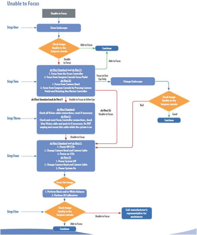
Troubleshooting when unable to focus. With permission from Intuitive Surgical Inc., Sunnyvale, CA
Similar to problems with white balancing, a surgeon seated at the Surgeon Console at the beginning of a robotic surgical case may alternatively experience a sensation of double vision. The image presented to the surgeon appears to have two separate or somewhat overlapping images of objects within the visual field. This problem occurs due to incomplete or inaccurate three-dimensional (3D) calibration of the camera. Difficulties with 3D calibration result in incomplete alignment of the images produced by the separate right and left lenses of the camera. To resolve this issue, the camera should be removed from the Patient Cart, and the alignment target should be attached to the end of the endoscope. The vision setup button on the camera should then be pressed allowing the vision setup menu to be displayed on the Vision Cart touchscreen, and the Auto 3D Calibration option should be selected. At this point, separate magenta and green crosses will appear on the Vision Cart touchscreen, and the computer of the da Vinci Surgical System will automatically focus the images and align the two crosses. Once calibration is complete, an onscreen prompt will ask if calibration appears correct. If the image is not correct, the calibration process can be repeated. It is also important to note that if an angled endoscope is employed, 3D calibration will have to be repeated in both the angled up and angled down positions [1, 2].
Stay updated, free articles. Join our Telegram channel

Full access? Get Clinical Tree


