Fig. 12.1
Fluoroscopic image of balloon dilation of distal right ureter
Dilation may also be carried out with small caliber (10–14Fr) ureteral dilators, with a 10Fr dual lumen catheter (which are often 6Fr at the tip), with 8/10Fr coaxial dilators and with the 10Fr tip of the Amplatz Renal fasical dilator set. The internal obturator of a ureteral access sheath can also serve as a ureteral dilator if initial attempts at placement of the combined ureteral access sheath-internal dilator meet resistance. Ureteral dilation is often best accomplished over a stiffer wire (i.e. Amplatz Superstiff wire) as it helps prevent buckling of the access sheath. Copious lubrication should be used with any dilator to facilitate ureteral passage, and caution must be used upon passage of any ureteral dilator as aggressive advancement may result in ureteral injury.
In addition, performing semirigid ureteroscopy prior to any planned flexible ureteroscopy can also serve to dilate the ureter and ease passage of subsequent instruments. Despite these tactics, if the ureter remains too narrow or does not easily accommodate the ureteroscope or ureteral access sheath, then placement of an indwelling ureteral stent for at least 1 week can allow for passive ureteral dilation. Thereafter, subsequent attempts to access the ureter are frequently successful.
Furthermore, not all ureteroscopes are the same. Size of the flexible ureteroscope is an important parameter with regard to access. While digital flexible ureteroscopes have recently seen increased utilization due to clear improvements in visualization, they are often greater in diameter than analog ones. Despite a difference of less than 2Fr in diameter, this difference was found to be enough to require a significantly greater proportion of ureteral access sheath utilization [19].
In cases of tortuous ureters, the combination of an angled wire and angled ureteral catheter can negotiate almost any series of ureteral twists and turns. The use of different caliber wires (0.038″, 0.025″, and 0.018″) can be helpful as well. In addition, if initial attempts to advance a non-hydrophylic wire (Bentson wire) are met with resistance, then changing to a hydrophilic wire (i.e., Glidewire or Sensor wire) can ease wire placement. Once the wire has been successfully placed into the renal pelvis, exchanging it for a stiffer wire (i.e. Amplatz Superstiff) through a ureteral catheter will better straighten out the ureter and simplify subsequent ureteroscopy attempts.
Ectopic ureteral orifice location, such as with cross-trigonal ureteral reimplants and renal transplant ureters, can pose a distinct challenge to adequately access for successful ureteroscopy. Gentle balloon dilation with subsequent placement of a stiff guidewire can be helpful and has been reported with success [20].
Poor Visualization
Several parameters may be adjusted to optimize visualization during ureteroscopy. The first of which is proper focus ahead of placement of the ureteroscope into the ureter. In non-digital systems, there are two items to focus; the scope itself and the camera, whereas in digital scopes the digital chip is located within the scope and hence focus is singular.
Another factor critical to good visualization during ureteroscopy is adequate flow of irrigant. The small working channels of most ureteroscopes and the long distance over which irrigation travels from source to ureteroscope creates significant resistance to flow necessitating some form or pressurized irrigation. A variety of instruments are available to assist with this task.
Pressure bags applied to the irrigation source are one option and are reusable, and hence cost-effective. Pressure can be modified by opening or closing the inflow valve near the ureteroscope. Hand pumps controlled either by the surgeon or by an assistant (i.e., Single-Action-Pump or Pathfinder) are alternatives. These systems are single-use and pressurize only a small volume of irrigant but do so at surgeon desired intervals and allow for temporary generation of higher pressures than those created with pressure bags. Foot-pedal activated pumps can also be utilized, but can be cumbersome if also using a lithotripter foot pedal. With any of these systems, care must be taken to avoid retropulsion of the stone.
Ensure there is no kinking along the irrigation tubing and eliminate air bubble introduction within the line to avoid air locks. If visualization is poor with an instrument through the working channel, consider using a smaller diameter instrument. In most instances in which a laser fiber is indicated a smaller size fiber between 200 and 365 μm will be sufficient. Similarly, many of the new stone baskets are of smaller caliber (1.5–1.9Fr) and provide adequate basket size, grasping strength and durability.
Sometimes during ureteroscopy a significant degree of light glare and artifact is present and can obscure the view. This can be noticed in particular with some of the contemporary digital and high definition optical systems. Decreasing the light source intensity or brightness helps minimize glare by decreasing the light reflectivity from the urothelium.
Patient Positioning
The ureter does not follow a straight course from kidney to bladder, and a couple of positioning strategies can facilitate ureteroscopy. The lithotomy position is preferred for routine ureteroscopy as the surgeon has ease access to the urethra and can ergonomically pass instruments along the same axis as the general course of the ureter. Reducing the degree of hip flexion and slightly abducting the hip ipsilateral to the side of the ureteral stone can straighten the course of the ureter as it travels across the psoas muscle and ease ureteroscope passage in this area [21].
Always be cognizant of frozen lower extremity joints, as it may not be possible to safely position the patient in such a way as to easily accommodate rigid or semirigid instruments. In these cases, flexible cystoscopes and ureteroscopes are necessary. In addition, flexible instruments may be necessary in patients with considerable pelvic scar tissue, such as those with a history of prior pelvic surgery, pelvic radiation, and pelvic malignancies.
Stone Basketing
One of the most common causes for iatrogenic ureteral injury during stone surgery occurs at the time of stone basketing, and usually results when attempting to extract a stone or fragment that is too large. Myriad baskets exist for stone retrieval, and as a general rule tipless baskets should be used, rather than baskets with tips, as they are less likely to directly perforate the ureter. When extracting a stone or fragment it is important to keep the basketed stone close to the tip of the ureteroscope and in direct vision throughout. If too large a gap exists between the basketed stone and ureteroscope tip, the ureteral wall can catch in this space and become injured or intussuscepted during stone removal.
Minimal resistance should be encountered during stone basket extraction, and the basket should never be forced or yanked. When an area of resistance is encountered, adjusting the stone’s orientation within the ureter or basket should be attempted. This is accomplished by opening the basket approximately half way and gently manipulating the basket proximally and distally, side-to-side, or rotationally. This allows the long axis of the stone to naturally realign with the long axis of the ureter. Thereafter, the basket can again be fully closed to secured the stone in the most favorable orientation and reattempt basket extraction.
Occasionally it becomes difficult or impossible to disengage a basket from a large stone fragment or fragments. In these instances, a few tactics can be tried. If there is room to pass a small laser fiber (i.e. 200 μm) alongside the basket, a small amount of energy applied directly to the stone is often sufficient to fragment the stone further and allow for basket disengagement. Alternatively, the laser can be used to cut one of the prongs of the basket, but one must ensure no basket pieces are left behind as these can serve as a nidus for further stone nucleation and growth.
Another option is to disassemble the basket at its handle, as some baskets have a screw mechanism that will free the handle from the rest of the basket. As a next step, withdraw the ureteroscope leaving the stone and basket in place within the ureter. Then reintroduce the ureteroscope alongside the basket and fragment the stone with laser. The basket can then be removed and a new one reinserted through the working channel of the ureteroscope.
Impacted Ureteral Stones
Impacted ureteral stone often present a challenge. They can be associated with an intense inflammatory reaction and ureteral edema around the impaction site which can obscure vision and interfere with safely positioning the laser or other lithotripsy fiber onto the stone. Once a guidewire has been safely advanced past the stone to the pelvicalyceal system, gentle attempts to maneuver the stone proximally into the often more dilated and non-inflammed ureter can be attempted. This provides additional space for irrigation and safe stone lithotripsy. Again, this should be done carefully and with only minimal force.
If you cannot pass the stone with a wire, options must be carefully assessed. In experience hands, an attempt to perform ureteroscopy to the stone and laser it without a safety wire in placed can be considered. With this approach, judicious laser use is recommended until the stone disimpacts, after which reattempts at wire placement should ensue. However, ureteroscopy and laser lithotripsy in the absence of a safety wire can be associated with significant complications, and have a nephrostomy tube or antegrade ureteral stent placed are reasonable alternatives.
If this stone does not easily move proximally, then judicious lithotripsy of the distal most aspect of the impacted stone is recommended. Dusting the distal edge of impacted ureteral calculi should minimizing the creation of large fragments and can be accomplished by using high frequency and low energy settings of the laser generator. Sometimes rotating the ureteroscope clockwise or counterclockwise along its long axis will change the position at which the lithotripter fiber contacts the stone and can be particularly helpful if a portion of the stone is covered by edematous urothelium. Then once the stone becomes disimpacted, it can be manipulated proximally to the more open and dilated ureter.
Preventing Proximal Stone Migration
The use of anti-retropulsion devices within the ureter can help reduce the proximal migration of stones and improve stone free rates. The Stone Cone (Boston Scientific, Natick, MA) and the NTrap (Cook Medical, Bloomington, IN) are two such devices, however each is attached to its own additional guidewire, which narrows the working space within the ureter, and comes with an additional disposable instrument cost.
To avoid the need for an additional wire, thermoactive liquid gels have been utilized and are deposited just proximal to the ureteral stone thereby creating a temporary barrier to proximal stone migration (BackStop liquid gel, Boston Scientific, Natick, MA). Surgical lubrication has also been described as a suitable and cheap agent for preventing proximal migration of fragments [22].
When the Laser Fails
It is reasonable to have a backup plan should the laser fail. If no substitute laser is available, consider pneumatic lithotripsy as an alternative (i.e. EMS Swiss Lithoclast or Olympus LUS 2). Though laser has a better safety profile, intraureteral pneumatic lithotripsy has been well documented as a viable technique with acceptably low complication rates [23]. Given the rigidity of the pneumatic probes, this is only feasible with a semirigid ureteroscope with a straight-entry working channel and offset lens. Keep in mind, ureteral stent insertion and returning for completion ureteroscopy at a later date is sometimes the most prudent option.
Medical Expulsive Therapy
Alpha blockers have been shown to increase SFR after ureteroscopy for ureteral stones [24]. In addition, alpha blockers help alleviate ureteral stent symptoms, pain, sexual life symptoms and overall quality of life following ureteroscopy [25]. As such if there are no contraindications, alpha blockers are reasonable to prescribe following ureteroscopy.
Semirigid Ureteroscopy
Semirigid ureteroscopy is the preferred treatment modality for most distal ureteral stones. In some patients, semirigid ureteroscopes can be safely advanced into the proximal ureter and renal pelvis. Compared to flexible ureteroscopes, semirigid ureteroscopes are more steady and maneuverable within the distal ureter and generally offers better optical quality than most flexible ureteroscopes. Furthermore, semirigid ureteroscopes usually have larger working channels allowing for improved irrigation even when using larger instruments and multiple instruments.
When performing semirigid ureteroscopy, the intial step is to place a safety wire. The safety wire allows for ureteral stenting should any difficulty arise during ureteroscopy. The safety wire also assists with ureteral access. Floppy tip, hydrophilic wires are recommended as the initial wire most likely to advance easily and least likely to perforate the ureter when navigating around a ureteral stone. Once positioned in the renal pelvis, these wires can be exchanged to a stiffer wire (i.e. Amplatz SuperStiff) through a small ureteral catheter (as previously discussed, a stiffer wire can better straighten the ureter).
Once the safety wire is placed, access into the ureter can be achieved through a number of different techniques. Passing the ureteroscope directly under the wire toward the ureteral orifice helps further tent the orifice open allowing the scope to pass under the wire and into the ureter (Fig. 12.2). Very small controlled movements should be used with the ureteroscope as readjusting even a few millimeters can be the difference between entering and missing the ureter.
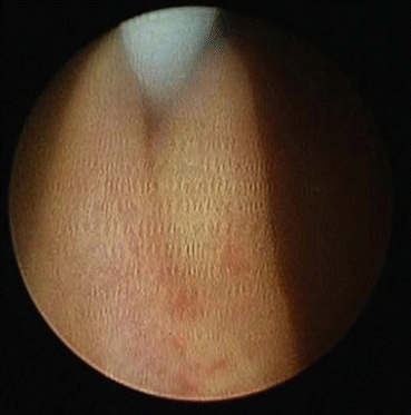
Fig. 12.2
Passing semirigid ureteroscope directly underneath guidewire that is tenting open ureter
If the ureteral orifice cannot be entered by passing the ureteroscope underneath the wire, try riding on top of the wire and using the scope to push down on the wire just as it enters the ureter. This can allow entry into the ureter when advancing below the wire fails.
If these techniques are unsuccessful, using a two-wire, or “railroad,” technique should be tried next (Fig. 12.3). Once the safety wire is in place, pass a separate wire through the working channel of the ureteroscope and intubate the ureteral orifice with the new wire. Next rotate the semirigid ureteroscope such that the beak of the scope is aligned between the two wires and advance the scope through the ureteral orifice in this manner. A stone basket, rather than a second guidewire, may be used to perform the railroad technique, but care should be taken as the stone baskets are more likely to kink and have a greater tendency to perforate the urothelium.
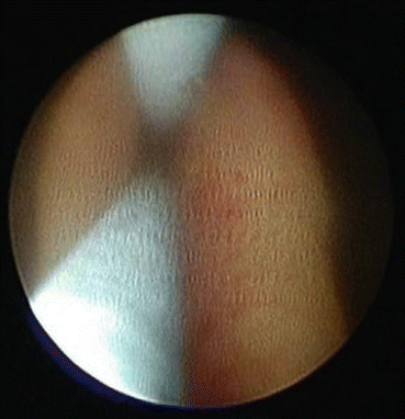
Fig. 12.3
“Railroad” technique for accessing the ureter, using one guide wire to tent open the ureter and a second guide wire passed through the working channel of the ureteroscope
Once in the ureter, the wire through the working channel of the ureteroscope can be used in a similar fashion to negotiate past any additional narrow areas. Alternatively, partially opening a stone basket just in front of the tip of the ureteroscope often straightens this segment of ureter and allows for segmental advancement of ureteroscope. Another option is to softly manipulate the safety wire in or out by approximately 1 cm. This can further straighten out areas of ureteral kinking and reduce areas of ureteral redundancy. Contemporary semirigid ureteroscopes have a beveled beak and this feature can be exploited by rotating the handle of the ureteroscope clockwise or counterclockwise. In this manner, the beak of the ureteroscope can be aligned better to the contour of the ureter improving the ability to safely navigate some of the narrower portions of the ureter such as the pelvic brim.
Related posts:
 Radiation Exposure to the Patient and the Urologist
Shock Wave Lithotripsy for the Treatment of Ureteral Stones
Selecting the Appropriate Treatment Modality for Ureteral Calculi
Complications in the Treatment of Ureteral Stones: Prevention and Management
Radiation Exposure to the Patient and the Urologist
Shock Wave Lithotripsy for the Treatment of Ureteral Stones
Selecting the Appropriate Treatment Modality for Ureteral Calculi
Complications in the Treatment of Ureteral Stones: Prevention and Management
 Semirigid Ureteroscopy for Ureteral Calculi
 Adjunctive Equipment for Ureteral Stone Management
Stay updated, free articles. Join our Telegram channel

Full access? Get Clinical Tree


