Thoracoscopic Lung Resections

Chapter 2 Thoracoscopic Lung Resections


imageThe videos associated with this chapter are listed in the Video Contents and can be found on the accompanying DVDs and on Expertconsult.com.


Minimally invasive thoracic surgery was introduced almost 100 years ago, when Jacobeus first inserted a cystoscope into the pleural space. Indications for thoracoscopy at that time consisted of drainage of pleural effusion or tuberculous empyema. Another 80 years were to pass, however, before thoracic surgeons embraced video-assisted thoracic surgery (VATS) as their standard approach to intrathoracic disorders. Increasing numbers of pulmonary, esophageal, and mediastinal resections are performed by VATS, and most experts would consider this the optimal approach to the pleural space. As in general surgery, many procedures in thoracic surgery are labeled “minimally invasive” but in actuality are done through incisions larger than the typical trocar, using retractors to access the chest cavity. For the purposes of this chapter, a VATS lobectomy will be defined as one having no chest retractors placed and including individual ligation of the hilar structures.


Operative indications


In patients undergoing thoracic surgical intervention, lung cancer is the most common indication for lobectomy. Other options for surgical resection include bilobectomy, pneumonectomy, and sleeve lobectomies. With the increased use of low-dose computed tomography (CT) scans for lung cancer screening, more subcentimeter lung cancers are being detected. This has stimulated discussion regarding the option of performing an anatomic segmentectomy to conserve lung function, while still achieving an acceptable oncologic resection. A nonanatomic wedge resection can be performed in high-risk patients with severely limited pulmonary function but generally is deemed a suboptimal cancer operation.


It is imperative that all cases of lung tumors be discussed at a multidisciplinary thoracic oncology conference to determine which treatment options are applicable for a given patient. Although surgery remains the best treatment option for patients with early-stage lung cancer, not all patients choose surgery or will be deemed suitable surgical candidates. In these patient groups, radiation therapy with or without chemotherapy will be the main alternative therapy offered. Radiofrequency ablation (RFA) is a newer modality available to treat pulmonary tumors. This modality is more applicable to patients with peripheral tumors and no associated adenopathy. This is due in part to a few case reports of fatal massive hemoptysis occurring a few days after RFA of more centrally located tumors.


Other, less common indications for lobectomy include carcinoids, mucoepidermoid tumors, adenoid cystic tumors, and sarcomas. Lobectomy also may be necessary to resect pulmonary metastases from other primary sites; however, if resectable, pulmonary metastases usually are treated with wedge resections. A lobectomy may be required to manage benign lung diseases that result from an underlying inflammatory or infectious etiology, such as an aspergilloma. These patients often are immunosuppressed and require resection due to the development of massive hemoptysis, bronchopleural and other fistulas, and empyema. Because of the presence of severe comorbidities, however, lobectomy for infectious etiology may be associated with high morbidity and mortality. In these situations, the use of antibiotics and antifungals, coupled with the use of percutaneously placed catheters or stents, may be used to temporize the patient until definitive surgical intervention can be accomplished with lower risk.


Preoperative evaluation, testing, and preparation


Patients scheduled to undergo a lobectomy should undergo a complete preoperative evaluation. Essential information regarding the patient’s physiologic ability to safely undergo lobectomy will help risk-stratify the potential operative candidate. Pulmonary function testing should include a forced expiratory volume in 1 second (FEV1), diffusion capacity of carbon monoxide (DLCO), and arterial blood gas measurement. Patients determined to be marginal candidates based on the postoperative predicted values (e.g., FEV1 <800 to 1000 cc and/or DLCO <40% predicted) also may benefit from information provided by a quantitative perfusion scan or a cardiopulmonary exercise stress test, or both. Those patients still deemed at high risk after obtaining these tests may be better served by a sublobar resection or nonoperative therapy. Because cardiovascular disease may coexist in this patient population, additional cardiac evaluation also may be obtained, as indicated by the history and physical examination.


Patient positioning in the operating suite


A multitude of options are available for performing minimally invasive thoracic surgery. Although most complex procedures are performed using general anesthesia and a double-lumen endotracheal tube to achieve single-lung ventilation, simple diagnostic procedures can be performed using local anesthetics, with the patient awake and spontaneously breathing. Most of these will be done through a single port site, but additional instruments can be added if required.


Depending on the surgeon’s preference, patients undergoing a VATS lobectomy may have a thoracic epidural placed by the anesthesia pain service to manage postoperative pain. Other options include the use of local anesthesia, administered by injection or an indwelling catheter. This is usually combined with some type of patient-controlled analgesia. Once adequate general anesthesia is obtained, a double-lumen endotracheal tube or a bronchial blocker is placed to obtain selective single-lung ventilation. In some cases, mainstem bronchial placement of a single-lumen endotracheal tube can be used, but this typically is not ideal management of the airway. Bronchoscopy should be performed routinely in all patients before an anatomic lung resection, both to assess for endobronchial disease and to confirm placement of the tube. This can be done through a single-lumen tube before the placement of the double-lumen tube.


Monitoring devices are placed at the discretion of the anesthesiologist but usually include an arterial line and one or two peripheral large-bore intravenous catheters. Central venous catheters are not mandatory in all patients undergoing lobectomy but may be helpful in selected higher-risk patients. In patients deemed to be at a higher cardiac risk, transesophageal echocardiography also can be used for real-time cardiac monitoring. An indwelling bladder catheter should be placed after the induction of anesthesia and usually will remain in place as long as the epidural is present, but it can be removed earlier in patients with other pain management strategies. Because many lobectomy patients are operated on for cancer or have multiple comorbidities, deep venous thrombosis prophylaxis (e.g., heparin or lower extremity sequential compression devices) should be used.


A VATS lobectomy most commonly is performed with the patient in the lateral decubitus position. The position can be maintained with the use of a surgical bean bag or blankets. Placement of an axillary roll is mandatory. The head should be supported so that the cervical spine is in a neutral position. The upper arm may be supported with blankets or an arm holder. The lower arm and both legs need to be carefully cushioned to prevent peripheral nerve injury. Once the patient is securely positioned, a body warming device should be placed.


Positioning and placement of trocars


With the patient in the lateral decubitus position (Fig. 2-1), the initial port site is in the seventh or eighth intercostal space at the midaxillary line (Fig. 2-2). This port will be used for the camera in most cases; ideally, the port will be just above the level of the diaphragm. This corresponds to point A in Figure 2-2. In general, the greater the body mass index (BMI) of the patient, the higher the level of the diaphragm; this requires cephalad movement of the camera site so that the diaphragm does not impair thoracoscopic visualization. The diaphragmatic position can be determined by reviewing the preoperative chest radiograph. This port site should be created under direct visualization to avoid passing through the intercostal space and diaphragm simultaneously, which would result in intra-abdominal camera placement.
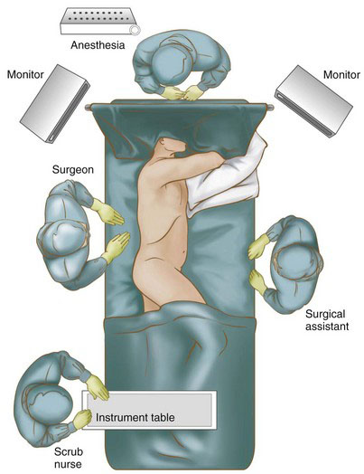


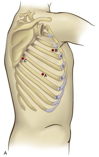
Before placing the camera port, digital examination of the pleural space should be performed to assess for the presence of adhesions or even pleural tumor implants. Many of these adhesions can be cleared by digital sweeping, but denser adhesions may need sharp dissection. Once there is an adequate space to insert the thoracoscope, the pleural space can undergo further evaluation. Because there is no need for insufflation, a simple reusable port typically is sufficient. A 30-degree scope provides excellent visualization of the upper mediastinum, the subcarinal area, the diaphragm, and the pericardium. Two additional port sites are placed under direct vision. The anterior port site (point B in Fig. 2-2) is placed in the fourth intercostal space when an upper lobectomy is planned, whereas the fifth intercostal space is used for a middle or lower lobectomy. The third (posterior) port is placed in the seventh intercostal space, anterior to the scapular edge (point C in Fig. 2-2). Unsuspected pleural metastases can be resected for biopsy if they are identified, and any adhesions are lysed at this time.


Operative technique


Right Upper Lobectomy


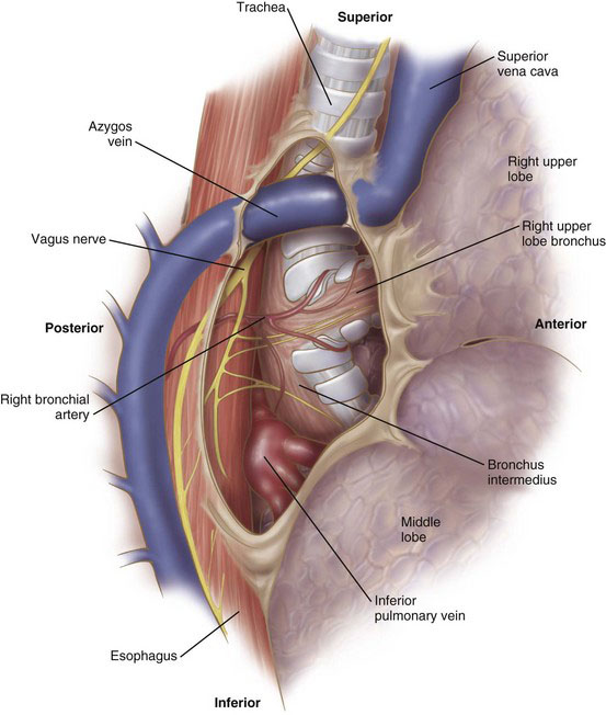
After placement of the ports, the lung is retracted anteriorly. The pleura along the posterior aspect of the hilum is opened with the electrocautery device. With gentle blunt dissection, the confluence of the right upper lobe bronchus and the bronchus intermedius is identified (Fig. 2-3). There usually is a lymph node located at this bifurcation. Clearing this area at the beginning of the operation will expedite completion of the fissure later in the procedure. The lung then is retracted posteriorly. The pleura on the anterior aspect of the hilum is opened with the cautery. The location of the phrenic nerve needs to be monitored at all times during the dissection of the anterior hilum. Clearing this portion of the pleura will identify the trunks of the superior pulmonary vein, which drain the right upper and right middle lobes. This dissection also will demarcate the fissure between the right upper lobe and right middle lobe. In some patients, a branch of the right middle lobe vein crosses the fissure and drains into the posterior segment vein. Whenever possible, this crossing branch should be spared, taking the upper lobe vein proximal to this branch.


The upper lobe vein is isolated by a combination of blunt and sharp dissection (Fig. 2-4). A large, blunt right-angle clamp can be used to clear the soft tissue behind the vein. This must be done carefully because the pulmonary artery is located immediately behind the vein. The vein then is divided with the vascular stapler. One option for positioning the stapler is to place a red rubber catheter on the stapling device to guide the blade behind the vein. Placement of the stapler blade behind the vein also can be facilitated with the use of a large right-angle clamp.
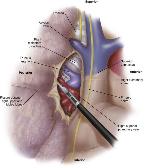


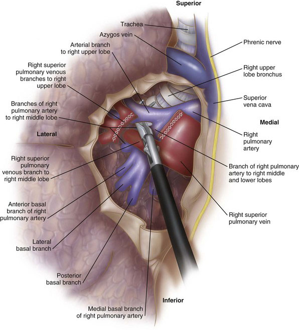
Division of the vein exposes the right pulmonary artery (Fig. 2-5). There usually is a single large arterial branch that supplies the upper lobe, although occasionally there will be two to three smaller branches. The right upper lobe pulmonary artery branch, once cleared, is divided with the stapling device. At this point there usually are two remaining structures to be divided: (1) the branch of the pulmonary artery supplying the posterior segment of the right upper lobe, and (2) the bronchus. It often is easier to clear the bronchus and divide this structure before stapling the posterior segment branch of the pulmonary artery (Fig. 2-6). Alternatively, depending on how complete the fissure is between the right upper and lower lobes, the remaining arterial branch can be divided first, followed by the bronchus. It is imperative to ensure correct placement of the stapler across the upper lobe bronchus by ventilating the middle and lower lobes before firing the stapler. After division of all hilar structures, the fissures can be completed with a laparoscopic stapler-cutter (e.g., Endo GIA, Covidien, Norwalk, Conn).


< div class='tao-gold-member'>

Stay updated, free articles. Join our Telegram channel

Jul 20, 2016 | Posted by in GASTOINESTINAL SURGERY | Comments Off on Thoracoscopic Lung Resections

Full access? Get Clinical Tree

Get Clinical Tree app for offline access