(1)
Department of Endoscopy, Fukuoka University Chikushi Hospital, Fukuoka, Chikushino, Japan
12.3 Explanation
12.5 Explanation
12.7 Explanation
12.8 VS Concordance
12.10 Explanation
12.12 Interpretation of M-NBI Findings Using the VS Classification System in Cases of Early Gastric Cancer
Summary
VS classification system for the differential diagnosis of cancer vs noncancer
V: regular/irregular/absent MV pattern
S: regular/irregular/absent MS pattern
Keywords
ClassificationEarly gastric cancerMagnifying endoscopyStomachVessel plus surface (VS)12.1 Principles of the VS Classification System
As described in Chap. 1 “Basic Principles for the Interpretation of Magnified Endoscopic (ME) Findings: Vessels (V) plus Surface (S) Classification System,” we should interpret ME findings by first assessing the mucosal subepithelial microvascular pattern (V) and mucosal microsurface pattern (S) separately and then integrate both findings using a uniform set of diagnostic criteria to arrive at a diagnosis. The reason is that V and S are in concordance (capillaries are observed in the intervening part: VS concordance) in the normal mucosa, and V and S dissociation (divergence) occurs as the state becomes more pathological, or the degree of atypia increases, or the disease stage progresses [1] (VS discordance).
More macroscopic and histological heterogeneity is seen in gastric cancers than any other cancers of the gastrointestinal tract, so production of a pattern classification of VS combinations would run into many, possibly hundreds, of combinations and likely be of little practical use. In general, findings of the V component alone are sufficient for magnified examination with WLI, and with M-NBI we can visualize a variety of microanatomical findings of the mucosal surface layers. In addition to the V findings, we now have the S component as a diagnostic marker for interpretation.
In this chapter, I will set out the markers and definitive features, as well as findings requiring interpretation, and the diagnostic criteria used in the VS classification system I use to make the differential diagnosis between cancer and noncancer.
12.2 V: Microvascular (MV) Pattern (Fig. 12.1a–c)
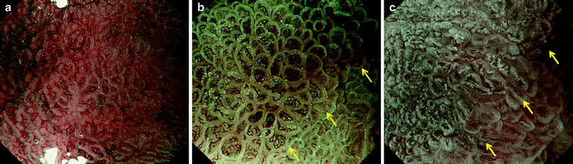
Fig. 12.1
V: microvascular pattern (a) Regular, (b) Irregular, (c) Absent (reprinted from [4] by permission of Endoscopy)
12.2.1 Summary
Microanatomical units and diagnostic markers
1.
Capillaries
2.
Collecting venules (CVs)
3.
Microvessels
12.3 Explanation
We analyze capillaries, collecting venules (CVs), and microvessel in terms of individual morphology, presence of heterogeneity, and overall distribution and arrangement. We classify the pattern as regular, irregular, or absent.
We can identify capillaries and venules, normal anatomical microvessels, in the normal gastric mucosa and in mucosa affected by chronic gastritis. New blood vessels, associated with pathology such as neoplasia or inflammation accompanying mucosal defects, cannot be definitely classified as capillaries or venules, however, so we use the overall term “microvessel,” encompassing capillaries, venules, and new blood vessels.
12.3.1 Regular MV Pattern
1.
The individual microvessels have a uniform morphology, with a regular closed- or open-looped shape.
2.
The individual microvessels do not show inequality in size or diameter.
3.
The microvessels show a regular arrangement and symmetrical distribution.
12.3.2 Irregular MV Pattern
1.
The individual microvessels have a nonuniform morphology, exhibiting a variety of shapes (closed looped, open looped, tortuous, branched, bizarre). Nonuniformity or heterogeneity of the microvessels, even within the same lesion, is characteristic. This nonuniformity of the individual microvessel can be expressed as “no two vessels with the same morphology can be seen within the lesion.”
2.
The individual microvessels show inequality in size or diameter.
3.
The microvessels show an irregular arrangement and asymmetrical distribution.
12.3.3 Absent MV Pattern
This refers to an inability to visualize the microvas-cular pattern due to reduced transparency of the mucosal surface layers, associated with the presence of white opaque substance (WOS) or something else adherent to the mucosa.
12.4 S: Microsurface (MS) Pattern
12.4.1 Summary
Microanatomical units and diagnostic markers
1.
Marginal crypt epithelium (MCE)
2.
Crypt openings (COs)
3.
Intervening part (IP)
4.
White opaque substance (WOS)
5.
Light blue crest (LBC): brush border
12.5 Explanation
First we assess whether each of the above features is present. When each individual feature is visualized, we analyze its morphological characteristics, evaluating heterogeneity, distribution, and arrangement. We classify the pattern as regular, irregular, or absent. In this section, I will refer mainly to the MCE (for explanations of the other features, please refer to the other chapters and presentations of actual cases).
It is worth noting that when the MCE is visualized, irrespective of whether it is classified as regular or irregular, linear, curved, or polygonal, the MCE tends to be histologically of the tubular type. When the MCE shows a circular or oval morphology, and the structure shows IPs isolated by MCE closed curves, the tubular histological type is common with a broad IP stroma, and the papillary type is common with a narrow IP stroma.
12.5.1 Regular MS Pattern
1.
Each MCE shows a regular linear, curved, or polygonal morphology or a regular circular or oval morphology. Within the lesion, the MCE morphologies are uniform (no heterogeneity is seen).
2.
The lengths and widths of the MCE are not uniform.
3.
The MCE shows a regular arrangement and symmetrical distribution.
12.5.2 Irregular MS Pattern
1.
Each MCE shows an irregular linear, curved, or polygonal morphology. Sometimes, it shows an irregular circular or oval morphology. Within the lesion, the MCE and IP structure does not show a repeating similar pattern, but is instead nonuniform and heterogeneous. The MCE paths and directionality are also nonuniform. Breaks and interruptions are seen in the MCE.
2.
The lengths and widths of the MCE are not uniform.
3.
The MCE shows an asymmetrical distribution and irregular arrangement.
12.5.3 Absent MS Pattern
This refers to an inability to visualize any features of the mucosal microsurface pattern, i.e., MCE, WOS, or LBC. This tends to occur in cases with little unevenness of the epithelial surface, associated with mucosal atrophy or small shallow irregular cancer glands.
12.6 Boundary Between Lesion and Surrounding Mucosa
12.6.1 Summary
Demarcation line (DL)
Definition: border between lesion and nonlesion areas, discernible through an abrupt change in the microvascular pattern (V) or mucosal microsurface pattern (S).
Intraepithelial microinvasion (IEMI)
1.
Subepithelial invasion
2.
Superficial invasion
12.7 Explanation
12.7.1 Demarcation Line (DL)
The presence of a DL is a useful diagnostic marker for the differential diagnosis between microcancers or 0 IIb cancers and chronic gastritis [2] and for tumor margin delineation [3] (see Chap. 6).
< div class='tao-gold-member'>
Only gold members can continue reading. Log In or Register to continue
Related posts:
 Basic Principles for the Interpretation of Magnified Endoscopic (ME) Findings: Vessels (V) Plus Surface (S) Classification System
The Proposed Vessels Plus Surface (VS) Classification System: Principles for Interpretation of Magnifying Endoscopy with Narrow-Band Imaging (M-NBI) Findings
Basic Principles for the Interpretation of Magnified Endoscopic (ME) Findings: Vessels (V) Plus Surface (S) Classification System
The Proposed Vessels Plus Surface (VS) Classification System: Principles for Interpretation of Magnifying Endoscopy with Narrow-Band Imaging (M-NBI) Findings
 Clinical Applications of Magnifying Endoscopy with Narrow-Band Imaging (M-NBI) of the Stomach
 Microanatomies as Visualized Using Magnifying Endoscopy with Narrow Band Imaging in the Stomach: Which Microanatomical Structures Can We Visualize in the Glandular Epithelium Using Narrow Band Imaging, and How Is This Achieved?
 Light Blue Crests (LBCs) and White Opaque Substance (WOS)
 Analysis and Interpretation of Magnifying Endoscopy with Narrow Band Imaging (M-NBI) Findings in Gastric Epithelial Tumors (Early Gastric Cancer and Adenoma) Stratified for Paris Classification of Macroscopic Appearance
Stay updated, free articles. Join our Telegram channel

Full access? Get Clinical Tree


