Fig. 13.1
Diagrammatic illustration of the Collarsling Musculature. ©2001 Corinne Sandone. Reproduced with permission of illustrator
Hill Repair Technique
Our standard positioning is low dorsal lithotomy with both arms out, the surgeon between the legs, the assistant on the patient’s left and the camera operator on the right, or using a robotic arm for camera fixation (Stryker Endoscopy; Wingman Scope Holder; 240-240-000). Prior to beginning the operation, the manometric equipment is prepared. The catheter is a water perfused, single use 8 channel esophageal manometry catheter, with four pressure ports at 0 cm from the tip, and subsequent ports at 5 cm intervals (Sierra Scientific Instruments, Catalog # 9012P1222). The manometric catheter is placed through a clear 48 Fr dilator (Cook Medical, Winston-Salem, NC), with the pressure port 10 cm beyond the tapered tip of the dilator, and taped together at the upper end. This arrangement is passed through the esophagus to 30 cm at the beginning of the case by the surgeon or an experienced anesthesiologist, taking care that the manometric catheter leads the way and does not fold under. The distal channel is connected to a transducer and anesthesia monitor at pulmonary artery catheter settings, or to a dedicated esophageal manometry system.
Five trocars are used for the operation. It is noteworthy that a 10–11 mm assistant port is placed just below the left costal margin in the mid-clavicular line, or more medially when the costal margin is narrow. This requires placement of the surgeons’ right-hand work port more inferiorly than would be typical for a Nissen repair, but the assistant port location facilitates management of the upper two untied repair sutures (Fig. 13.2). A sixth optional port for downward traction of lesser curvature fat may be added in the left lower quadrant to gain better exposure to the preaortic fascia. The left lobe of the liver is elevated with a 5 mm retractor [Nathanson: Cook medical, G26912, C-NLRS-1001 and G26913, C-NLRS-1002] and fixed to a self-retaining table-mounted system [Thompson Surgical, Cat # 90011B].


Fig. 13.2
Trocar placement for laparoscopic Hill repair
Dissection is performed with ultrasonic shears. Care is taken to dissect along the anterior aspect of the phrenoesophageal fat pad (Hill’s phrenoesophageal bundle) and bring it down with the dissection, keeping it attached to the GEJ. Following dissection the anterior and posterior fat pad/bundles are trimmed as necessary to eliminate hernia sac and redundancy, while avoiding the lesser curvature and the Vagus nerves. Short gastric vessels are not routinely taken, but it is essential to free the entire posterior fundus by opening the lesser sac from left gastric artery to GEJ. This is accomplished from the lesser curvature aspect under view from the angled laparoscope and through critical exposure provided by the assistant, who lifts the posterior phrenoesophageal tissue immediately posterior to the posterior Vagus nerve. The location of the celiac trunk should be roughly identified (though not dissected) and the preaortic fascia and overlying diaphragmatic muscle should be exposed down to this level. The preaortic fascia is a dense connective tissue layer that lies deep to the posterior fusion of the right and left crura and extends inferiorly to the celiac axis, where its inferior edge forms the median arcuate ligament.
Dissection is continued into the mediastinum to obtain adequate length of intra-abdominal esophagus. A Penrose drain is not used and would be in the way of subsequent suture placement. Following dissection the hiatus is closed posteriorly with 0-braided non-absorbable suture using an extracorporeal knot pusher or the Ti-Knot device [LSI Solutions; Ti-Knot, Catalog # TK-5]. For larger hernias, some of the repair may need to be completed anteriorly, since too much angulation of the esophagus may be created from excess posterior closure and posterior fixation of the GEJ to the preaortic fascia.
The Hill sutures are placed through the collar sling musculature of the GEJ. This lies immediately beneath the phrenoesophageal fat pad/ligament (Hill’s phrenoesophageal bundles), commencing just to the patient’s left/anterior of the anterior vagus nerve, extending over the angle of His, and ending just to the patient’s right/posterior of the posterior vagus nerve. Thus the vagus nerves are important landmarks and must be identified. The anterior vagus nerve is found under tension by pulling down on the lesser curvature tissue. The posterior vagus nerve is found by lifting the posterior fat pad upward/to the patient’s right, as it consistently lies in the groove created by this maneuver.
Following hiatal closure, four Hill sutures of multi-colored 48 inch 0-Ethibond (Ethicon Endosurgery multipack, Cat. #22970D8684) are placed through the tissue and left untied and clamped externally. The first two sutures are introduced through the surgeon’s right-hand working port, while the third and fourth are introduced through the assistant port in the left upper quadrant. This is the most critical part of the repair, and exact placement is important (Fig. 13.3). There are three separate and distinct bites of tissue with each suture, the first being placement through the anterior bundle/collar sling musculature from inferior to superior; the second being placement through the posterior bundle/collar sling musculature from superior to inferior; and the third being transverse placement through the inferior aspect of the preaortic fascia.


Fig. 13.3
Illustration of the Hill sutures. ©2001 Corinne Sandone. Reproduced with permission of illustrator
The first bite of the first suture is placed immediately to the patient’s left of the anterior vagus nerve. Grasping the bundle with the left hand and maneuvering the tissue over the needle facilitates this. This bite must go deeply enough to grab the collar sling musculature. It is usually necessary to trim away part of the anterior fat pad to expose this anatomy (Fig. 13.4).


Fig. 13.4
First Hill suture, first bite: intra-operative view
The second bite is placement through the posterior bundle. The assistant retracts the lesser curvature tissue between the vagus nerves anteriorly and to the left, exposing the posterior vagus nerve. The surgeon grasps and manipulates the posterior bundle with the left hand. Beginning just posterior to the posterior vagus, the suture is passed through the bundle in a superior to inferior direction, including the underlying collar sling musculature. To do this successfully the needle will frequently need to be nearly upside down at the time of entry. Again, exposure is usually facilitated by trimming excess adipose tissue overlying the anatomy (Fig. 13.5). Posterior bundle suture placement may be aided by preliminarily fixing the superior aspect of the posterior fundus to the left crus and left aspect of the preaortic fascia with 1 or 2 sutures.


Fig. 13.5
First Hill suture, second bite: intra-operative view
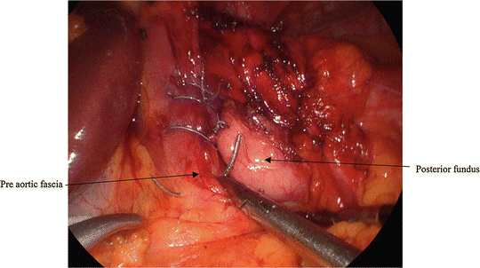
The third bite is transverse placement through the preaortic fascia. The assistant retracts the GEJ to the left and inferiorly for exposure. The suture is passed through the preaortic tissue inferiorly, immediately superior to the fatty tissue overlying the celiac axis. The location of this suture determines the final length of intra-abdominal esophagus, so it is important to be sufficiently inferior. The aorta lies 5–10 mm deep, and may be avoided by lifting the tissue upward with a grasper and driving the needle transversely from left to right, rather than too deeply (Fig. 13.6). Finally the suture is brought out again through the right-hand port, taking care to buttress it with a grasper where it exits the tissue to avoid excess tissue trauma, and is clamped to itself with a hemostat (Fig. 13.7).


Fig. 13.6
First Hill suture, third bite: intra-operative view

Fig. 13.7
First Hill suture, completed but not tied: intra-operative view
This same process is repeated with three more sutures, advancing each suture 2–3 mm further up the two bundles (e.g., in the direction of the angle of His) and the preaortic fascia. With excess advance the repair will be too snug, whereas with inadequate advance the repair may be too loose. The uppermost suture should enter the anterior bundle at approximately the left lateral border of the esophagus. It should enter the posterior bundle at its upper extent without going behind the esophagus. The third and fourth sutures are introduced and withdrawn through the assistant port. Colors should alternate to aid in preventing tangling. Care should be given to the angle of entry of the sutures through the ports, to also prevent crossing. Three-eighths inch Teflon pledgets may be added to either end of the sutures; it has been our standard to add pledgets to the second and fourth sutures.
Stay updated, free articles. Join our Telegram channel

Full access? Get Clinical Tree


