The “First Quarter:” The First Three Months After Transplantation


The “First Quarter:” The First Three Months After Transplantation


Alan Wilkinson


Kidney transplantation has reached a comforting state of success and maturity with well-defined stages that require special attention to particular aspects of patient care. These are usually defined as the early post-transplantation period, the first 3 months, and the later post-transplantation period, which encompasses all that happens for the remainder of the life of the transplant. The period discussed in this chapter ends at the beginning of the fourth month, although statistical analyses usually define the end of the first year as the onset of the late post-transplantation period. This division makes sense because many of the more acute events occur within the first 3 months, whereas from the end of the first year, patients are more stable. Rejection is most common in the early period, as are some of the more significant infections. Relatively high levels of immunosuppressants are used at this time, and the side effects are more marked than they are later on. The late period is discussed in the next chapter.

For most patients, this is one of the most exciting and yet anxious times in their life, and it is important to recognize this as we perform what for medical and transplant professionals have become quite routine tasks. The care of transplant recipients should ideally be a combined effort by medical and surgical teams that brings both their experience and expertise to the care of the patient. The best circumstance is for a single cooperative team to follow each patient together, making joint rounds and decisions about patient care. A relatively well-defined postoperative care pathway facilitates care, efficiency, and cost savings during this time of complex decision making. It is most useful to document all the events during the first admission in a manner that can easily be transmitted to the outpatient clinic. Some patients require readmission in this early period, and verbal and written communication between those caring for the patient in the clinic and the hospital is crucial to good care. The first 3 months are a time of relatively rapid change in management and also a time when surgical and immunologic complications are most common. It is sometimes tempting to focus most particularly on concerns related to graft function, immunosuppression, and rejection, but many of the medical issues discussed more completely in Chapter 10 are already present at this stage, and these too should be managed aggressively.


THE DAY OF THE OPERATION


Postoperative Assessment

Responsibility for the immediate postoperative care in the recovery room should be specifically designated to a member of either the surgical or medical team. This person must be familiar with the patient’s preoperative medical history and evaluation as well as details of the source of the donor kidney. Because urine output is an important measure of early graft recovery, it is
necessary to know whether the patient had any preoperative urine output. The assessment should include ensuring hemodynamic and respiratory stability and assessment of volume status. The operative record and early laboratory results should be reviewed, paying particular attention to confirming that immunosuppression was given as ordered. The surgeon should discuss any unusual aspects of the operation with those taking over the patient’s care. Most transplantation programs have a set of standard postoperative orders, and these should be completed. A sample set of orders is shown in Table 9.1. Institutions vary as to whether they transfer patients from the recovery room to an intensive care unit, a “step-down” unit, or a general ward. Most patients may be safely transferred directly to a general ward, but no matter where the patient goes, it is important that the staff there is experienced in the postoperative care of transplant recipients and familiar with the importance of measuring urine output, establishing volume replacement, and maintaining homodynamic stability. Strict control of blood glucose concentrations in all patients, not only diabetic patients, is important to facilitate recovery of the allograft and to promote healing.


TABLE 9.1 Suggested Postoperative Orders on Transfer of Kidney Transplant Recipient from the Recovery Room


Postoperative Nursing Orders*


1.


Vital signs checked every hour for 12 hours, then every 2 hours for 8 hours, then every 4 hours for stable patients


2.


Intake and output every hour for 24 hours, then every 4 hours


3.


Intravenous fluids per physician


4.


Daily weight


5.


Turn, cough, deep breathe every hour; encourage incentive spirometry every hour while awake


6.


Out of bed first postoperative; ambulate daily thereafter


7.


Head of bed at 30 degrees


8.


Dressing changes daily as needed


9.


Check dialysis access for function every 4 hours


10.


No blood pressure; venipuncture in extremity with fistula or shunt


11.


Foley catheter to bedside drainage, irrigate gently with 30 mL normal saline as needed for clots


12.


Catheter care every 8 hours


13.


Notify physician if urine output drops to less than 60 mL/h for 2 consecutive hours or greater than 300 mL/h for 4 hours or greater than 500 mL/h for 2 consecutive hours


14.


Notify physician if systolic blood pressure, >180 mm Hg or <110 mm Hg


15.


NPO until changed by surgical team


16.


Chest radiograph immediately postoperatively


Postoperative Laboratory Orders


1.


Complete blood count with platelets, electrolyte, creatinine, glucose, and blood urea nitrogen every 6 hours for 24 hours, then every morning


2.


Calcineurin inhibitor level each morning


3.


Chemistry panel including liver function tests; urine culture and sensitivity, twice weekly


With acknowledgement to Angela Phelps, RN and Elizabeth Hands, RN.


Fluid Replacement

There are probably almost as many protocols for intravenous fluid replacement as there are transplantation programs. The most important aspect is to ensure sufficient fluid replacement to maintain homodynamic stability and urine output, while avoiding making it necessary to dialyze patients who have received more fluid than the new allograft can adequately excrete. Patients are usually admitted for surgery somewhat above their “dry weight” and should be slightly hypervolemic at the end of surgery. If they are dialyzed preoperatively, it is best not to bring them to their dry weight, but to leave them about 1 kg over this. If all the urine passed is replaced, this overhydrated state will persist, and the protocol should allow for this by progressively reducing the volume replaced, provided urine output and blood pressure are adequate. It is useful to separate fluid replacement into “maintenance fluid” and “replacement fluid.” Maintenance is used to replace insensible loss, about 30 mL per hour. This is usually provided as 5% dextrose and water. Urine output and any nasogastric fluid losses are replaced by replacement fluid using half-normal saline because the urine sodium concentration in the early postoperative period is usually 60 to 80 mEq/L. One protocol is to replace all of the initial 200 mL, and then to replace 50% of any volume greater than 200 mL. If the patient is hypovolemic, a greater volume of the urine is replaced, and occasionally if the urine output is low, an additional 500 to 1000 mL of isotonic saline is given as a bolus. Where necessary, potassium, bicarbonate, or calcium replacement should be given in a separate infusion. Potassium replacement should be gradual in oliguric patients, and even some patients with good urine output may not excrete significant amounts of potassium. Serum electrolytes should be ordered at least every 6 hours and more frequently if there are clinical indications to do so such as a very high urine output, or if potassium is being replaced.


Hemodynamic Evaluation

Frequent hemodynamic evaluation is important because an adequate blood pressure and volume status is necessary to establish good graft function. The adequacy of urine output has to be assessed in the context of these two parameters. This may require the use of central venous pressure or a pulmonary pressure or pulmonary wedge pressure measurement. However, for most stable patients, this is not necessary, and a simple regular clinical assessment should be sufficient. Many patients are hypertensive after surgery. This may resolve spontaneously or with adequate pain control, and over-aggressive intervention may lead to the pressure falling too low, increasing the risk for acute tubular necrosis (ATN) and delayed graft function (DGF). In the acute setting, a mildly elevated blood pressure (systolic pressure < 180 mm Hg) is acceptable because blood flow to the newly transplanted organ is dependant on an adequate mean systemic blood pressure. Intravenous hypotensive agents such as labetalol or hydralazine can be used, or if the patient is able to take oral medications, one can use drugs such as clonidine and nifedipine. Unless these are used deliberately to affect calcineurin inhibitor (CNI) concentrations, both diltiazem and verapamil should be avoided because of their potential interactions that increase CNI concentrations.


Assessment and Management of Urine Output

It is a good idea to warn patients before surgery that their urine output after the operation may vary widely. Knowledge of the patient’s pretransplantation urine output is important because those receiving preemptive transplants, and even some dialysis patients, may have daily urine volumes of 1500 to 2000 mL, and this urine from the native kidneys must be accounted for in assessing post-transplantation urine output. In most patients who receive living donor
transplants, urine output is quite high, partly because of their relatively hypervolemic state, and also because of the diuretic effects of an elevated urea nitrogen and of mannitol if this is used intraoperatively. In patients who receive a deceased donor organ, and who had minimal or no urine output before surgery, urine volumes may range from complete anuria, through various degrees of oliguria, to polyuria with very high hourly urine volumes. The use of furosemide, dopamine, or fenoldopam infusions postoperatively is routine practice in some programs, although their benefit has been hard to prove.

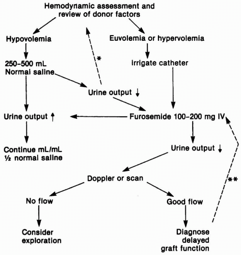
In anuric patients or those with a low urine output, a Doppler ultrasound may be ordered in the in the recovery room to assess the blood supply to the newly transplanted kidney (Fig. 9.1). The urgency with which the ultrasound is performed depends somewhat on whether oliguria is anticipated. Recipients of a living donor transplant are always anticipated to have a brisk urine output, and oliguria must be managed in an emergent fashion. If DGF is anticipated, it is reasonable to delay imaging studies. Imaging studies also serve to establish that there is no evidence of ureteric obstruction or a urine leak, although this is usually made obvious by the presence of urine flowing out through the perinephric drain, confirmed by an elevated creatinine concentration in that fluid. The routine use of a double-J stent from the urinary pelvis to the bladder makes ureteric obstruction uncommon. We also recommend an ultrasound for any patient who had a significant preoperative urine output. It is possible that even with the most carefully placed allografts, blood supply may be compromised as the various layers of the incision are closed, and one should not be dissuaded by an overconfident surgical opinion that the blood supply “is fine.” This should be
done concurrently with assessment of volume status and patency of the bladder catheter. If the blood supply is compromised, this is a surgical emergency, and the patient should be returned immediately to the operating room. If the Doppler study is inconclusive, an isotopic study using diethylene pentaacetic acid (DTPA) or a MAG3 renal scan can be used to further define any abnormalities of flow, obstruction, or a urinary leak (see Chapter 13).


FIGURE 9.1 Algorithmic approach to post-transplantation oliguria. *The volume challenge can be repeated, but only after careful reassessment of the volume status and fluid balance. **Repeated doses of intravenous furosemide “drips” may be valuable in patients whose urine output fluctuates. Persistent oliguria usually does not respond to a repeat dose.

When the urine volume is low, less than 50 mL per hour, or in the presence of anuria, additional initial evaluation includes careful assessment of the patient’s volume status and fluid balance, and confirmation that the bladder catheter is not obstructed, by irrigating it gently to avoid excessive pressure on the newly fashioned ureteric anastomosis. If there are blood clots affecting the patency of the catheter, gentle irrigation may be continued to flush them from the bladder. If this fails, the catheter should be replaced. If the patient appears hypovolemic, a bolus of isotonic saline should be ordered, and when the patient’s volume status is acceptable, or when initial assessment confirmed adequate hydration, a 100- to 200-mg dose of furosemide is given intravenously. If there is no response at this stage, there is little value obtained from repeated doses of diuretic, and the patient should remain on the standard fluid replacement orders. In the rare instance of overzealous fluid replacement, it will be necessary to use dialysis to remove sufficient fluid to correct edema, hypoxia, or congestive heart failure. Here, too, care should be taken to maintain an adequate mean systemic blood pressure at all times, to avoid adding to the degree of ATN.


Postoperative Bleeding

Any combination of the triad of hypotension, a decreasing hematocrit, and pain should raise the suspicion of significant postoperative bleeding. The perinephric drain may fill with blood, or there may be a visible or palpable hematoma. If the hematoma is contained, the buildup of pressure will usually be sufficient to stop further bleeding. If this hematoma appears to be placing pressure on the ureter or vascular bundle, it may be prudent to evacuate it to lessen the risk for ureteric or vascular necrosis. If bleeding continues, and especially if it is not possible to maintain the blood pressure with intravenous fluids or blood replacement, exploration may be required to try to find the source of persistent bleeding. Occasionally, the bleeding is retroperitoneal, and this may be associated with significant pain. In patients with coronary artery disease and in diabetic and older recipients, it is important to maintain a hemoglobin above 10 g/dL.


Postoperative Dialysis

It is usually possible to dialyze patients preoperatively to reduce the requirement for postoperative dialysis. Postoperatively, if the serum potassium concentration is elevated or if the patient is compromised by overhydration, dialysis should be performed to correct this. Except in emergencies, it is possible to use both hemodialysis and peritoneal dialysis if the peritoneal dialysis catheter is still in place. Some centers now routinely remove peritoneal dialysis catheters in all living donor transplant recipients and in those deceased donor recipients in whom it is thought that DGF is unlikely to occur. If hemodialysis is used, the blood pressure must be maintained at adequate levels, and no heparin should be used. In the case of peritoneal dialysis, the volumes used for each cycle should be reduced to about 500 mL because larger volumes may be associated with significant pain.


THE FIRST POSTOPERATIVE WEEK

In most transplantation centers, patients are discharged before the end of the first week. This period is usually one of an improvement in allograft function. Immunosuppressive therapy is adjusted, as described in Chapter 5, and
attention is paid to the control of hypertension and hyperglycemia. As soon as possible, patients should be advised to sit out of bed and to take short walks.

Although the urine output is still measured, the frequency of measurement may be reduced and patients switched to a standard intravenous fluid regimen rather than adherence to replacing the urine output. As soon as patients are able to take in sufficient fluid by mouth, the intravenous fluids may be discontinued. Any significant decrease in urine output should be investigated and managed along similar lines to the recommendations on the immediate postoperative day. The bladder catheter is usually retained for 3 to 4 days, although some surgeons remove it earlier. After it has been removed, patients should be advised to attempt to void on a frequent basis even when they do not feel an urgency to do so. Patients who have been anuric for many years may have quite small contracted bladders and may find that they have to void frequently. This may alarm them, and they should be reassured that this will correct itself quickly as the bladder starts to stretch and accommodate. In older men who may have undiagnosed prostatic disease, and in patients at risk for neurogenic bladder dysfunction, a postvoid residual urine volume should be assessed, and if this is greater than 100 mL, the catheter should be replaced. If this problem persists, these patients will require training in self-catheterization either during this admission or later as an outpatient. In older men, an α-blocking drug, such as terazosin, doxazosin, or tamsulosin, should be started before re-removal of the catheter to facilitate voiding at the next trial. The perinephric drain should be retained until after the bladder catheter is removed, and even then kept in place if more than 100 mL per day of fluid is drained through it. This can be left in at discharge and removed in the clinic.

Most patients recover bowel function rapidly over the first 2 days and may be advanced from a liquid to a solid diet. Occasional patients have a more prolonged ileus, and this may be associated with marked bowel distention. There is the possibility of them developing Ogilvie syndrome, a dilated cecum, with a significant risk for perforation. If this occurs, passing a rectal tube may decompress it, but some cases require preemptive surgical intervention. Postoperative incisional pain is managed initially with opiates, and patients should be transitioned to less constipating analgesics as tolerated. The sudden onset of more severe pain, or pain that appears to be aggravated by voiding, suggests the possibility of a urine leak but may also be caused by the development of a new hematoma. Although it is now rare for rejection to cause pain, this also has to be considered in the differential. Fevers are not uncommon and not infrequently caused by atelectasis of the lung. If they persist, they may indicate rejection.


Urine Leak

Urine leaks usually occur at the ureteric anastomosis, most frequently in the first 72 hours, and their management depends on local surgical practice. In cases in which there is both a perinephric drain and a double-J stent in place, in addition to a bladder catheter, it is reasonable not to intervene except by keeping all of these in place until the leak has healed. A voiding cystogram should be performed to document the leak. If there is no drain, one should be placed percutaneously to drain the urine. It is occasionally necessary to place a percutaneous nephrostomy tube to divert the urine and facilitate healing. This also allows for the performance of an anterograde study of the ureter to better delineate the site of the leak and to exclude ureteric stricture formation. These drains and other tubes are left in place until the leak appears to have healed by virtue of the fact that all the urine is draining through the bladder, at which time the absence of a leak is confirmed by a cystogram. If the leak persists, or if it is considered unlikely to heal, then re-exploration is indicated to reimplant the ureter.


Allograft Function

The recovery of the transplanted kidney dictates much of the early management. If the kidney is not functioning well enough to maintain an acceptable volume and solute homeostasis, intermittent dialysis will be required until sufficient function has recovered. Almost all recipients of a living donor kidney, and about half of recipients of a deceased donor kidney, will rapidly develop excellent kidney function, and the serum creatinine concentration will decline to normal range. These patients do not require any further imaging studies except in the circumstances of altered kidney function described previously. The creatinine and the urine output can be used as measures of changes in kidney function and as markers of the development of new problems, such as rejection, problems with blood flow to the kidney, or obstruction to urine flow. Deterioration in function may result from CNI toxicity, rejection, or development of other pathologic events such as a thrombotic microangiopathy (TMA). Most of the remaining patients will have a more gradual decline in kidney function, but unless it is thought that the rate of improvement in function is slower than expected (the term slow graft function, or SGF, is sometimes used), these patients can be managed in the same way. If the creatinine plateaus, or if it increases, the CNI level should be adjusted if it is elevated; and if the creatinine remains high, an ultrasound and a biopsy should be performed. In patients with DGF, in the event that there are no markers that suggest the onset of rejection or other problems (Table 9.2), it is recommended that they have an ultrasound and biopsy every 7 to 10 days, until there is evidence of an improvement in function. It is not necessary that they remain in the hospital for dialysis or biopsy because these can be arranged in the outpatient setting.


Acute Tubular Necrosis

Most cases of DGF are a consequence of ATN. Other causes of DGF include accelerated cellular or humoral rejection, vascular insufficiency, or an undiagnosed obstruction or a urinary leak. There have been reports of the ureter being mistakenly anastomosed to the peritoneum, and in anuric patients who develop abdominal distention and “ascites,” this needs to be considered. ATN is an ischemic injury that results from acute kidney injury (AKI), which occurs in the donor before the removal of the organs, the impact of ischemia during the period the organ is maintained in preservation fluid, a prolonged warm ischemia time during surgery, reperfusion injury, and any episodes of hypotension following implantation. Animal models have demonstrated that brain injury leads to the release of inflammatory cytokines that cause injury to the

Only gold members can continue reading. Log In or Register to continue

Stay updated, free articles. Join our Telegram channel

Jun 17, 2016 | Posted by in NEPHROLOGY | Comments Off on The “First Quarter:” The First Three Months After Transplantation

Full access? Get Clinical Tree

Get Clinical Tree app for offline access