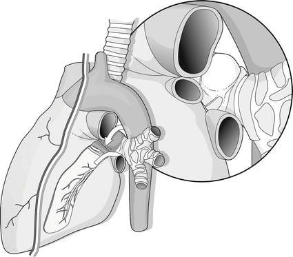
Fig. 7.1
(a) Anterior view. (b) Posterior view. The diagram shows the utilization of a pedicled pericardial patch in the repair of a wide gap in the wall of the right main stem bronchus (From Boaron et al. [3] with permission)
The proximal thoracic trachea is approached by means of a cervicotomy which can be extended using an upper sternal split.
Limited lesions can be stitched or occasionally resected. In the case of blunt trauma or a contused laceration due to hemorrhagic infiltration of the tissues, it may be difficult to identify the structures, and the damage can be overestimated. In these cases, a tracheostomy can be performed on the damaged wall, and the insertion of a Montgomery tube will enable the surgeon to make a more precise evaluation and provide secondary repair. This is even more advisable for critical patients or inexperienced surgeons.
Ruptures of the trachea distal to the thoracic inlet or of the main stem bronchi are approached through a fourth space posterolateral thoracotomy. It is wise to preserve the intercostal or the serratus muscle to be used as a patch. Tracheal lesions are managed as previously described.
The anterior wall of the mediastinal trachea can also be approached by means of a median sternotomy which can be extended to a laparotomy in order to simultaneously treat abdominal lesions and mobilize the omentum. However, managing the carinal region through the posterior pericardium is technically difficult, and furthermore, the posterior wall of the trachea is out of vision [4]. A clamshell incision is indicated where complex endothoracic lesions are suspected.
7.1.3 Bronchi
Severe damage or complete disruption of the main bronchi is not uncommon, mainly on the right side. On the left side, mobilization of the aortic arch is required to reach the proximal tract of the main bronchus. Conservative surgery with 3-0 interrupted sutures and buttressing is usually indicated; pulmonary resection is rarely needed. The intercostal muscle is usually adequate, but a pedicled pericardial flap is best for repairing the bronchial wall or wrapping around a risky suture (Fig. 7.2).


Fig. 7.2
A wide pericardial flap prepared for enforce the left bronchial wall
Patients should be extubated as soon as possible after surgery to avoid both the pressure of the cuff and its ischemic effect on the suture line.
The repair is checked in the postoperative course by fiberoptic bronchoscopies to detect any dehiscence or granulomas.
7.2 Thoracic and Abdominal Esophagus Injuries
Due to the deep mediastinal location of the esophagus, its lesions, in both blunt and penetrating traumas, are rare and regularly associated with other endothoracic lesions which can frequently be lethal. From a clinical point of view, tracheobronchial and esophageal lesions have symptoms and, at imaging, are occasionally superimposed, requiring specific study. When suspected preoperatively, 100 % of the damage is diagnosed by means of esophagoscopy, swallow contrast study, and contrast CT with Gastrografin using a nasogastric tube. Occasionally, in the course of emergency thoracotomy, esophageal damage is detected and can be studied in detail with an intraoperative esophagoscopy.
Stay updated, free articles. Join our Telegram channel

Full access? Get Clinical Tree


