Surgical Robotics

Chapter 42 Surgical Robotics


The videos associated with this chapter are listed in the Video Contents and can be found on the accompanying DVDs and on Expertconsult.com.image


Surgical robotics is a rapidly developing field in which new prototypes are developed on a regular basis by laboratories worldwide. The practical applications have remained relatively few in number. In 2011, surgical robotic devices approved by the U.S. Food and Drug Administration (FDA) and available for hospital use in the United States included the following: (1) the da Vinci Surgical System (approved in 2000); (2) the orthopedic systems ROBODOC and the Mako Rio System (approved in 2008 and 2009, respectively); (3) the CyberKnife radiosurgical system, (approved in 2001); and (4) the cardiac ablation system Sensei (approved in 2009). In addition, a variety of laparoscope holders also have become available. In the near future, however, the array of approved devices should increase. Currently, there are devices under development that expand robotic capabilities for some subspecialties or address the basic limitations of current robotic applications.


Limitations of robotic surgery have included space requirements imposed by the robots, both on the floor of the operating room and in the working space above the operative field. In addition, the generally poor haptic feedback that characterizes contemporary robotic technology has required the surgeon to rely on the interpretation of visual clues when applying force to tissue. Another limitation has been the number of degrees of freedom (DOF), or motion capabilities, that the surgical robot has possessed. As a standard of comparison, the human arm has 7 DOF. Simpler robots with 3 DOF or less are limited to simpler tasks such as camera holding. More complex robots with greater DOF can mimic the human hand and also eliminate the tremor that can accompany hand articulation.


Emerging trends in robotic surgery have included the development of systems with reduced size, improved haptic feedback, expanded operative capability, and greater independence. The last trend may eventually eliminate the need for a surgeon in some cases. Although an exhaustive review of the complex engineering, computer science, and material science required to make these machines is beyond the scope of this chapter, we do discuss some of the newer trends in robotic surgery as they apply to care providers in general and to cardiac, urologic, otolaryngologic, and gynecologic surgery.


General surgery


In the future, the general surgeon may find an operating room robot capable of (1) assisting in procedures that formerly required human assistants, (2) augmenting surgeon capabilities, and (3) performing autonomous procedures. A surgical assist device is defined as a tool that aids a surgeon without offering increased capabilities or augmenting surgical skill. Much as a human assistant does, a surgical assist device frees the surgeon to operate at peak skill level by performing tedious tasks ancillary to the primary procedure. Unlike their human counterparts, robots do not suffer from tremor, fatigue, or inconsistency. A surgeon augmentation device is defined as a tool that enhances the performance of the surgical procedure. Probably the most well-known example of an augmentation device is the da Vinci Surgical System (Intuitive Surgical, Sunnyvale, Calif.). Augmentation devices are designed to provide improved visualization, increased precision, and reduced invasiveness.


CoBRASurge


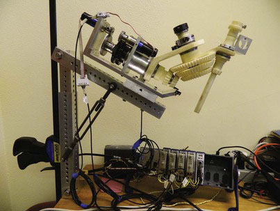
The University of Nebraska Medical Center for Advanced Surgical Technology (our laboratory), in association with the University of Nebraska—Lincoln Department of Mechanical Engineering, has created a robot for surgical assistance. The CoBRASurge (Complex Bevel-geared Robot for Advanced Surgery) is a small device for laparoscope tool movement and placement that consists of a compact table-mounted robot with multiple gears and a joystick for movement and control (Fig. 42-1). It uses a bevel-geared mechanism and a spherical mechanism that allow the tools and laparoscope to center on a specific point in space, namely, the trocar. The device mounts to the operating room table, so it does not require floor space. The robot can control a laparoscopic camera and articulated robotic graspers. In its current application, it is controlled from a joystick across the room. Our plans are to engender surgeon control of the robot with a foot joystick. The compact design of this robot allows increased room around the operating table for personnel and instruments.


Laparoscope Soft Tissue Scanner


The laparoscopic soft tissue scanner developed by Vanderbilt University’s Medical and Electromechanical Design Laboratory permits preoperative scanning of soft tissue for three-dimensional (3D) image generation that would facilitate instrument placement and needle guidance. This technology could be used for biopsy or tumor excision from deep tissue beds with minimal damage to normal tissue. Previous systems with the capability of soft tissue scanning required an open incision to obtain accurate tissue imaging. The small size of the Vanderbilt robot permits surface registration to occur through a single laparoscopic port. The scanner consists of a laser distance device and an optical tracking device and uses conoscopic holography to match scanned tissue point clouds with preoperative 3D computed tomography. This combination allows the surgeon to know organ and tissue placement without requiring substantial soft tissue excavation. It also allows for tissue surface scanning without the need for a large incision or contact with the organ in question, thus eliminating the possibility of contact deformation. Testing with artificial organs (e.g., for liver biopsy) has demonstrated the accuracy of the Vanderbilt robot. This device also may be useful for liver-directed therapy, such as radiofrequency ablation for liver metastasis from colorectal cancer. The Vanderbilt robot furthers the integration of radiologic imaging with virtual operative planning using a minimally invasive approach.


VECTOR Capsule Endoscope


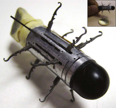
The aim of the VECTOR project (Versatile Endoscopic Capsule for gastrointestinal TumOr Recognition and therapy) is to develop a mobile endoscopic robot with future surgical applications. The project currently includes 19 university laboratory participants in Europe, including the Center for Research in Microengineering (CRIM) in Italy, with input from Vanderbilt University’s MED lab. The original VECTOR prototype consisted of a cylindrical body with a camera and two motorized sets of six legs that unfolded from the body of the robot to interact with the intestinal wall (Fig. 42-2). These legs allowed the robot to “walk” insect-like along a small distance in the intestine. The legs are made of heat-treated nitinol, a flexible nickel-titanium alloy with elastic properties designed to reduce the risk for perforation. The robot was engineered to permit passage through a deflated colon in order to reduce one of the main sources of pain from gastrointestinal imaging procedures (i.e., an insufflated colon). This ability is partially facilitated by the robot’s unique leg design. The 12 legs are capable of independent motion, which can aid in tight turns, such as the splenic flexure. The robot has been successfully tested in a porcine model. The VECTOR robot may represent an improvement on current swallowable endoscopes because of its movement control and possible future augmentation with accessories, such as for biopsy.


As part of the VECTOR project, the CRIM laboratory has developed other prototype WCEs with the ability to steer and even deploy hemostatic clips. One of their WCEs is equipped with internal permanent magnets that can be steered externally by a magnet on an industrial robotic arm. Although this device also can be controlled manually, the researchers found that robotic control offered greater precision, albeit at the expense of longer procedure times. The WCE has been shown experimentally to be capable of maneuvering in the porcine colon, including against colonic peristalsis. The current prototype measures 14 × 38 mm (too large to swallow), but the laboratory expects the next generation to be miniaturized sufficiently to allow a patient to ingest. Unlike the VECTOR, the magnet-steered device does require some degree of insufflation, but nevertheless, the latter has shown promise as a practical steered WCE device. A similar device also has been developed that can deploy a nitinol clip to a bleeding site; this has been tested in the porcine intestine.


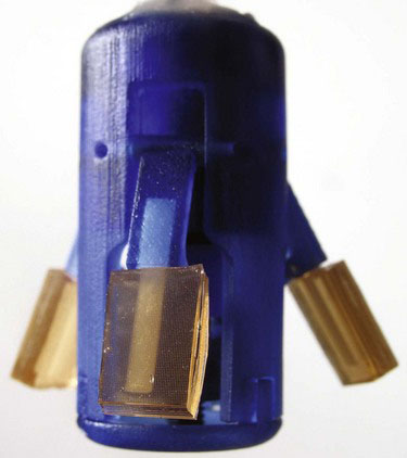
Other steering mechanisms under development at the CRIM include a swallowable capsule that can navigate the fluid-distended stomach through the use of four propellers. All four propellers are contained at the “rear” of the capsule in order to minimize the risk to the esophagus during swallowing. Simultaneous activation of two ipsilateral propellers is the basis of the steering mechanism. The capsule has been tested successfully in the porcine stomach. The novelty of the propeller-driven “submarine” device relates back to the fact that a typical camera capsule cannot fully investigate gastric disease before the capsule passes through the pylorus. Current capsule cameras cannot be steered to areas of interest in the stomach. The propeller-driven device eventually may provide a workable solution to gastric capsule endoscopy.


SILS and NOTES Applications


Efforts to accomplish single-incision laparoscopic surgery (SILS), laparoendoscopic single-site surgery (LESS), and natural orifice transluminal endoscopic surgery (NOTES) have presented new challenges to the surgeon. Restricting surgical instruments to one incision, whether in the abdominal or intestinal wall, limits triangulation and visualization. In addition, a NOTES procedure requires flexibility to snake through a natural lumen, but then stiffness to provide an operative platform once the target site is reached. The operating instrument remains bound by the fulcrum effect of one insertion site. Articulating instruments can help, but current iterations allow only modest and relatively stiff articulation; new and improved instrumentation is necessary in this discipline. The robots described later in this chapter attempt to address these needs and to facilitate the performance of single-incision minimally invasive surgical procedures.


ARES (Assembling Reconfigurable Endoluminal Surgical System)


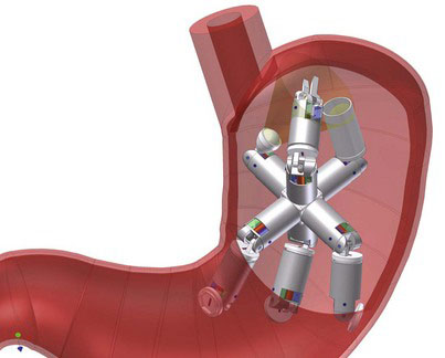
The ARES robot (Fig. 42-4), developed by a consortium of European universities and laboratories, was designed for transgastric surgical applications and represents an attempt to address some of the limitations of endoscopic or natural orifice procedures. The robot consists of multiple modular units molded from an acrylic photopolymer. The current module size is 36.5 mm in length and 15.4 mm in diameter, or about the size of a current “pill cam.” These dimensions are designed to allow the patient to swallow the modules. Once in the stomach, the robot undergoes magnetic self-assembly. The researchers have demonstrated a 74% success rate for self-assembly in a model stomach. The robot’s placement potentially could be tracked by ultrasonic emissions from one of the modules. Power would be supplied by an onboard battery or with electromagnetic induction. Currently, the design requires a number of modifications to be usable in human surgery.

< div class='tao-gold-member'>

Stay updated, free articles. Join our Telegram channel

Jul 20, 2016 | Posted by in GASTOINESTINAL SURGERY | Comments Off on Surgical Robotics

Full access? Get Clinical Tree

Get Clinical Tree app for offline access