Fig. 7.1
Calcium oxalate dihydrate stone (Courtesy of Louis C. Herring & Co., Orlando, FL)
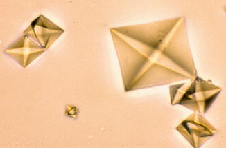
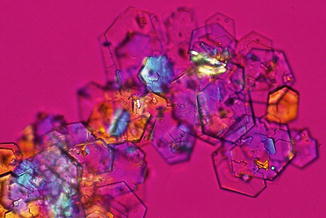
Fig. 7.2
Urinary calcium oxalate dihydrate crystals under polarized light microscopy (M. Daudon, CRISTAL Laboratory)
Calcium Oxalate Monohydrate
These stones are also called Whewellite. These stones are usually knobby shaped and have few to no spiked crystals on their surface (Fig. 7.3). These stones are typically dark brown in color. Similar to diamonds, these stones are very tough and do not break up with lithotripsy. Calcium oxalate monohydrate crystalluria vary in size and may have a spindle, oval or dumbbell shape (Fig. 7.4).
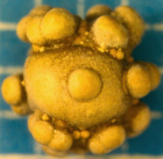
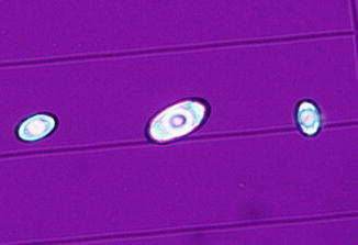
Fig. 7.3
Calcium oxalate monohydrate stone (Courtesy of Louis C. Herring & Co., Orlando, FL)
Fig. 7.4
Urinary calcium oxalate monohydrate crystals under polarized light microscopy (M. Daudon, CRISTAL Laboratory)
Calcium Phosphate
These stones are also known as Hydroxyapatite. These stones are usually smooth and round (Fig. 7.5). The urine crystals of these stones are amorphous shaped found on microscopic examination (Fig. 7.6).
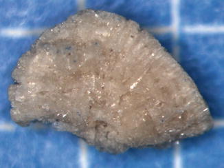
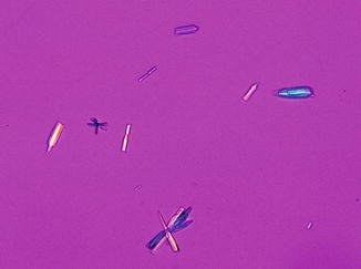
Fig. 7.5
Calcium phosphate stone (Courtesy of Louis C. Herring & Co., Orlando, FL)
Fig. 7.6
Urinary calcium phosphate crystals under polarized light microscopy (M. Daudon, CRISTAL Laboratory)
Uric Acid
These stones are usually orange-colored (Fig. 7.7). The urine crystals of these stones are diamond or rhomboid shaped discovered on microscopic examination (Fig. 7.8).
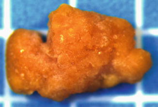
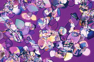
Fig. 7.7
Uric acid stone (Courtesy of Louis C. Herring & Co., Orlando, FL)
Fig. 7.8
Urinary uric acid crystals under polarized light microscopy (M. Daudon, CRISTAL Laboratory)
Struvite
These stones are also known as magnesium ammonium phosphate or infection stones. These stones can become very large, called staghorn calculi. These stones are tan in color (Fig. 7.9). The urine crystals of these stones are coffin-lid shaped identified on microscopic examination (Fig. 7.10).
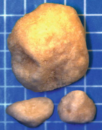
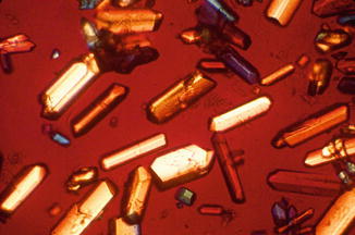
Fig. 7.9
Struvite stone (Courtesy of Louis C. Herring & Co., Orlando, FL)
Fig. 7.10
Urinary struvite crystals under polarized light microscopy (M. Daudon, CRISTAL Laboratory)
Cystine
These stone are amber in color (Fig. 7.11). The urine crystals of these stones are hexagon “stop sign” shaped seen on microscopic examination (Fig. 7.12).
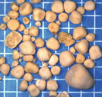
Fig. 7.11
Cystine stone (Courtesy of Louis C. Herring & Co., Orlando, FL)